SlideShare ist ein Scribd-Unternehmen logo
1 von 36
POWER RANGERNOTES ANATOMYOF FINFISH
1
ANATOMY OF FINFISH
1. Anatomy of FinFish
1.1 External Anatomy ofFinfish
Anatomically (Biologically) speaking a fish is composed of
ten systems of bodily organs that work together to make up
the whole individual. These ten systems cover the fish,
handle its food, carry away wastes,they integrate the life
processes of the fish and relate it to conditions in the
environment. They also provide for breathing and for
protections against injury. They support the body and
enable movement and finally they work to perpetuate fish
as species and through evaluation, as a major group of
animals.
The living species of fish are usually divided into three
classes: the Agnatha, the jawless fishes, comprising the
hagfishes and lampreys; the Chondrichthyes, the
cartilaginous-skeleton fishes, such as sharks and rays; and
the Osteichthyes, the bony-skeleton fishes, comprising all other living fishes. The skeletons of these three groups vary in
fundamental ways. In the hagfishes and lampreys the backbone is basically a notochord, a rod like structure composed of
unique notochordal tissue. In sharks and rays the notochord is surrounded and constructed by spaced rings of cartilage, the
vertebrae, to form a backbone.
1.2. External Anatomy ofFishes
1.2.1. Body forms
Commonly the fish body is Torpedo – shaped (fuci form) and often slightly to strongly avoid in cross section. In a perfectly
“stream lined” body form (if head pointed trunk broadened and gradually tapering towards the tail) the greatest cross
section is about 36% of the length back from the anterior tip and gently sweep back to the tailed.
However,all fishes do not have this typical body form. The shape of the body is variously modified depending on their
habitat and made of living. There changes in shape are Globe shapes (Globiform – pufters of family Tetraodontidae)
Serpentine (Snake like Angulli form – eels of family Anguillidae) Thread like in form (filiform – snipe eel Nemichthyidae).
Some are strongly flattend from side to life (compressed butterfly fishes, chactodentidae and flounders – pleuronectidae)
others flattened but greatly elongated trachipteriform – ribbon fishes, Trachipteridae) and till others flattoned form top to
bottom/ depressed the skates and rays (Rajidae).
In spite of the many variations the ground plan of body organization in fishes is bilaterally symmetry. The left and right
halves of the body are basically mirror images of one another. The tail is an integral part of the body rather than an
appendage only the fins are distinctly peripheral anatomical parts in most fishes.
1.2.2. Body Covering
Fish is generally covered by a tough skin. It is continuous with the lining of all the body of the entire body opening and is
transparent as it runs over the surface of the eye. Much of the diverse coloration of fishes is due to its colour cells and the
slimy coating is due to its mucus cells.
The skin in many fishes is devoid of scales,but in them it is armored by scales that develop in it. Scales range in size from
microscopic to large. In thickness from tissue thin to very thick (in ornamentation from simple to complex in extent of body
coverage from portal to complete). In structure from non-bony to bony loosely attached to very firmly attached. The types
of scales may characterize the major fish groups. Scale morphology and the number of scales rows along or around the
POWER RANGERNOTES ANATOMYOF FINFISH
2
body frequently serve as specific and genetic characters. Living Agnathaus are scale less, shark and their relatives have
dentinal placoid scales,Bony fishes have various types of bony scales.
1.3. Internal Anatomy of finfish
Internal anatomy of a bony fish: finned aquatic vertebrates animal with skin covered with scales. It lives in water
and is usually oviparous.
Brain: seat of the mental faculties of a fish.
Esophagus: part of the digestive tract connecting the
mouth to the stomach.
Dorsal aorta: vessel in the back that carries blood
from the heart to the organs.
Stomach: part of the digestive tract between the
esophagus and the intestine.
Air bladder: pocket in which urine collects.
Spinal cord: part of the nervous system that connects
the brain to all other parts of a fish.
Kidney: blood-purifying organ.
Urinary orifice: opening for eliminating urine.
Genital Orifice: opening related to the genital organs.
Anus: end of the digestive tract.
Gonad: hormone-secreting sexual gland of a fish.
Intestine: last part of the digestive tract.
Pyloric cecum: cul-de-sac related to the intestine.
Gall bladder: small sac containing the bile.
Liver: bile-producing digestive gland.
Heart: blood-pumping organ.
Gills: respiratory organ of a fish.
Tooth: hard organ of a fish used to shred food.
Eye: sight organ of a fish.
Olfactory bulb: bulging part of the smell organ of smell of a fish
1.4. Structure of fins
Appendages of fishes comprise of fins and cirrti (flaps of flesh) which attain extreme development in the
sargassum fish (pterophryne) and the leafy sea dragan (phyllopteryx). The fins are classified as median or paired.
Median fins (Unpaired fins)
Rayed fins in line with the median axis of a typical fish are these of the back (dorsal fin or fins) the tail (caudal
fin) and the lower edge of the body just behind the vent (Anil fin) Although most commonly all of the median
fins may be present, but some may be modified or absent in same fishes.
Also developed in the median axis may be a rayless fatty adipose fin (as in the trouts – Salmonidae) off fins
reduced to a few disconnected spines as in the stickle backs – Gasterosteidae) The anal fin may be modified into
an intermittent organ, the gonopodium for use in copulation (as in the live bearers – Poecilidae).
POWER RANGERNOTES ANATOMYOF FINFISH
3
Paired fins
Paired fins are the Pectorals and Peluics (ventral) the pectorals are
supported by the pectoral girdle that joins the skull. Although ordinarily
present in fishes, the pectorals are wanting in such kinds as the lamprey
(Petromyzonidae) and hag fishes (Myxinidae) They are greatly enlarged in
the (Soaring) flying fishes (Exocoetidae) and flying characins
(Gastropelecinae). In some cling fishes (Gobiesocidae) and Asiatic suckerbelly loaches (Gastromyzan) the
pectorals serve as a part of the ventral hold fast organ.
The pelvic fins vary substantially in position and in adoptive modification typically their support is by a pelvic
girdle anchored in the bully musculature.
In soft rayed fishes (Malacopterygians) the peluics are abdominal in position, Eg. Clupeoids, Salmonids,
cypriroids, but they also be situated arterially, just below the pectorals in a thoracic position (as in many spiny
rayed species, the Acanthopterygians) or even under the throat in a jugular position (as in blennies). The pelvic
are lacking in the lampreys and hag fishes and is lost in various other fishes, rlotably the cels (Angullidae etc) In
the sharks and some relatives they are modified into claspers. In cling fishes (Gobiorocidae) sucker belly loaches
(Gastromyzon) and certain other fishes, the pelvic from part of a hold fast organ that resembles a ruction cup on
the belly.
1.5. Skin and Scales
The skin of a fish consists of two layers. The outer layer is epidermis and the inner, the dermis or
corium. The epidermis is composed superficially of several layers of flattened, moist epithelial cells.
The deepest layers are a zone of active cell growth and multiplication. Here cell multiplication goes on
all the time to replace from with in the outermost layers of cells as it is worm off and provide for growth.
These epithelial cells from the epidermis are the first to close a surface wound.
The dermal layer of the skin contains blood vessels nerves and cutaneous sense organs and
connective tissue. The dermis plays the main role in the formation of sales and related integumantory
structure.
Scattered among the flattened cells of the epidermis are numerous openings of the tubular and flask
shaped mucous gland cells that extent into the dermis.
These cells secrete the slippery mucus that cover most fishes. The mucus lessons the drag an a fish
when it swims through water.
As in other vertebrates, the skin of a fish is the envelope for the body and is the first lines of defense
against disease. It also affords protection from, and adjustment to environmental factors that influence
life, for it contains sensory receptors tunes to the surroundings of a fish. Further more, the skin has
respiratory, excretory and osmoregulatory functions.
POWER RANGERNOTES ANATOMYOF FINFISH
4
1.6. Scalation
Outstanding among the special feature of the skin are the very prominent scales that most fishes have.
However some do not have scales. Examples include the lampreys (petromyzonidae) and a number of
cat fishes North America (F.W) (Ietaluridae). An intermediate structural category have seals only on a
few palces of the body for example: Paddle fish (polydon) three spines stickle bask Gasterosteus
aculeatus and mirror carp. Then there are a few fishes that have very small deeply imbedded scales
that they look scale less for example Fresh Water Eels (Anguilla) Brook trauts (Salvelius fontinalis) and
the barbot (lota) of Cod family (F.W) (Gadidae)
In arrangements scales are most often “imbricated” and thus overlap like roof tiles with free margin
directed towards the tail in a manner that minimize friction with water. In some fishes such as barbot
(lota) and F.W eels (Anguilla) the pattern is mosaic rather than overlapping one another, the scales are
minutely separated or meet their neighbors only at their margins
Scales shapes
Although they comprise only a few basic structural types, Scales exhibit many modifications that are
often characteristic of groups or species on the basis of shape one type is plate like (Placoid), with
each plate carrying a small cusp, as common among the sharks (Elasmobranchil) a Second type is
diamond shaped (rhombic) and found in gars (Lepirosteidae) and the bichirs (pdypterus). A third type
of scale is called Cycloid because it is typically smooth disc like and more or less circular mostly found
in soft rayed fishes. (Malacopterygi). In fourth type is ctenoid the posterior surface or margin is toothed
or comb like and a characteristic of spiny rayed bony fishes (Acunthopterygil)
However, some of the soft rayed fishes have ctenoid like contact organs on their scaes, examples
characins (characid) Killi fishes (cyprinodontidac). Some spiny rayed fishes have cepeloid scales
exclusively. For example the brooke silverside labidestus. Many spiny rayed species exhibit both
cycloid and ctenoid scales.
Example: Common basses (Micropterus).
Structural types
Structurally, there are two types of fish scales, placoid and Non-placoid. Non-placoid scales are
basically of the three kinds – cosmoid, Ganoid and Bony ridged scales.
I) Placoid scale
Placoid scales also called dermal denticles have an ectodermal cap or covering of enamel like
substances (as an human teeth) termed “Vitrodentine” beneath this is a thicker layer of entine with a
pulp cavity and dentinal tubules emanating form it. Each scale has a disc like basal plate in the dermis
with a cusp projecting outward from it though the epidermis. Placoid scales occur among the sharks
POWER RANGERNOTES ANATOMYOF FINFISH
5
and their relatives (chandrichthyes).
II) Non – Placoid scales
1) Cosmoid scales: The cosmoid scales have a thinner, harder outer layer than the placoid ones. It is
also termed as vitrodentine. The layer below this hard and non cellular and is called cosmine. The
layer beneath is vascularised mid layer of perforated bony substances called “Iropedine” The growth of
the scale is at the edge and beneath no ligivng cell layer covers the scale. The cosmoid scales found
in (latimeria).
2) Ganoid Scale: In ganoid scale the outer layer is a hard inorganic substances called ganoine which
is different from Vitrodentine. The layer below the ganoine is a cosmine like layer. The innermost layer
is a bony layer called isopedine. The growth takes place at the edge, beneath and on the surface.
Three types of scales are found in bichirs (pdypterus) and gars (Lepisosteidae).
3) Bony Ridges Scales: Bony –ridged scales are typically thin and transclucent lacking both dense
enameled and dentinal layers of the three other kinds. Bony ridged scales characterizes the many
living species of bony fishes (Osteichthyes) that have either cycloid or ctenoid scales. The outer layer
of there scales is marked with bony ridges alternate with depression. The inner part or plate of the
scale is made up of layers of criss – crossing fibrous connective tissues. The growth of the scales is
both on the outer surface and from beneath.
For ridges on the scale two terms are used ridges and circuli changes in the growth pattern of the
individuals fish is reflected in the character and distribution of ridges. “Breeding” and “Yearmarks” can
be identified on this basis in many species. In both cycloid and ctenoid scales, a nuclear central zone
can be recognized. This zone may be called as “focus” the scale. However this central position shifts
due to differential growth of fore or after part of the scale, shifting the focus to more posterior and
anterior margin. In many species grooves or radii radiate from or near focus towards one or more
margins of the scale. The ctenoid scales have teeth on its posterior margins.
2.Cellstructure, tissue and body organization
2.1. Organization: kind of construction of the body
A cellular or protoplasmic level of organization: Body is not differentiated into cells. Example: Protozoa
(subkingdom: Protozoa); All other animals except Protozoa come under the subkingdom: Metazoa (Body is
composed of numerous cells)
Cellular level of organization: The cells of the body are more or less loose and Independent. Example: Amoeba
Tissue level of organization: In some animals, body cells form tissues. Example: Coelenterata and Ctenophora
Organ system level of organization: In most of the animals, tissues combine into organs, which, in turn,
combine to form organ systems. Example: Animals like Human being, Fish, Reptiles etc.,
Germ Layers:
The fundamental cell layers laid down in an early embryo of the Metazoa are called germ layers. The
Coelenterata have only two layers: Ectoderm and Endoderm. They are termed the diploblastic animals. Most
other Metazoa possess three germ layers: Ectoderm, Mesoderm and Endoderm. They are known as the
triploblastic animals. Example; Animals except Coelenterata
2.2. Symmetry
It referstothe similarityin size,shape andnumberof partsonthe opposite sidesof amedianline.Partsof ananimal
bodyare oftensoarrangedthat it ispossible tocutit intotwosimilarhalvesbyone or more planes.Suchanimalsare said
to be symmetrical.Some animals,likeAmoebaandSnail,cannotbe dividedintoequal partsbyanyplane.Theyare saidto
be asymmetrical.The symmetricalanimalsshow one of the three typesof symmetry:spherical,radial andbilateral.
POWER RANGERNOTES ANATOMYOF FINFISH
6
Section– 1 Spherical (universal) symmetry:
Bodyis divisible intosimilarhalvesbyanyplane passingthroughthe center.Example:Seaurchin
Section– 2 Radial symmetry:
Bodyis witha numberof similarpartsradiatingoutfroma central axis.The boycan be splitupintoequal halvesbyany
plane passingthroughthe middle fromtoptobottom.The animalswithradial symmetryare calledRadiata.Examples:
Spongesandmostcoelenterates.
In some animals,some partsof the bodyare single orpairedratherthanradial,so that onlyone or twoplanesthrough
the longitudinalaxisdivide the animalintosimilarhalves.Suchasymmetryiscalledbiradial.Examples:Comb-jellies,
seanemonesandseastars.
Section–3 Bilateral symmetry:
In restof the Metazoa,chief organsof the bodyare pairedandare arrangedon the sidesof a central axisconnectingthe
headwiththe tail.Withthe result,the bodycan be dividedintotwosimilarhalvesonlybyone plane.Suchare termedas
bilateral.The bilaterallysymmetrical animalsare termedthe bilateria.
A plane orsectionpassingdorso-ventrally(vertically)throughthe middleof the anterior-posterior(longitudinal) axisis
knownas the Sagittal plane orsection.Thisisthe plane thatdividesthe bilateral animal intosimilarandrightand left
halves.
A plane orsectionrunningantero-posteriorlyatrightanglestothe sagittal plane iscalledthe frontal plane orsection.It
dividesthe bodyintodorsal andventral halves.
A plane orsectionpassingdorso-ventrallyatrightanglestothe antero-posterioraxisisknownastransverse plane or
section.Itdividesthe bodyintoanteriorandposteriorhalves.
Section– 4 Digestive tract:
The Mesozoa andparazoa lack a digestive tract.The Eumetazoapossessadigestive tract,andare termedthe Enterozoa.
Certainenterozoa,viz.,coelenterata,CtenophoraandPlatyhelminthes,have incompletedigestive tract,whichhasasingle
external opening,the mouth,thatservesforintake of foodaswell aseliminationof faeces.Allotherenterozoahave
complete digestivetract,whichhastwoexternal openings,the mouthforintake of foodandthe anusfor eliminationof
faeces.
Section– 5 Coelom:
Some bilateriahave nocavityintheirbody,exceptthatinthe digestivetract.Suchformsare knownas the acoelomates.
All others possessacavityinadditiontothe one in the digestive tract.The space betweenthe twocavitiesiscalledthe
bodycavitythe nature andoriginof thiscavity variesindifferentgroups.Whenlinedbyamesodermal epithelium,the
peritoneum, itisknown asthe true coelom,andwhenunlinedbyperitoneum, itistermedfalsecoelomorpseudocoel.
The animalswithfalse andtrue coelomare respectivelycalledthe pseudocoelomatesendeucoelomates.InMolluscaand
Arthropodathe coelomisgreatlyreduced,andthe bloodfillsthe spacesbetweenthe internal organs.Suchabody-cavity
iscalledthe haemocoel.The coelomisof greatsignificance inanimal evolution.Itprovidesspace forthe developmentof
variousorgansystems.
Section– 6 Metamerismor Segmentation:
Segmentsoccurinthree phyla:Annelida,ArthropodaandChordata.Eachsegmentiscalledametamere orsomite.
Metamerismaffectsexternal andinternal structures.Itissaidto be homonomous,if the somitesare similar,asin
earthworm(annelida);andheteronomous,isthe segmentsare dissimilarasinprawn (Arthropoda).Thesegmentationin
Chordatesshowsupclearlyinthe embryonicstage inte segmentallyarrangedmesodermalsomites.Inadultchordates
segmentationislargelyinternal andisseenintheirvertebrae,ribs,nervesandbloodvessels.
3. Oral region and Associated structures
POWER RANGERNOTES ANATOMYOF FINFISH
7
3.1. Adaptations for feeding
(The feedinghabitsorfeedingbehaviorof fishesisthe searchforand ingestingof food) The diversityinfeedinghabits
that fishesexhibitisthe resultof evaluationleadingtostructural adaptationforgettingfoodfromthe environments.The
structural adaptationof feedingorganssuchas 1) positionandshape of the mouth2) Presence orabsence of “Teeth”.3)
Gap of the mouthetc.,greatlyhelpinpredictingthe nature of foodandmode of feedingof the fish(inquestion).
Section1: Major feedingtypes:
On the basis,fishescanbe classified,accordingtotheirfeedinghabitsas1) Predators2) Grazers 3) Foodstrainers4) Food
suckersand 5) Parasites.
1) Predators
Fishesthatfeedonmacroscopicanimals,theyusuallyhave welldevelopedgraspingandholdingteethasinmanysharks
(Elasmobrabch) the Barracuds(Sphyraena),the pikes(Esox) gars(Lepisosteus).
2) Grazers
In Grazing,the foodis takenbybitesor continual browsing(Grazingcharacterizemanyfishesthatfeedonplanktonsoron
bottomorganisms) (sometimesorganismsare takensingle oratother timesinsmall groups) mayfeedonbottomor
column – a bluegill (Lepomismacrochirus),Parrotfishes(scaridae) Butterflyfishes(chaetodontidae) browsingoncoral
reef.
3) Strainers(Filterfeeders)
Filterthe waterforplankton,here foodsare selectedbysize andnotby kind.Herringclupidae,Gizard,shads(Dorosoma),
Paddle fish(polyodon) whaleshark(Rhincodon) alsohave efficientfoodstrainingorfilteringadaptation.The principal
adaptationforfilterfeedingorstrainersisthe developemtof numerous,closely –setand elongatedgillrackers.
4) Suckers
The suckingintothe mouth of foodor foodcontainingmaterial istakenpracticedbybottomfeedingfishessuchasthe
sturgeons(Acipenseridae) andsuckers(catostomidae)
5) Parasites
Parasitisumisperhapsthe mostunusual andhighlyevolvedfeedinghabitsamonganimals.Theysuckbodyfluidafter
raspinga hole inthe sidesof the body.Parasiticlampreys(Petromyzonidae),Sealamprays(Petromyzonmarinus)Pacific
lampreys(Lamptratridentata)
3.3. Oral adaptation for feeding
Lips
Suctorial feeders have an “inferior mouth” and fleshy modification of lips. Notable among these are the
sturgeons (Acipenseridac) and suckeres (catastomidae). The lips of strurgeons and suckers are mobile and
described as “Plicate” (having folds) or papillose (having small tufts of skin or papillose). Many sectorial feeders
also have well developed barbells bordering the mouth.
Sucker mouth : Moxostoma (Redhorse) suctorial lips of free – living fishes may also serve as holdfast organs in
fast flowing mountain streams. For example (Glyptorternum). In loach – like Gyriocheilid (Gyrinocheilus) has an
extreamly specialized suctorial adaptation, in addition to suctorial lips the opercular structure has separate
inhalent and exhalent device for respiration.
3.3.1. Modificationsin the shape of mouth
Amongthe grazersand suctorial feeders,thereexistnotonlyspeciallydevelopedlipsbutalsoadaptationsof othermouth
parts. The Trumpetfishes(Aulostomidae) the cornetfishes(Fistularidae)andthe pipe fishes(syngnanthidae) aswell as
manybutterflyfishes(chaetodontidae) of coral reefs,have mouthsthatresemble elongatedbeaks.Thisadaptationis
achievedbya protractionof the hyomandibularbone ratherthanbya lengtheningof the lowerjaw bones(denteries)
themselves.
POWER RANGERNOTES ANATOMYOF FINFISH
8
Amongthese fishes,the methodof feedingmaybe bysuctionasin the case of trumpetfishes,cornetfishesandpipe
fishesoritmay be a selectivegrazingactionwithsharpteethwhenthe longsnoutenablesthe butterflyfishtoreachinto
small crevicesof the coral.
A peculiarstructure amongmouthmodificationhasariseninthe half beaks(Hemiramphidae)where the lowerjaw
projectsintoa beakoftena thirdof the lengthof the fishitself,withthe mouthopeningabove it.Half beaksare usually
surface feedingfishes.
3.3.2. Teeth
Outstandingamongthe obviousoral adaptationforfeedinginfishesare the teeth.Theyare thoughttohave arisenfrom
scalescoveringthe lips,asrepresentedinlivingsharks(squliformies) where the placoidscalesof the skinvisiblygrade
intoteethonthe jaws.
In bonyfishes(Osteichthyes) Teethare of three kinds,basedonwhere theyare foundJaw,Mouthand Pharyngeal.
Jaw Teeth
Jaw teethare variouslythose onthe maxillaryandpremaxillarybonesabove andonthe dentariesbelow.
In the roof of the oral cavityteethare variouslyborne bythe medianvomerandbythe palatine andectopterygoidbones
on eachside.Inthe floorof the mouththe tongue oftenhasteethonit. Pharyngeal teethoccuraspads on variousgill
arch eolementsinmanyspecies.Inthe carps(cyprinedae)andsuckers(cjcatastromidae) the onlyteethare those indeep
inthe pharynx (gut,mouthandeoesophagous) thatdeveloedmodificationof lowerelementsafterlastgill arch,inclarius
and labeothe teethare modifiedforgrasping,tearing,grindingandrazorlike cuttingteethhave developedinpredacious
fishes.
Kinds ofjaw teeth
Basedon theirformmajorkindsof Jaw teethare
1) Cardiform2) Villiform3) Canine 4) Incisorand5) Molariform.
Cardiform Teeth
Cardiformteethare numerous,shrotfine andpointedsuchdentitionwithvariationsisfoundinmanyfishesthathave
multiple rowedteeth.Forexample Americancatfish(Ictaluridae)perches(percidae) andmanyseabases(serranidae)
Villiformteeth
Villiformteethare more orlesselongatedcardiformteeth.Forexample:Needlefishes(Belonidae) andLionfishesas
(Pterois).
Canine teeth
Caninesare dogtooth like orfange like (longpointedtooth) theyare elongatedandsubconical,straightorcurvedandare
adaptedforpiercingandhodlingforexample walleyes(Alskapollock) (Stizostediosn).Incertainfishessuchasmoreys
(muraenidae) the caninesare hinged(thehook) yieldotbackwardpressure butlockwhenmovedforwardandadaptation
to retainlivingmovingpreyinsidethe mouth.
Incisors teeth
Incisorsare sharp edgedcuttingteeth.Insome fishesincisorsfuse togetherincutting beakasin parrot fishes(scaridae).
Molariformteeth
Molariformteethare forcrushingand grindingthusflatwithprodrudingdenticlesonthe surface.These teethare found
inbottomdwellingfisheslike skatesandraysand some sciaenidae (drums).
Within a single groupthe diversityof dentitiononidentical bonesmayvarylargely.Forexample Incarpsand minnows
(cyprinidae)the pharyngealteethrange fromsharpincarnivoressuchas insemotilustomolariforminthe commoncarp
(Cyprinuscarpio).
In general teethare absentinplanktonfeedersandinsome of the more generalizedomnivore.Theyare presentin
increasingnumbersof bonesinmore andmore relative toitspredatorybehaviors.The premaxillarybonesare toothed
whenjawboneshave little ornoteeth.Thisistrue for manysoftrayedspeciessuchas bowfin(Amia) the gars
(lepisosteus) the salmonsandtrouts(Salmonidae).
POWER RANGERNOTES ANATOMYOF FINFISH
9
The maxillae are typicallytoothedinthose softrayedfishesasthatcarry premaxillaryteeth.Howevertoothlessin
otherwise tooth –bearingspinyrayedfishes.
3.4. Gill Rakers
Besides protecting the tender gill filaments form abrasion (a rubbing off or scrap) by ingested materially that are
coarse in texture, gill rakers are specialization in relation to food and feeding habits. They are very stubby (short
and thick) and unornamented in most omnivores for example sunfish (Lepomis cynellus).
In many plankton feeders, the gill rakers are elongated, numerous are variously lamellate or ornamented to
increase efficiency of filtering. Simple but very numerous rakers are possessed by gizzard shads (Dorosoma) and
paddlefish (polyodon) Ornamentation of gill rackers are of taxonomic value. In pleuronectdae the gill rakers
resembles feather having a main axis with lateral processes these lateral processes are branched. The adjacent gill
rakers overlap to form a very fine sieve.
4. Gastro Intestinal Tract OR Digestive system and associated digestive glands
4.1. Gastro Intestinal Tract
Esophagus
Esophagus forms the beginning of the gastro intestinal tract/ In
general the oesophagus is no distensible that it can
accommodate anything the fish can get into the mouth and
sometimes even accommodate the item if it happens to double
on itself two or three times, on its way to the stomesh. The
ocsgtagus is a short and narrow to be in a no. of Herbivorus and
omnivorus fishes. (Cyprinus corpio, L.rohito,Tor tor etc. A
large no. of mucus – secreting cells are scattered in the mucosa
at taste buds are also present in some or species.
4.1.2. Stomach
Frequently in literature the fishes are classified as stomach or
stomach less fishes. 85% telecast have stomach 15% had no stomach.
The stomach too shows various adaptations one of which is shape. In carnivore the stomach typically is straight,
elongate for example in gars (lepisosteus), bowfins (Amia), Pikes (Esox) and barracudas (sphyraena).
In omnivorous species, the stomach is most often sac-shaped. A very special adaptation is the modification of the
stomach into a grinding organ as in the sturgeons (Acipenser) gizzard shads (Dorosoma) and mullets (Mugil).
Here the stomach is reduced in overall size but its wall greatly thickened and muscularized. The lining too is
heavily strengthened with connective tissue and the lumen is very small. The organ is not for storage, mixing and
primary digestion but rather a food grinder.
Great dispensability is the adaptation of the stomach in the predatory deep sea swallowers (saccopharyngidae)
and gulpers (Eurypharyngidae) enabling these fishes to take relatively huge prey.
A remarkable modification of the stomach exists in the puffers (Tetradontidae) which can inflate themselves with
water or air to assume often on almost globular shape. The adaptive value of this modification of the digestive
tract is probably mainly one of defense, for many puffers and porcupine fishes have spines all over the body
which can thus be created.
Not all fishes have a stomach that is a portion of digestive tube with a typically acid secretion and a distinctive
epithelial lining different from that of the intestine.
In most of the herbivores and phytoplankton feeders the epithelial tissue of the esophagus grades directly into
that of the intestine, thus termed stomach less. Though the primary criterian for being without stomach does not
seem to be whether a fish is an herbivore or a carnivore but whether accessory adaptations for trituration and fine
grinding of food exist either in the form of teeth or a grinding apparatus such as a gizzard. Where stomach exist
POWER RANGERNOTES ANATOMYOF FINFISH
10
most pronouncedly (strongly) in carnivores. They are characterized by a low pH and the prominent presence of
pepsin (enzyme) among other digestive juices.
4.1.3. Intestine
The intestine too has many variations. It is shortened in carnivores such as in the pike (Erox), perhaps because
meaty foods can be digested easily. Where as in herbivores the intestine is highly elongated and several times the
body length of the fish and in carps and certain catfishes. In sharks and other elasmobrachs the intestine has a
coiled layer of absorptive tissue called spiral valve which increase absorptive surfaces for the relatively in short
intestine.
4.1.4. Pyloric caeca
(Fishesare onlyvertebratesthathave appendages(Caeca) atthe gastro-intestinaljunctions). Onthe intestine of most
bonyfishesatthe pyloricendof the stomach.There maybe fromone to manyblindsacs or pyloniccaecaor intestinal
caeca. A fewgroupssuchas cat fishes(Ictaluridae) topminnows(Cyprinidae) andpikes(Esocidae)lackthese structures.
In suchgroups as flatfishes(Pleuronectiformies)the pyloriccaecaare few usuallynotmore thanfive (5).Inotherssuchas
Mackerels(Scombridae),Salmons(Salmonidae)the numberof these caecamayrange to 200 or more.Generallycaecaof
differentspeciesvaryconsiderablyinsize,state of branchingandthe connectionwiththe gut.Insturgeons
(Acipenseridae) the manycaecaformalarge mass,but onlya single ductleadstothe intestine.Insalmon,eachcaecum
communicatesdirectlywiththe gut,the functionsof pyloriccaecaprobablyinvolve bothdigestionandabsorption.
4.1.5. Rectum
Morphologicallythere isnodistinctionbetweenthe rectumadintestine,the ilio-rectal valveispresentinsciana,tetradon
and muracnosox.
4.1.6. Anus
It isthe posterioropeningof the alimentarycanal ordigestive system.The internalsurface of the regionnearthe rectum
iscoveredwithan epitheliumrichmucouscells.The anusismade upof an innercircularand outerlongitudinal muscle
layer.The circularmuscle isthickdevelopedformingsphincter.The regionof anusfacingthe anteriorhasthe epithelium
continueswiththe skin.
4.2.1. Associated digestive glands
Liver
Like othervertebrates,liverisanimportantorganinfisheswhichhasboth“secretary”and “storage”function.Thisisa
large glandinall fishes,butsharksandraysmay have extremelylarge liverscomprisingabout20% of the bodyweight
especiallyinsome pelagicsharks.The liverusuallyliesoverorpartiallysurroundsthe stomach.Itistypicallybilobed,but
may have onlyone lobe asinsalmonor three as inmackerel.InHag fishesthe liverisintwodistinctparts,withseparate
ducts leadingtothe gallbladder.Adultlampreyhave nobile ductsorgall bladder,gall bladder isalsoabsentinburbot
(lota) butinmost otherfishesthe gall bladderispresentandfunctiontostore liversecretions.Ordinarilyone hepatic
ducts originatesfromeachlobe of the liverandjoinsthe cysticductsfromthe gall bladderto formthe “bile ducts”
Liverfunctionincludesbile secretionandglycogenstorage inadditiontoseveral otherbiochemical processes.Apartfrom
theirfunctionfishliveralsostoresfats,vitamin“A”and“D” and the wearyRBCs releasinghemoglobinforrecyclinginto
the bodyliveralsohelpsproductionof ureaandothernitrogenouscompounds.
4.2.2. Pancreas
Hag fisheshave asmall – pancreaswithseveral ductsthatemptyintothe bile duct.Lampreyshave pancreatictissue
locatedthroughoutthe liverandintertinal wall.Amongbonyfishesthe pancreatictissue isusuallydiffusedinoraround
the liver.Thisisespeciallytrue of spinyrayedfishes,inwhichthe pancrease andliverincorporate ina“hapatapancrease”,
inmany of soft rayedbonyfishesPancreasis adistinctorgan,insharks andrays alsothe pancrease isa compact organ
withtwolobes.The pancreaticductmay reach the small intestineseparatelyfromthe bileductasin the sharksor may
POWER RANGERNOTES ANATOMYOF FINFISH
11
discharge intothe bile ductas inthe gar (lepisosteus).The pancreassecretsseveral enzymes,thatare active indigestion.
In additionthe pancreaticisletshave the endocrinefunctionof producinginsulin.
4.2.3: Spleen
The spleenisusuallyrecognizedasadark redstructure lyingon or behindthe stomachto whichit attachesbya bandlike
ligament.Althoughitisassociatedwiththe digestive organsithasno digestivefunction,butratherisinstrumentalin
bloodcell formation(The functionof redbloodcell distractionisascribedtospleenof higherbony fishes).Inlampreys
and Hag fishes,whichdonothave a compact spleen,spleenliketissue isdiffusedalongthe intestine.
4.2.4. Gas bladder
The Gas bladderisa thinwalledsactypicallyfoundinthe upperpartof the bodycavity immediatelybelow the kidney.In
manyfishesthe shape issimple,usuallysomewhattorpedoshaped,butthere are manyvariations,minnowsandcarps
(cyprinidae)have anteriorandposteriorchambers,connectedbyanopeningcontrolledbyasplinter.Featherbacks
(Notopteridae) have airbladderdividedlaterally,buttwochamberscommunicate anterioritymostcroackers(scieanidae)
have unusual airbladdersinthatvariouslyshapedsacsor branchingcaeca maybe arrangedalongeach side of the organ.
In Herrings(chupeidae) the gasbladderhasaposterioropeningtothe exteriornearthe anus.
In the embryologyof bonyfishesthe gasbladderoriginatesasan outgrowthof the alimentarycanal andremains
attachedto the esophagusorstomach viathe pneumaticductssuch fishesare knownasphysostomes.Some groupsof
fisheslose the connectiontothe alimentarytract.These fishesare knownasphysoclistous.Some of suchfishesmay
retainthe connectiontill larval orjuvenilestages.
Some bottom-dwellingstreamfishessuchasdarter (Etheostoma) andSculpin(cottus) lackthe gasbladder.Variousother
bathypelagicfisheshave alsolostgasbladder.Agnathusandcartilaginousfishesalsolackgasbladder.
5. Circulatory System
5.1. Introduction
Fishesare cold-bloodedaquaticvertebratesandcanbe foundinbothsaline andfreshwater.The circulatorysystemof
fishesisresponsiblefortransportingbloodandnutrientsthroughoutthe body.Ithasa closedcirculatorysystem,i.e.
bloodtravelsacrossthe bodythroughthe networkof bloodvessels.Unlike humans,fishesexhibitsingle cycle circulation,
where the oxygendeprivedbloodcomestothe heart,fromwhere itispumpedtothe gillsandthencirculatedtothe
entire body.Onthe otherhand,inmammals,the deoxygenatedbloodentersthe heart,fromwhere itispumpedintothe
lungsforoxygenation.Then,the oxygenatedbloodreturnstothe heartfromthe lungs,tobe transportedthroughoutthe
body.
5.1.1. Circulatory System of a Fish
The circulatorysystemof fishisquite simple.Like mammals,the circulatorysystemof fishconsistsof aheart,bloodand
bloodvessels.The heartof a fishisa simple muscularstructure thatislocatedbetweenthe posteriorgill arches.Itis
enclosedbythe pericardial membrane orpericardium.Inmostof the fishes,the heartconsistsof anatrium, a ventricle,a
sac-like thinwalledstructure knownassinusvenosusandatube,knownas bulbusarteriosus.Inspite of containingfour
parts,the heartof a fishisconsideredtwo-chambered.
The bloodcontainsplasma(the fluidportionof blood) andthe bloodcells.The redbloodcellsorthe erythrocytescontain
hemoglobin,aproteinthatfacilitatesthe transportof oxygentothe entire body,whilethe white bloodcellsare an
indispensable partof the immune system.The thrombocytesperformthe functionsthatisequivalenttothe role
executedbythe plateletsinthe humanbody,i.e.theyhelpinbloodclotting.Bloodiscirculatedthroughoutthe bodywith
the helpof bloodvessels.The blood vesselsare of twotypes,arteriesandveins.The arteriesare responsibleforcarrying
oxygenatedbloodfromthe hearttothe restof the body,while the veinsreturndeoxygenatedbloodfromthe different
parts of the bodyto the heart.
POWER RANGERNOTES ANATOMYOF FINFISH
12
5.1.2. Heart
The heart ismodifiedbloodvessel exhibitingthe three layerscharacteristicof arteriesaninnerlining(endocardium) of
endotheliumandelastictissue onmuscularlayer(myocardium) thatisverythick,especiallyinthe ventricularregionand
an outerfibroustunic(epicardium) onthe surface of whichisthe visceral pericardium.The heartpulsatesasaresultof
the response of the muscle cellstothe electrolytes(Bloodelectrolytes) thatinfuseit.The rhythmicityof the pulsationsis
regulatedbyflexlybythe autonomicnervoussystemexceptinhagfishes,inwhichnonerve fiberssupplythe muscle.The
heartoccupiesthe pericardial cavity(asubdivisionof the coelomanteriortothe septumtransversem).The heartof all
vertebratesisbuiltinaccordance withabasic architectural pattern.Itisdemonstratedinitssimplestforminhagfishes
where itsexhibitsaseriesof fourchamberssinusvenous,atrium, ventricle andconusarteriousthroughwhichfloodflow
inthat sequences.
Chondrychthyeshascontractile,muscularandvalvedbase the conusarteriosustothe ventral aortawhere itleavesthe
ventricle.Inhigherbonyfishesthe planislike thatof the sharkand itsrelatives,butthe firstsectionof the ventral aortais
typicallythin-walledandvalvedbulbusarteriosusandnotcontractile butelastic,alternatelyenlargingandshrinkingin
response tochangesinbloodpressure fromalternate ventricularcontraction,systole(contraction) anddiastole
(relaxation)
The ventral aorta ina fishismedianinposition,beneaththe gillsfromitsbranchthe afferentbronchial arteriestoeach
gill pouchor arch. Within the gills,afferentbronchial breakdownintocapillariesandcollectagaininto efferentvessel
that formthe dorsal aorta mainvessel fordistributionof bloodtothe body.
5.2.2. Arterial Systemin Elasmobranchs
Afferentbranchial arteries
The ventral aorta arisesformthe conus arteriosus andveinsforwardsalongthe ventral surface of the pharynx rightupto
the posteriorborderof the hyoidarch where itbifurcatesintotwobrancheseachbranchdivide intofirstandsecond
afferentbranchial arteries.The firstafferentbranchial arteryrunsalongthe posteriorborderof the firstbranchial arch
and suppliedarterialbranchestoanteriorandposteriorgill lamellacof the firstbranchial archof the third,fourthand
fifthafferentbranchialarteriesarise formthe ventral aortaalmostequidistantfromone anotherandrunsalongthe outer
borderof the secondthirdandfourthbranchial arches.Thissupplythe bloodtothe 3rd 4th and5th gill arches
POWER RANGERNOTES ANATOMYOF FINFISH
13
respectively.
Efferentbranchial arteries
The bloodfromthe capillariesof gilllamellacis collectedbyaseriousof bloodvessel calledthe efferentbranchial
arteries.There are nine efferentbranchialvesselsoneachside (maybe lessdependinguponspecies)of these the first
eightjoininpairsto formfour complete toopsaroundthe 1stfour gill clefts.The 9thrunsalongthe anteriorborderof the
fifthgill cleft.
The four loopand5th efferentbranchialarteriesare joinedbyshortlongitudinalconnectivesrunningacrossthe inter
branchial septa.The efferentbranchial subsequentlyjointoformthe mediumdorsal aorta.
Arteriesof the head
The firstefferentbranchial andasmall part fromthe dorsal aorta supplybloodtothe head.The firstefferentbranchial
vessel givesrisetothree branchesthe external carotidafferentspiracular andhyoidean.
The afferentspiracularrunsforwardandgivesrise toophthalmicartery.Thisarterymovesforwardjoinswithexternal
carotidto form cerebral artery.
-
5.2.3. Dorsal aorta and its branches
The dorsal aorta runsbackwardsalongthe whole lengthof the bodylyingbeneaththe vertebral column.Inthe tail region
it continueswithinthe haemal canal ascaudal artery.Anteriorlyitgivesoff several small branchestothe roof of buccal
cavity.The followingarteriesthatarise fromdorsal aorta,the celiacsuppliesbloodtostomachand liver,the anterior
mesentericsuppliesthe pancreasthe intestineandthe rectumthe posteriormesenterestothe gonad.The parietal tothe
bodywall the renel tothe kidney(andthe femoral tothe griddle region).
5.2.4. Arterial systemin Bony fishes
Heart issimilartothat of shark.But there isno “conusarteriosis”.However,the base of the ventral aorta isenlargedto
forma non-contractile “bulbusarteriosis”.
Arterial system
The ventral aorta iscomparativelyshorter.Itgivesoff 4pairsof afferentbrachial arteriesthroughwhichvenousbloodis
suppliedtothe gills( 4 pairs) forpurification.The bloodfromthe gillsiscollectedby4 pairsof “efferentbranchial
arteries”Inthe bonyfisheseach gill archhas a single efferentarteryunlike the conditioninshark.The fourefferent
branchial arteriesunite toformthe lateral aortaon each side.The twolateral aortaare joinedtogetherbothanteriorly
and posteriorlyformingaringshapedvessel knownasthe “circularcephalicus”.Inthe anteriorend,the external and
internal carotidsarise oneachside formthe circular cephalicusposteriorlythe twolaterial aortafuse togethertoproduce
the dorsal aorta whichsuppliesall the organsof the abdomenandendsinthe tail.
5.2.5. Venous system
The blood distributed to the different parts of the body by the arteries and their braches through capillaries and
returned to the heart by the veins. The veins differ in structure from the arteries in possessing thin walls and in
forming wide irregular spaces called sinuses (during then curve)
The Venus system can be devided into the following systems.
 Anterior cardinal system.
 Posterior cardinal system
 Hepatic portal system
 Ventral sinus
 Cutaneous system.
Anterior cardinal system: the anterior cardinal system consists of the following viens, the internal juglar veins
it has the alfactory sinus, orbital sinus, post orbital sinus and anterior cardinal sinus. The orbital sinus finally
opens into large anterior cardinal sinus. It opens into ductus cuaveri.
The posterior cardinal system: This system consists of medium caudal vein two renal veins and two large
posterior cardinal sinuses. The renal portal vein gives of branches to kidney recollect into renal veins to form the
posterior cardinal sinuses.
POWER RANGERNOTES ANATOMYOF FINFISH
14
Hepatic portal system: This system has the hepatic portal vein anterior and posterior intestinal veins, gastric
vein and the anterior and posterior gastric. These subsequently form hepatic sinuses which open into sinus
venosus.
Ventral veins: The ventral vein comprises two groups. 1) the anterior veins which discharges the blood into
ductus curvier through the inferior jugular sinuses and the posterior veins which empty their blood through the
subclavians.
Cutaneous systems: The cutaneous system includes a dorsal, a ventral and two paired lateral veins. The ventral
joins the abdominal veins. The laterals empties into the branchial vein.
6. Respiratory system in fishes
6.1.1. Gills
Gaseous Respiration in Fish
Just like you and me fish need a constant supply oxygen in the form of O2 in
order to run their metabolism. Without oxygen they can't turn their food into
energy or make any new fish body. All the free oxygen on this planet on this
planet, was, or is being, released into the air by plants, the atmosphere at the
moment is about 21% O2. However oxygen will dissolve in water, in a
similar sort of way that the bubbles in your Coca Cola are dissolved into the
liquid, which is mostly water, that makes up the drink.
Fish could of course breathe air like Seals and Whales, and some do, but if
they wish to stay safe under the water for longer periods of time it would be
much easier if they could get the oxygen they need from the water, and this is exactly what they do. In fact they
were doing this long before any vertebrate animals learned how
to breath the air.
So fish live in the water and they breathe the water, to do this
they have special organs called gills. Gills are wonderfully well
designed, and they have to be because although the water does
hold some oxygen it never holds any where near as much as the
air, and to make things more difficult the amount of oxygen a
body of water can hold decreases the warmer it becomes, and
also, salt water holds less oxygen than fresh water. If the air
above a body of water is 21% O2, this means that 210 parts per
thousand are O2, but if we do the math for one of the figures
below, such as cold salt water we see that it contains only 7.58
parts per thousand O2. What this means to the fish is that their
gills need to be a lot more efficient at extracting O2 from the
water than our lungs need to be at extracting it from the air.
Fish solve the problems of extracting the O2 they need from the water they live in in a variety of ways. Firstly
they have different life styles, obviously a fish that spends most of its life resting on the bottom of the ocean
waiting for its dinner to swim by needs less O2 than a fish which actively chases smaller fish for its dinner.
However most of the problem is solved in the design of the gills.
A fish's gills are situated one set on either side of the body and near the back of the head They are open to the
gullet at the front, and open to the external environment behind. They are designed so that water can flow
continually passed them, coming in through the mouth, and/or the spiracle in sharks and their allies, and passing
out through the single external gill opening in fish or through one of the 5 to 7 gill clefts in sharks and rays. In
fish there is a bony plate protecting the gills, this is called the operculum, and it is hinged and has muscles
attached to it so it can be regularly opened and closed.
This ability to have water continually passing over the gills is one of the major factors making gills more
efficient than lungs. With lungs the air comes in, fills the space, and then has to be expelled before any more O2
POWER RANGERNOTES ANATOMYOF FINFISH
15
rich air can be brought in. With gills there is no time wasted getting rid of the old air/water and no energy wasted
reversing the direction of the flow.
In sharks and rays the number of gills is usually 5, but there are some species with 6 or 7 sets, in fish the number
of gills is 4 on either side of the body. Each gill is supported by a gill arch and protected by gill rakers. Each gill
arch supports one set of paired gill filaments. The gill rakers help make sure that no extraneous material gets into
the gill filaments to clog them up. Each paired gill filament in turn supports numerous lamellae (sing. lamella),
extending out from both sides of the filament body. It's here in the lamellae that the uptake of O2 actually occurs.
The lamellae are very fine structures, however there exact dimensions depend on the normal activity levels of the
fish in question. The more active the fish the thinner they are and the less distance there is between them. Also
the absolute thickness of the individual lamellae walls varies, this is important in considering to facility with
which O2can diffuse from the water to the fish's blood, the thinner the membrane the more quickly. and easily the
O2 can pass across it.
Thus in sluggish fish like
the American Brown
Bullhead (Amerius
nebulosus) the lamellae
are 25 µ thick, 45 µ apart
and the lamellae walls are
10 µ thick giving you 14
lamellae per mm, whereas
in a highly active fish such
as the Atlantic Herring
(Clupea harengus) the
lamellae are 7µ thick, 20
µ apart and the lamellae walls are >1µ in width, giving you 32 lamellae per mm.
The presence of all these lamellae greatly increases the surface area of the gills, meaning that a large amount of
water is available for gaseous exchange at any particular moment of time. In active fish, such as the Atlantic
Mackerel (Scomber scombrus), which has nearly the same gill dimensions as the Atlantic Herring, there may be
as much as 1,000 square mm of lamellae surface for every gram of body weight. Such a fish weighing 1 kg will
have approximately 1 square metre of gill surface area. Having such a large amount of gill area obviously helps
the fish in its battle to extract enough O2 from the water it lives in.
6.2.1. Structure ofgills
6.2.2. Gill slits
There are six or sevenpairsof gillsincartilaginousfisheswhile fourpairsinbonyfishesdue tothe lossof spiracle.Gill slits
of bonyfishesare coveredbyoperculumwhileoperculumisabsentincartilaginousfishes.Insharksgill slitsare situated
while inraystheyare ventrallyplaced.A pairof spiracle ispresentinElasmobranchiianteriortofirstgill which
POWER RANGERNOTES ANATOMYOF FINFISH
16
correspondstoa vestigial primitivefirstgillslit.Althoughspiracle isabsentinbonyfishes,inActinopterygiiitisreplaced
by a pseudobranchwhichisfree insome fishesbutskincoveredinothers.
6.2.3. Pseudobranch
In carp and rainbowtroutthe pseudobranchisembeddedinsubmucosal connective tissue of pharyngealwall andshows
a glandularappearance due tocomplete conglutinationof branchial filaments.Insome species,apseudobranchwith
hemibranchsstructure islocatedinsidethe operculum.However,ineelthe pseudobranchisnotpresent.itisalsoabsent
incat fishes(Siluroidae) andfeatherback(Notopteridae).
In glandularpseudobranch,abundantdistributionof bloodcapillariesisfoundinthe parenchymaenclosedbyconnective
tissue.Itcontainsacidophiliccellsinmitochondriaandendoplasmicreticulumandisrichin enzyme carbonicanhydrase.
The pseudobranchregulatesthe flow of the arterial bloodtothe ophthalmicarterytoincrease the amountof blood
carbon dioxide.Inrainbowtroutextirpationof pseudobranchindusedmelanophore expansionandbodycolourchange,
suggestingthe secretionof amelanophore-aggregatinghormone frome tissue.Italsohelpsinmetabolicgasexchange of
retinaand fillingof gasbladder.Because of itsdirectvascularconnectionwithchoroidglandonthe eyeball,the
pseudobranchhasbeenimplicatedinthe regulationof intercellularpressure.The structure of gillshasbeenstudied
extensivelyinIndianfishesbylight,transmissionandscanningelectronmicroscopy.The gill comprisesgillrakers,gill
filaments(primarygill lamellae) andlamellae (Secondarygill lamellae).
A complete gill isknownasholobranch.Itconsistsof abonyor cartilaginousarch.The anteriorandposteriorpartof each
gill arch possessesplatelikegill filaments.Eachholobranchconsistsof ananterior(oral) anda posterior(aboral)
hemibrnch.The architectural planof teleosteangillsshowshetergenityintheirfunctional unitwhichisdue tovaried
osmoregulatory,feedingandrespiratorybehaviourandtothe physcochemical statusof theirenvironment.
In teleostfishes,five pairsof branchial archesare presentof whichfirstfourbeargill lamellae butthe fifthisdevoid of gill
lamellae andtransformedintothe pharyngeal boneformasticationof food.Itdoesnotplayanyrole in respiration.The
gill arch isan importantunitand bearsprimary(gill filament) andsecondarylamellae.The branchial archtypicallyconsists
of pairedpharyngobranchials,epibranchials,ceratobranchials,hypobranchialsandamedianunpairedbasibranchial.The
epibranchial andthe ceratobranchial elementsof eachbranchial arch bearstworows of gill filamentsof the two
hemibranchsof the holobranch.,whichare the seatgaseousexchange.Itenclosesafferentandefferentbranchialarteries
and veins.Itisalsoprovidedbynerves.The branchesof 9th(glossopharyngeal)cranial nerve innervatethe firstgill,while
II,III and IV archesare suppliedbythe branchesof vages(10thcranial nerve).Italsocontainsabductorandabductor
muscles.Inside itcontainesgill rakers,taste buds,mucousglandcellsandsensorypapillae.
6.2.4. Gill raker
It occurs intwo rowson the innermarginof each gill arch.Each gill arch isshort stumpystructure supportedbybony
elements(fig3,a& b).The gill archprojectsacross the pharyngeal opening.Theyare modifiedinrelationtofoodand
feedinghabits.The mucouscellsof the epitheliumhelptoremove sedimentsfromthe coveringepitheliuminorderto
enable the taste budstofunctioneffectivelyandtosense the chemical nature of foodpassingthroughthe gill sieve.
6.2.5. Gill Filaments(Primarygill lamellae)
Each hemibranch consistsof bothprimaryandsecondarylamellae.The primarygill filamentsremainseparatedfromthe
branchial septumattheirdistal endmakingtwohemibranchinoppositionwhichdirectthe waterflow betweenthegill
filaments.Amongstduel breathersthe heterogeneityinthe gill systemismore pronouncedparticularlyinthe swampeel,
Monopterus,Amphipnouscuchiaandclimbingperch,Anabastestudineus.InMonopterusgill filamentsare stumpyand
are presentonlyinsecondpairof gill andlack gill lamellae.AccordingtoMunshi andSingh(1968,a) and Mnshi et
al,(1990), the remainingthree pairsare withoutfunctional lamellae.Itisthe modificationforanotherwayof exchange of
gases.
The gill filamentsare blad-like structuressupportedbygill rays.The headsof the gill raysof boththe hemibranchsare
connectedbyligaments(Yadavetal.1993). Theyprovidedwithtwotypesof adductormuscle unitsinteleosts.The gill
filamentsare alsolinedbyepitheliumreferredtoasprimaryepithelium.The epitheliumhasglandularandnonglandular
part.
POWER RANGERNOTES ANATOMYOF FINFISH
17
6.2.6. Lamellae (secondary lamellae)
The each gill filamentsismade upof secondarylamellaewhichare actual seatof exchange of gases.Theyare generally
semicircularandlinedupalongbothsidesof the gill filaments.The lamellae frequencyisdirectlyproportional tothe
dimensionandresistance of the gill sieve.The secondarylamellaeare havingtwosheetsof epitheliumwhichare
separatedbyspace and throughthese spacesbloodcirculates.The epithelial sheetsare separatedbyaseriesof pillar
cells.Eachcell consistsof a central bodyand is providedwithextensionateachend.
6.3.1.Functions of Air bladder or Gas bladder
As a Hydrostatic organ: It isimportantfunctionof airbladderishydrostatic.Thisisbecause of the fact that the bonyfish
can fill upor emptiesthe bladderatwill.Thishelpthemtomaintaintheirbodydensitycloselytothatof the surrounding
environmentthusthe airbladderactuallyactas “float” inthe bodycavity.Whenthe
gas contentinthe bladderisincreased,the fishbecomeslighteranditrisestohigher
levelsandconverselywhendiminishedthe fishsinksdeeperinwater.
As a respiratoryorgan: fisheswhichhave tolive indraughtconditionorfoul water,
the bladdersometimesactsas a subsidiaryoraccessoryrespiratoryorgan.Insome
deepwatermarine fishesthe airbladderactsasa store house of oxygen.Insome gill
breatheralsostore or intheirair bladderforemergencypurpose.
As a sound,producing organ: A large numberof fishspeciesusesthe airbladderfor
soundproduction.Byproducingcharacteristicssound,the fishprobablycommunicates
withthe apart sex.Soundisalsoproducedwhenfish“apprehendsany danger”.
As a sensoryorgan: Itis viewedthatthe wall of the airbladderishighlysensitiveto
waterpressure aswell astemperature fluctuations.Thusairbladderactsas a
barometerormonometerora Hydrophone.
7. Nervous system in fishes
7.1. Introduction
As inall vertebrates,the nervoussystemof fishesisthe primarymechanismcoordinatingbodyactivities,aswell as
integratingthese activitiesinthe appropriate mannerwithstimuli fromthe environment.
The central nervoussystem,the brain, andspinal cord,are the primaryintegratingmechanisms.The peripheral nervous
system,consistingof nervesthatconnectthe brainand spinal cordto variousbodyorgans,carriessensoryinformation
fromspecial receptororganssuchas the eyes,internal ears,nares(senseof smell),taste glands,andotherstothe
integratingcentresof the brainandspinal cord.The peripheral nervoussystemalsocarriesinformationviadifferent
nerve cellsfromthe integratingcentresof the brainandspinal cord.This codedinformationiscarriedtothe various
organs andbodysystems,suchas the skeletal muscularsystem, forappropriate actioninresponsetothe original external
or internal stimulus.Anotherbranchof the nervoussystem, the autonomicsystem, helps tocoordinate the activitiesof
manyglandsand organs andis itself closelyconnectedtothe integratingcentresof the brain.
The nervoussystemconsistsof Brain,Spinal Cordandthe nervoustoco-ordinate variousactivities(Peripheral nervesand
Autonomicnervous) of the body.Fishbrainisanenlargedanteriorendof the spinal cord.Itisdividedintoseveral
anatomical andfunctional parts,all closelyinterconnectedbuteachservingasthe primarycentre of integratingparticular
kindsof responsesandactivities.Several of these centresorpartsare primarilyassociatedwithone type of sensory
perceptionsuchassight,hearing,orsmell (olfaction).Itspartsprogresslinearlyfroma“forebrain”region(enlarged
cerebral hemisphere andthe connectingtweenbrain),throughthe “Midbrain”withitsswellings(the opticlobes) tothe
“hindbrain”(cerebellumandmedulla) andcontinue towardthe caudal finwiththe spinal card(Inembryonic) the
forebrainiscalled“Prosencephalon”the midbrain“mesencephalon”andhindbrain – Rhombencephalon,the brainand
spinal cordare whitishandsoft.(The brainishousedincraniumof the skull).The spinal cordrunslengthwise of the fish
inneural canal of the vertebral column.The cerebral hemisphere (forebrain) andcerebellumare more prominentin
sharksand relatives(Chondrichthyes) andbonyfishes(Osteichthyes) thaninthe lampreysandhagfishes(Cyclostomes).
The “mid brain”prominentinthe cyclostomesisalsoprominentinthe Chondrichthyens.However,thatof the higher
bonyfishes(Actinopterygi)isoftenverylarge.The cavitiesof the brainare continuouswiththatof the spinal cord.
Beinghighlycomplexlifeformsfishneedabrainand a nervoussystemtocontrol theirbody'sactions.The nervous
systemof fish,muchlike ours,iscomposedof a central co-ordinatingbrain,aspinal cordandmany,many nerves.
POWER RANGERNOTES ANATOMYOF FINFISH
18
7.1.1. The Brain
Generallyspeakingfishhave smallbrainsinrelationshiptotheiroverall bodyweight.Elasmobranchs(SharksandRays) in
general have aslightlylargerbrainforthe same bodymass as Teleosts(BonyFish),howeverthere isgreatvarietywithin
the teleostsscientistshave learnedsomethingquitesurprisingaboutthe Elephantnose Fish(Gnathonemuspetersii).
The brainsof cyclostomes(HagfishandLampreys) are simple butspecificallyevolvedtosuittheirlifestyles.Forinstance
the opticlobe iswell developedinthe visuallyorientedLampreysbutindiscernibleinthe blindHagfish.Inbothhowever
the medullaislarge andthe cerebellumsmall.Togetherthe cerebellumandthe medullamake upthe hindbrain.
The medullacontrolsthe operationsof the innerorganssuch as heartrate,bloodpressure,digestionandwaste disposal.
It isalso a relaycentre formany nervessendingmessagestoandfromthe midandor forebrain.
The cerebellumcontrolsmotorco-ordination(butitdoesnotinitiatemotoractivities).Thismeansitcontrolsthe timing
and interactionof musclesonce amuscularactionhas beeninitiated.The cerebellumisalsoimportantinmaintaining
equilibrium.
The mid-brain of a fishconsistsmostlyof the opticlobes,whichvarygreatlyinsize betweenspeciesinaccordance with
theirdependance onsight,andinsome speciesthe opticlobesmaybe solarge theycompletelycoverthe forebrain.In
fishthe mid-brainisimportantinsortingoutincominginformationanditisalsothe maincentre of learning(whereasin
mammalsitis the forebrainthatisthe maincentre of learning).
The forebrain of fishisdominatedbythe olfactorylobeswhichextendforwardsandmaybe placedatthe endof stalks.
These olfactorylobesare large inthe cyclostomesandverylarge inthe elasmobranchsreflectingthe importance of smell
to these togroupsof fish.The teleosts, forwhomsightisoftenthe mostimportantsense have smallerolfactorylobes.
In manyelasmobranchsandsome teleoststhere existsacerebrumorpairof cerebral hemispheresThese alsoseemtobe
predominantlyinvolvedwiththe senseof smell (inmammalsthe cerebrumismuchlargerandinvolvedinplanningand
learning).The pituitaryalsoarisesoutof the forebrain,itplaysandimportantrole inthe regulationof metabolism.
A fish'sbrainnevercompletelyfillsthe cranium,the cavityinthe skull where itliesprotected.The remainingspace is
filledupwithagelatinousmaterial.Finallyasinall vertebratesthe brain,plusthe gel,are surroundedbyamembrane
that helpskeepforeignmatterandmicro-organismsfromcontactingthismostimportant organ.
The forebraininfishesdenotedtothe receptionelaborationandconductionof smell impulses.The midbrainconsistsof
the optictectumandall visionsensesare receivedhere.Thisoptictediumisgreatfunctionalimportance inthe central
nervoussystem(CNS) andhasseveral layersof nerve cells.
Hindbraincontrolsthe swimmingequilibrium,maintenance of musculartomesandorientationinspace inthe anterior
mostregions.The braindivisionshave developedasthe sensoryintegrations.Itis the centre to whichleadthe sensory
nervesexceptthose of smell(I) andsight(II)
7.2. Nerves
Apart fromthe brainand the spinal cord the fishbodyissuppliedwithavastnetworkof nerves,the electricwiresof the
bodyalongwhichmessagestravel.Nervesare builtof of numerousneuronsandneuronsare aone-waysystem,
messageseithertravel toor fromthe brain or the spinal cordalonga particularneuronal path,butneverbothways.
Those nervesthatarise fromthe spinal cord are calledspinal nervesandthose whicharise fromthe brainare called
cranial nerves.
Normallythere isone pairof spinal nerves(leftandright) foreachvertebrae,thuslongthinfishwithmanyvertebrae such
as eelswill have manymore pairsof spinal nervesthan amuchshorterfishsuch as a gobi.In fishthere are 10 pairsof
cranial nervesall withwell definedroles.
7.2.1. Peripheral nervoussystem
There are twokindof nerves,spinal andcranial nerves.The formertake theiroriginfromthe spinal cordandare
metamericallyarranged,thatisto lay,theirnumberissame as thatof the vertebrae.
Spinal Cord:
The spinal cord,or nerve cordis similarinall fish.Itisa thicksheathof nervousmaterial thatrunsfrom the base of the
brainback alongthe fish'sbodythrough,and protectedby,the neural canal of the spinal column.Normallyitextendsthe
full lengthof the fish'sbody,buta notable exceptiontothisisthe giantSunfish(Molamola) whereinthe spinal cordis
actuallyshorterthanthe brain.It servesas the basisof many simple responsesandasthe majorlinkto the brainfor
POWER RANGERNOTES ANATOMYOF FINFISH
19
sensoryinputandbrain-mediatedresponses.
Cranial nerves
The cranial nervesarise fromthe brainandTen (10) pairsof themare typicallypresentinateleost(twelve i nhigher
vertebrates).
Theyare as follows:
1) Olfactory2) Optic3) Oculomotor4) Trochlear5) Trigeminal 6) Abducens7) Facial 8) Acousticor Auditory9)
Glorrophoryngeal 10) Vagus.
Olfactory nerve:It is purelysensoryone connectingnasal organwitholfactorylobe.Itisaspecial sensorynerves
conveyingsmellimpulsestothe brain.
Optic nerve:Likewise sensoryandsuppliestothe eyes.Inbonyfishes,the twocrosseseachotherbelow the brain
immediatelyafterleavingthe opticlobes.The nerve fromthe leftlobejoinstothe righteye andvice versa.Itsuppliesthe
visual impulses.
Oculomotor nerve:Arisesfromthe lowersurface of the brainand innervatesfourof the six striatedmusclesof the eye
ball.
Trochlear nerve:Arisesformthe dorsolateral side of the brainbetweenthe opticlobesandthe cerebellumandsupplies
the superioroblique muscle of the eye ball.
Trigeminal nerve:Arisesfromthe lateral side of the medullaoblongataandsuppliesthe snoutandupperandlowerjaws.
It isa mixednerve andisdividedintothree importantbranches –the ophthalmic,maxillaryandmandibullar.
Abducensnerve:Arise fromthe ventral side of the medullaoblongata,alittle behindthe trigeminal nerve.Italsoenters
the orbitand suppliesthe posteriorrectusmuscle thatmovesthe eye ball.
The Facial nerve:has independentoriginfromthe side of medullaoblongatabehindthe trigeminal,butsoonjoinsthe
laterto formthe trigemino –facial complex whichdividesintothree branches,the supra–orbital,infraorbital and
hyomandibular.
The Auditory nerve or acoustic nerve: Arisesformthe side of the medullaoblongatabehindthe facial andsupplies
nervestothe “innerear”.
The Glassopharyngeal nerve:Arisesfromthe ventro –lateral aspectof the medullaoblongatabehindthe auditoryand
entersthe firstgill slit.Itisa mixednerve andsuppliesapartof the lateral line system, taste budsinthe pharynx and the
musclesof the firstgill slit.
The Vagus: The vagus nerve arisesbehindthe glasso-pharyngeal andhasanextensive distribution.Itdividesintobranchia
– visceral trunkanda lateralisbranch.The branchiovisceral trunkdividesintothree branchiolisnervesanda visceralis
branch.Each branchial branchgivesoff a slenderbranchtosupplythe musclesof the gills.The visceralisbranchsupplies
variousorgansof the viscera.The lateraliesisastoutnerve thatruns upto the endof the tail alongthe lateral line canal
and innervatesitbya several branches.
8. Urino-Genital System
8.1. Introduction
The fish,like mostanimals,beginslife asanegg andas in otherinvertebrates,the single cell eggcannot developunlessit
isfertilizedbyaspermproducedby a male.Fishspermismostcommonlyreferredtoasmilt.
Eggs may be fertilizedeitherexternallyorinternally.External fertilizationtakesplace whenthe eggispenetratedbythe
spermafterthe egg leavesthe female’sbody.Mostfishare reproducedbythissystem.Internal fertilizationoccurswhen
the male introducesthe spermintothe female’sbody,where itmakescontactwithandfertilizesthe egg.Some sharks
are ovoviviparous;thatis,the eggisfertilizedinternallyandheldwithinthe female withoutattachmenttoheruntil itis
readyto be extrudedalive.Inotherspecies,suchassome of the sharksand the sculpin,andthe skate,the egg is
penetratedbythe sperminside the female’sbody,butitdoesnothatchuntil some time afterbeingreleasedfromthe
female.Reproductionandassociatedactivitiesinfishare generallyreferredtoasspawning.The spawningseason,or
POWER RANGERNOTES ANATOMYOF FINFISH
20
breedingperiod,isthattime whenthe eggsof the female andthe milt,orsperm, of the male are ripe.Thisperiodmay
lastonlya fewdays or itmay extendintoweeksandevenmonths.Fishthatlive intropical watersof fairlyconstant
temperature mayspawnyearround.
8.2. Reproduction
The reproductioninfishesisbisexual,hermaphrodite orparthenogenicthe mostpredominate processisbisexual
reproduction.Insuchfishesthe sexesare separate.Eg:- The individualsare dioecious.Infew fishesbothsexesare
presentinsame individualsi.e.,suchfishesare hermaphrodite.Eg.Perca,Stizortadian,Micropterus.Insome fishes,
juvenilehermaphroditismhasbeennoticed.InPoecilliafermosaparthenogenesisoccurs,actuallythe correctprocessis
gynogenesisi.e.,the developmentof youngwithoutfertilization.
MembersbelongingtoclassPiscesshowavarietyof sexualityfromsynchronoushermaphroditism, protogynousand
protandroushermaphroditism(consecutivehermophroditism) togonochorism(Dioecious).
Types ofReproduction:
Synchronus hermaphroditism:
In synchronushemophroditism, bothtestisandovarymature at the same time witha possibilityof self fertilization.Eg.
Scrranus subligirus
Family – Serranidae,Cyprinadontidae,Maenidae,Labridae,Ipoopidae etc.
Consecutive hermaphroditism:
In Consecutivehermaphroditismthe fishmaybe firstafemale withfunctional ovariesandconsequentlyafunctional
male,suchhermaphroditismiscalled“Protogynoushermaphroditism”andwhenthe fishfirstafunctional male andthen
a functional femalesuchhermaphroditismis term“Protandroushermaphroditism”.
Protandrous - Gonostamatidae,Sparidae,Labridae.
Protogynous –Synbranchidae,Serranidae,Maenidae.
Gonochorism(Dioecious):
Gonochorismmeanswhenboththe sexes,maleandfemale are twoseparate functional individuals (unisexual the male
and female reproductiveorgansbornondifferentindividuals).
8.3.Sexual differences
The characteristicsof sexual differencesorsexual dimorphismthatunable identificationof the sexesisclassifiedas
primaryand secondary.Primarysexual charactersare those thatare concernedactuallywiththe reproductive processes.
Testisandtheirducts inthe male and ovariesandtheirductin the female constituteprimarysexual characters.
The secondarysexual charactersthemselvesare reallyof twokindsthose whichhave noprimaryrelationshipwiththe
reproductive actat all,andthose whichare definitelyaccessorytoreproduction.A genital papillaispresentinthe male
fishesof lampreys(Petromyzonidae) darters(Etheostomanigrum) white bass(Moronechrysops).
“Tubercles”appearonvariousareasof the bodysurface in manymalesduringsexual maturity.Forexample,smelts
(Ormerus),some minnows(Cyprinidae).The “finsoften”providecharacteristicdistinctiveof males:onanaverage they
are largerthan infemale.Insome fishes,the “caudal fin”mayshow sexual dimorphism,forexample the lowerlobe as
greatlyextendedinthe malesof the swordtail (Xiphopherushelleri) andsomewhatenlargedinwhitesucker(Catastomus
commersoni).
Obviously“colouration”infishesoftenservesasamark of sexual
distinctionandrecognition;itistermedsexualdichromatism.In
general,the malesare brighterormore intense incolourthanthe
females.Forexample,inorange spottedsunfish(Lepomishumilis)
Bowfin(Amia calva),the male hasa darkereye spoton the tail
region.Otherexampleare Wrasses(Labridae)andparrotfishes
(Scaridae).Several differentheadcharacteristicsalsoserve to
distinguishthe sexesamongfishesforexample,InSalmonsand
Trouts (Salmonidae) the breedingmakestypicallydevelopaknobby
hookor kype nearthe tipsof both the upperand lowerjaw.
POWER RANGERNOTES ANATOMYOF FINFISH
21
An accessorysexual charactersmarksthe malesof several speciesinwhichthe anal finbecomesenlargedintoan
“intermittentcopulatoryorgan”.Thisorgandesignatedasthe “gonopodium”,occursinsuch fishesasthe mosquitofish
(Gambusia affinis) the guppy(Lebistes) andothermalesof live bearingtopminnows(Poecilidae).
Pelvisfinsare variouslymodifiedinthe sharksandtheirrelatives(Elasmobranchi)asintermittentstructures,the
“myxopterygia”(Claspers) thathelptoensure internalfertilization,whichiswidespreadinthisgroupof fishes.A few
accessoryreproductive structuresamongfemalesserve assexual
characteristics.Anoutstandingexampleisthe egglayingtube or
ovipositorinthe female of the Europeanbetterlings –Rhodeusamarus.
8.4. Reproductive system
Gonads
The gonads of fishesare usuallyelongatedstructuressuspendedby
mesentriesfromthe dorsal aspectof the abdominal cavity.Their
relationshiptothe kidneyandtheirductsdifferswidelyamonggroups.
8.4.1. Lampreys
The gonads are single,suspendedfromthe midlineandreachmostof the
lengthof the bodycavity. Thoughno spermductsor oviductsare present
at spawningbutboththe eggsandspermare shedintothe bodycavityfrom whichtheyexitthroughpairedabdominal
poresto the urogenital sinus.A prominenturogenital papillaisdevelopedinmature specimen.
8.4.2. Hag fishes
As inlampreys,asingle elongategonadispresentinbothsexes;the testisisirregularandlobate,withoutaspermduct
and spermreleasedintothe bodycavity.Unlikethe lampreys,hagfishesproducelarge eggswithtoughshells.There isno
oviduct.Botheggsand spermreachthe exteriorthroughanabdominal pore justbehindthe anus.Inhagfishesthe
gonadsare suspendedbyamesenteryfromthe gut.
8.4.3. Sharks
The testes are paired and usually placed anteriorly in the body cavity suspended dorsally by means of a
“mesorchium”. Often right testis is larger than the left, sperm discharges into a central canal network that
communicates with the anterior part of the kidney through efferent ducts tranversing the mesorchium.
The front part of the kidney is modified into a glandular “epididimis” where the archinephric ducts receives the
efferent ductles and runs down posteriorly. Just behind the testis the kidney is modified into “Leydig gland” in
which the tubules secrete a “seminal fluid” into the archinephric duct.
As the archinephric ducts runs down along the kidney as the vas deferens, it enlarges into a seminal vesicle from
which a sperm sac opens dorsally. The vesicles and sperm sacs open into the urinogenital sinus, which in turn
empties into the cloaca. From the cloaca sperm enter the grooves of the claspers, through which they can be
transferred to the female.
Ovaries are paired, but the left one may be greatly reduced in size in some species. Like the testes, they are
placed well anteriorly in the body cavity; each is suspended by a mesovarium. The oviducts open anteriorly to
the ovaries and usually have a common mouth or funnel. Eggs are released into the coelom, proceed into this
funnel and then traveled down the oviduct to the region of the shellgland (Nidamental gland) where fertilization
POWER RANGERNOTES ANATOMYOF FINFISH
22
occurs and a horney shell or membrane as secreted. In oviparous species, the shell is tough and protects the
developing embryo. In viviparous species, the shell is slight or vestigial and the young develop in the posterior,
uterine portion of the oviducts.
8.4.4. Bony Fishes
In mostof the bonyfishestestisare whittish,lobulateorgans,lyingalongthe gasbladder,althoughinsome groupssuch
as Salmonids,the organsare smoothand entire withoutlobules.Inmostof the forms,there isnoconnectionatall
betweenthe reproductiveopeningstothe exteriorforthe twosystemswiththe urinarypore posteriortothe genital
pore.
In some the spermductsconnectswiththe urinarysystem ina urogenital sinuslocatedatthe posteriorendof the body
cavity.Ovariesof bonyfishesare typicallysaccularandcontinue withthe oviduct.
Interestingexceptionstothissysteminvolvesthe more primitive formsviz., Acipenser,PolydonandAmia all shedeggs
fromthe incompletelycoveredovariesintothe bodycavity.Eggsare caught bya coelomicfunnelopeningpartiallyfrom
the back of the ovaryand conveyedthroughthe oviduct.
Otherexceptionsare seeninthe Smelts,Salmonids,Eelsandfew others,none of whichhave saccular(A halloworbagor
a pouch and a flexiblestructure inthe body).Ovariescontinue withoviducts.Smeltshave coelomicoviductwithfunnels
openingbehindthe ovaries.Salmonidsshowincompletelyenclosedovariesandextrude eggsthoughaveryshortfunnel
leadingtoa pore justanteriorto the urinarypore.Eelshave no funnels;the eggssimplypassoutthrougha pore.The
ovariesof fishesare usuallywell separated,butfusionof the rightandleftorganscan be seenin some Percoids(fishesof
the familyParciformes).Inlarge mouthbars,the ovariesjoinposteriorlytoproduce a“V” shapedstructure.Ovariesof the
yellowperch,Percaflavaseensare socompletelyfusedsoasto give the appearance of a single organ.Thisovaryisfused
to the bodywall justposteriortothe anus and eggsare extrudedwhenthisarearuptures,sothose oviductsare not
functional.The rupture of the bodywall healssoonafteroviposition.
8.5. Excretory system
The primaryexcretoryorgan in fishes,asinothervertebrates,isthe kidney.Infishessome excretionalsotakesplace in
the digestive tract,skin,andespeciallythe gills(where ammoniaisgivenoff).Comparedwithlandvertebrates,fishes
have a special probleminmaintaining theirinternalenvironmentata constantconcentrationof wateranddissolved
substances,suchas salts.Properbalance of the internal environment(homeostasis) of afishisina greatpart maintained
by the excretorysystem,especiallythe kidney.
The kidney,gills,andskinplayanimportantrole inmaintainingafish'sinternal environmentandcheckingthe effectsof
osmosis.Marine fisheslive inanenvironmentinwhichthe wateraroundthemhasa greaterconcentrationof saltsthan
theycan have inside theirbodyandstill maintainlife.Freshwaterfishes,onthe otherhand,live inwaterwithamuch
lowerconcentrationof saltsthantheyrequire inside theirbodies.
Osmosistendstopromote the lossof waterfrom the bodyof a marine fishandabsorptionof waterbythat of a
freshwaterfish.Mucusinthe skintendstoslowthe processbut isnot a sufficientbarriertopreventthe movementof
fluidsthroughthe permeable skin.Whensolutionsontwosidesof apermeable membrane have differentconcentrations
of dissolvedsubstances,waterwill passthroughthe membrane intothe more concentratedsolution,whilethe dissolved
chemicalsmove intothe areaof lowerconcentration(diffusion).
The kidneyof freshwaterfishesisoftenlargerinrelationtobodyweightthanthatof marine fishes.Inbothgroupsthe
POWER RANGERNOTES ANATOMYOF FINFISH
23
kidneyexcreteswastesfromthe body,butthatof freshwaterfishesalsoexcreteslarge amountsof water,counteracting
the waterabsorbedthroughthe skin.Freshwaterfishestendtolose salttothe environmentandmustreplace it.Theyget
some saltfrom theirfood,butthe gillsandskininside the mouthactivelyabsorbsaltfromwaterpassedthroughthe
mouth.Thisabsorptionisperformedbyspecial cellscapable (likethose of the kidney) of movingsaltsagainstthe
diffusiongradient.Freshwaterfishesdrinkverylittle waterandtake inlittle waterintheirfood.
Marine fishesmustconserve water,therefore theirkidneysexcrete littlewater.Tomaintaintheirwaterbalance marine
fishesdrinklarge quantitiesof seawater,retainingmostof the waterandexcretingthe salt.Byreabsorptionof needed
waterin the kidneytubules,theydischargeamore concentratedurine thandofreshwaterfishes.Mostnitrogenouswaste
inmarine fishesappearstobe secretedbythe gillsasammonia.Some marine fishes,atleast,canexcrete saltbyclusters
of special cellsinthe gillsandintestine.
There are several teleosts-forexample,the salmon-thattravel betweenfreshwaterandseawaterandmustadjustto the
reversal of osmoticgradients.Theyadjusttheirphysiological processesbyspendingtime (oftensurprisinglylittle time) in
the intermediatebrackishenvironment.
Marine lampreys,hagfishes,sharks,andrayshave osmoticconcentrationsintheirblood aboutequal tothatof seawater
so do nothave to drinkwaternor performmuchphysiologicalworktomaintaintheirosmoticbalance.Insharksandrays
the osmoticconcentrationiskepthighbyretentionof ureainthe blood.Freshwatersharkshave aloweredconcentration
of ureainthe blood.
8.5.1. Structure of Kidney
“Kidney is the organ through which most of the metabolic wastes are excreted”. The kidneys of fishes are
reddish brown, soft elongated paired structures lying ventrally to the vertebral column. Though paired, the two
kidneys show multiple shapes in different fishes due to fusion at various positions. Ventrally the kidneys are
covered by coelomic epithelium. The teleostean kidney may be divided into a “head kidney” and a “trunk
kidney” but this differentiation sometimes is not possible by external observations. Anatomically fish kidney is
of two types – Pronephric and Mesonephric. In fishes with kidney distinguishable to a head and trunk kidney
show the pronephric part in the head region (generally non functional in terms of excretion and mesonephric part
in the trunk region. Generally there are no conspicuous differences in shape between the two sexes. “Ogawa”
(1961a) has classified the marine teleostean kidney into five (5) configurational classes.
 Type I: The two sides of the kidney are completely fused throughout. No clear distinction between trunk
and head kidney. Ex. Clupeidae (Herrings).
 Type 2: The middle and posterior portions only are fused. Clear distinction between head and trunk
kidney. Ex. Plotosidae (Marine catfishes), Angulidae (Eels).
 Type 3: Posterior portion only is fused, anterior portion represented by two slender branches, clear
distinction between head and trunk kidney. Most marine fishes have this type of kidney. Ex. Bellonidae,
Mugilidae, Scombridae, Carringidae and Pleuronectidae.
 Type 4: Extreme posterior portion only is fused and head kidney is not recognizable. Ex. Syngnathidae
(Pike fishes).
 Types 5 : The two kidneys are completely separate. Eg: Lophidae (Angler fishes)
Ogawa has observed that all the freshwater teleost species that he examined can be grouped into the first 3 or the
5 groups described above. Examples are as follows.
Type 1 – Salmonidae – Salmons and trouts.
Type 2 – Cyprinidae – Carps and minnows
Type 3 – Cyprinodontidae (Killi fishes), Gastrostidae and Cottidae.
Generally the anterior part of the kidney (Head kidney) is pronephric and possesses lymphoid, haematopoitic
internal and chromofin tissue. The nephron is generally absent in this region but when present it is devoid of
renal corpusele and glomerulis. In most fishes the pronephrons is traditional and it is taken over by the
mesonephros in the later period of the life (Adult stage). In other words the head kidney in majority of fishes is
not functional and in some other it is functional only in the early stage of the individual (life). Some teleosts,
however, show the extension of posterior kidney (trunk kidney) towards the anterior kidney. In this part of the
kidney typical nephrons are present with glomerulli, renal corpuscle, renal tubule and collecting ducts. The
variable amount of haematopoitic and pigment cells are distributed among the tubules and vascular spaces in the
POWER RANGERNOTES ANATOMYOF FINFISH
24
trunkate region. The two archineptric ducts are always fused. This fusion may occur at the posterior end of the
kidney or at some point between the kidney and the urinary papilla. Dilation of the orchinephric duct may found
a bladder like enlargement (Urinary bladder) in many bony fishes.
9. Endocrine system
9.1. Introduction
The vertebrate endocrine system is present in it essence already in Lampreys and Hag fishes,further similarities
with higher vertebrates appear in both the shark and bony fishes. Fishes possess a well developed endocrine
system comparable to that of other vertebrate .The recent demonstration of tissue with parathyroid like function
(Rasquin and Rosenbloom 1954) has filled a gap formerly thought to exist in the list of fish endocrine organs.
Major contributions in the fish endocrine have also been made in studies of physiology of migration
(Fontain,1954,Oliverean 1954)and reproduction (Bretsch-neider,Wit 1947,Docid 1955,Hoar,1957). Important
Endocrine Glands in Fishes.
Following are important endocrine glands found in fishes
 Pituitary gland.
 Thyroid gland.
 Chromaffin tissue (Supra renal body, medullary tissue represent the adrenal).
 Interrenal tissue (Adrenocortical tissue).
 Gonads.
 Ultimobranchial tissue.
 Intestinal mucosa.
Following are main tissue with possible endocrine function:
 Carpuscles of stanius.
 Pineal organ.
 Thymus
 Urohypophysis
 Pseudobranches.
9.2. Pituitary gland
The arrangement and organizations of the component parts of pituitary gland varies greatly in different
species.Olivereace,1954,has recently discussed the terminology and the homologus of the different area.the
pituitary gland or the hypophysis develops from two parts as follows:
 Neurohypophysis derived from the brain.
 Adenohypophysis which develops as an ectodermal up-growth the roof of the buccal cavity (Rathke’s
pouch).
The Hypophysis of the ventral side of the brain just behind the optic chaisma and is attached to the infundibulam
by means of stalk.In some fishes the stalk is absent and the gland is pressed closely against the floor of the brain.
Adenohyphophysis
POWER RANGERNOTES ANATOMYOF FINFISH
25
The adenohypophysis is divided into pars distallis and pars intermedia. Of these the parsis further divided into
rostral pars distalis (Pro-adenohypophysis)and posterior distalis (meso adenohyphophysis).the pars intermedia
corresponds to the meta adenohypophysis of the older
terminology.
Neurohypophysis
The region consists of a large number of nerve fibres that
arises from cells bodies in the hypothalamus and enter
the hyphophysis,where they branch repeatedly and
ramify in the pairs intermedia.
Pituitary in Elasmobranch
The neurohypophysis of this group is diffused and
intermingled with the pars intermedia.the two parts are
often been collectively referred to as neurointermediate
lobe.Neurosecretory axons are arose from the paraoptic
nuclei and lateral hypothalamus terminate in the
neurohypophysis but,these are absent from the anterior
infaundibulam floor.a few such fibres termed the saccus vasculosus are folded and highly vascularised structure
laying posterior to the neurohypophysis. The pars distalis lies below the infandibulam and is divisible in the
procentral and rostral zone.the ventral lobe is a peculiar feature of the Elasmobranches adenohypophysis and
varies greatly in size and shape among different species.
9.2.1. Pituitary in teleosts
In teleosts the adenohypophysis consists of a pars intermedia and an intermediate lobe which is sometimes
divisible topographically into a rostral and procentral region.This region of pars destalis appears to contain cell
and the cell types characteristic of the mammalian anterior lobe .some workers have homologues the rostral pars
distalis with the tetradopods pars tuberalis,but it is not certain that this is true. the neurohypophysis of teleost as
in Elasmobranch is diffused and interdigites with the cells of pars intermedia and to a lesser extentwith the cells
of pars distalis.Neurosecretory fibrous axons from the paraoptic nucleus and the lateral tubular nucleus terminate
in all parts of neurohypophysis but their secretions have also been found in both regions of pars distalis. The
Saccus vasculosus is well developed in many teleosts but it does not appear to be supplied by neurosecretory
tract.
9.2.2. Thyroid gland
In elasmobranchs,thyroidisaneucapsulatedorganusuallylocatednearthe pointwhere the afferentbranchial arteries
leave the ventral aorta.the thyroidistypicallyanunpairedorganlyingbelow the pharynx.Inteleoststhe thyroidfollicles
are scatterdalongthe ventral aorta.thyroidfolliclesare oftenfoundinthe headkidneysof the platyfi sh
(platypolcitus).tyhe numberincreasingwiththe age.inafew teleostssuchas‘Bermuda’Parrotfish,the folliclesare
arrangedto form a compact unpairedgland.
9.2.3. Chromaffintissue
Adrenal tissue are presentinall vertebratesfromcyclostomatatomammalsbutprofounddifferencesare encountedin
the arrangementof the functional componentsi.e.,steroidproducingcellsandcatecholamine producingcells.in
Elasmobranchesthe stereogenetictissueiscomposedintoseveral well formbodieslyingbetweenthe caudal endsare
kidneys.theseare interregnal glands.pairedaggregationof chromaffinpairsare presentbetweenkidneys.the two
componentsof the adrenal are typicallyseparatedthroughsmallcellsof chromaffincellsandhave beendescribedinthe
interregnal glandof the ray.Greatvariationisfoundamongactinopterygianfishes(teleost) withrespecttothe
condensationanddispersionof adrenal tissue.these are generallylocatedwithinorjustanteriortothe cephalickidney
and occur aroundthe postcordinal veinsandtheirbranches.
9.2.4. Interrenal tissue
‘Baecker’-1928 and ‘Dettus’-1940 have described the interregnal tissue of many fishes.Abion-1940 has made an
extensive review of the literature.In Elasmobranchs interregnal is located posteriorly in one or more compact
POWER RANGERNOTES ANATOMYOF FINFISH
26
masses between the opisthonephric and is discreate enough to permit surgical removal.In teleosts the interregnal
gland is located historically.here it is confined to the cranial regions of the head kidney.
Inter-renal tissue (a)
9.2.5. Gonads
In fishes, the maturation and possibly also the elaboration of gametes requires the presence of sex harmones in
addition to secondary sex characteristics such as coloration,breeding tubercells and the maturation of
gonopodium depends upon the pressure of endocrine substances from the sex glands.
Testis
Testis is made up of lobular units.in teleost lobular unit may be short and long tubules and internally divided
space with their apex at the center of the organ and their broader ends
directed towards the periphery.In most of fishes however the com-plex
network of elongated and intergreatly divided lobules, and
lobularspaces are bound together forming a compact organ. Septum
and covering contain much elastic but no muscle.the lobules open into
a spermatic duct which may be long and tortuous with a lining of
secretory epitheliumor may be straight and simple.
Interstitial tissue and serotoli cells.Seasonal variation and the amount
of interstitial and connective tissue cells of gonads have been
described in details.in this species the development of interstitial tissue
probably the source of testicular androgen considers with the
appearance of secondary sexual characters and complex breeding
behavior. Sertoli cells have been described in both Elasmobranch and
teleost and show cyclical changes in relation to spermatogenesis.this is
noted sperms are often discharged at the stage.
Ovary
The ovary is made up of numerous ovarian follicles embedded in connective tissue.the arrangement of ovarian
follicles within the supporting tissue varies greatly in different groups.The ovary in most teleost fishes is a
hollow sac-like organ into which extend numerous ovigerous folds lined by germinal epithelium. The germ cells,
the endodermally derived oogonia, multiply mitotically and get transformed into nonyolky primary oocytes
whose, nuclei are arrested at the prophase of the first meiotic division until maturation. These processes can take
place even in the absence of the pituitary (Barr, 1968; Hoar, 1969). Primary oocytes, covered generally by two
layers of follicle cells, an outer thecal layer and an inner granulosa layer, undergo vitellogenesis when yolk is
deposited in the. ooplasm. During maturation, the first polar body is given cut and the second meiotic division is
arrested at metaphase. The eggs are spawned by the fish at this stage and the second polar body is released only
after fertilization. In some fishes, ovulation and- spawning occur almost at the same time, whereas in ethers
POWER RANGERNOTES ANATOMYOF FINFISH
27
(rainbow trout and milkfish) ovulated oocytes are retained
in the ovarian or peritoneal cavity and spawning takes place
much, later.
9.2.6. Isletsof Langerhans
Isletsof Langerhans,alsocalledislandsof Langerhans,irregularly
shapedpatchesof endocrine tissue locatedwithinthe pancreas
of mostvertebrates.The isletsconsistof fourdistinctcell types,
of whichthree (alpha,beta,anddeltacells)produce important
hormones;the fourthcomponent(Ccells) hasnoknown
function.Withinthe pancreasof higherformsof sharks,isletsof
Langerhansdevelopfromthe tubulesof the otherwise called
digestive gland.isletstissue inthe actinopterygianfishestendto be concentrated.There isusuallyone isletorsometimes
twolarge isletsinthe regionof the bile duct.inagnathanfishespancreaticisletshave beenobservedinthe lampreybut
not inthe Hag fish.The endocrine pancreasispresentinmostfishasisletof Langerhansandisassociatedwiththe
exocrine pancreas.Insome speciesthe isletsare verylarge andmay be grosslyvisible(Brockmanbodies).Duringthe
spawningseasonthe size andnumberof isletwill increaseinsome fish.
9.2.7. Thymus
In the actinopterygian fishes a rather massive thymus is observed in the medial wall of the branchial cavity.this
thymns apparently arises from contributions of several of the branchial pouches.the thymus of Latimeria is a
comparable to that of the actinopterygian but is lobular.in the cyclostomes all of the gill pouches give rise to
thymus tissue dorsally.the anterior pouches also have ventral anlagen. In the adult only the dorsal parts remain.
9.2.8. Ultimobranchial Body
An ultimobranchial bodyissaidto be presentinthe sharks,holocephalans,andsome ray-finnedfishes.insome of the
teleosts,thereisflatdiscoidalmasslyinginthe connectivetissuebetweenthe floorof the oesophagousandthe sinus
venosus.Thisorgansecretescalcitonin(lowersserum calciumlevels)thatactswithhypocalcin(secretedbythe
corpusclesof Stannius) toregulate calciummetabolism.
9.2.9. Intestinal mucosa
The intestinal mucosabeingproducingharmoneswhichjoinwithnervouscontrolstoregulate the pancreatic
secretions.the intestinal harmonesare secretinandpancreozymin.secretinproducesflowof the enzyme carryingliquids
fromthe pancreasand pancreozyminenhancesflowof the zymogens.bothharmonesare foundinthe anteriorportionof
the small intestineandothertissue withpossibleendocrine function.
9.2.10. CorpusclesofStannius
The Corpusclesof Stannius,whichoccurinthe kidneyof manyactinopterygianfishes,have oftenbeenviewedas
potentiallyendocrine.these arise asmany(40 or) more)diverticulate fromthe nephricductsinthe anteriorpartof the
opisthonephros(segments9to 12) of Amiaand Lepisosteusor,inteleosts,asafew or evena single pairof diverticulaein
the posteriorregionof the kidney.Theseare islandsof eosinophilicgranularcellslocatedinpairedorgansonthe ventral
surface of the kidney.Thisorgansecretesaproteincalledhypocalcin(teleocalcin) thatactswithcalcitonintoregulate
calciummetabolism.The parathyroidglandsare absentinthe sharksandin the actinopterygianfishes;theirfunctionis
takenoverby otherendocrine organs(Corpusclesof Stannius).
POWER RANGERNOTES ANATOMYOF FINFISH
28
9.2.11. Pineal Gland
The pineal organ,outgrowthfromthe roof of the brain,isrecognizedasa photoreceptorinfishes(itisvestigial inthe hag
fish),butitsglandularstructure suggestsendocrine
activity.removal of the pineal organof the guppy(Lebistes) is
followedbyreducedgrowthrate,skeletal abnormalities,are
markedstimulationof bothpituitaryandthyroidglands.The
pineal glandisa lightsensitive neuroendocrine structure that
liesinthe anteriorbrainandis a well-vascularizedorgan.This
glandsecretesmelatoninthatmayplaya role incontrolling
reproduction,growth,andmigration.
10. Skeletal system
10.1. Introduction
The skeletal systemof vertebratesiscomposedof bone and/orcartilage.Bone tissueisfoundonlyinthe Subphylum
Vertebrata.Some of the lowervertebratesdonotpossessbone,butall the highervertebratesdo.Assuch,bone isoften
thoughtof as beingtypical of vertebrates.Invertebrates,bone functionsasa supportingtissue,acalciumreserve andas
a hemopoietic(bloodforming) tissue.
The skeletonisthe basisof formand supportof the vertebrate body.Musclesattachtothe skeletonandvital organsare
surroundedandprotectedbyskeletal elements.Asyouexamine the skeletonsof the perchandthe rat youshouldnote a
numberof basicchangesthat have occurred inthe evolutionof the vertebrate skeleton.Some of the changesinvolve a
reduction inthe numberof bonesinthe skull anda reductioninthe numberof ribs.Correlatedwiththe move froman
aquaticto a terrestrial environmentare the increase inthe complexityof the limbsandlimbjoints,the developmentof
the pectoral and pelvicgirdlesandthe strengtheningof individual bonestosupportthe weightof the organismonland.
The central structure of supportinthe lowervertebrates,the notochord,isprogressivelyreplacedfunctionallyby
elementsof the vertebrae.Althoughthe notochordrunsthe lengthof the vertebral columninfish,inmanyithasbeen
greatlyrestrictedbythe vertebrae.Inadulttetrapods,the onlyremnantof the notochordisthe gelatinousmaterial found
inthe intervertebral discsbetweensuccessivevertebrae of the vertebral column.
In the studyof the skeletal systeminthe perchanda more advancedvertebrate,the rat,youshouldtryto determine
whichskeletal featuresare signsof typical evolutionaryadvancementandwhichmaybe specializationsdue tothe
animal’swayof life.
The skeletonof vertebratesisbroadlydividedintotwoparts:the axial skeletonconsistingof the skull,vertebrae and
ribs; and the appendicularskeletonconsistingof the pectoral andpelvicgirdlesand the bonesof the appendages.
10.2.1. The Axial Skeleton
Skull
The skull of the perch isactuallya double structure consistingof two“boxes”of bone,one enclosedbythe other.The
POWER RANGERNOTES ANATOMYOF FINFISH
29
outerskull isan armourof dermal bone.Primitive
extinctbonyfishhaddermal bonyarmourcovering
mostof theirbodies.Inthe modernfish,the outer
skull isvirtuallyall thatremainsof thisarmour.Dermal
bone forms,as itsname implies,inthe dermisof the
skinand isnot proceededbyacartilage structure.The
innerskull iscomposedof endochondral bone.
Endochondral bone developsunderthe dermisand
replacesexistingcartilaginousstructures.Hence the
name “endochondral”denotesthe bonytissue
develops“within”existingcartilage structures.
Elementsof the innerskull formthe craniumorbrain
case.
The perch skull consistsof manysmall bones.Youare
not responsible forknowingthe identityof these
bones,butlookcloselyatthe skull tosee the inner
endochondral skull encasedbythe outerdermal skull.
10.2.2. Vertebral Column
A seriesof endochondral bonescalledvertebrae formthe vertebral column.Vertebraehave several commonfeatures.
The large spool-shapedcentral portionof eachisthe centrum.Extendingthroughthe middleof eachcentrumisa canal
for the passage of the notochord.As mentioned
previously,manyfishretainanotochordthroughout
life.
Above the centrum,anarch of bone surroundsand
protectsthe spinal cord.A dorsal projection,the neural
spine,extendsoutwardfromthe vertebral column.The
fishvertebral columnisdividedintotwosubdivisions:
the trunk andthe tail (caudal).
Althoughfishdonothave a neck,the firsttwo trunk
vertebrae are modified.Thesevertebrae lackribs.The
restof the trunk vertebrae possessribs.Caudal (tail)
vertebrae possessaventral portionwhichformsahemal archwhichsurroundsbloodvessels.
10.3. Appendicular Skeleton and Fins
Using the preserved perch and the skeleton, locate the following structures:
Median Fins
The dorsal fins of the perch have fin rays for support. The anterior dorsal fin has ossified fin rays which provide
POWER RANGERNOTES ANATOMYOF FINFISH
30
stiff support, while the fin rays of the posterior dorsal fin are not ossified and are flexible. Only the first two fin
rays of the anal fin are ossified. The caudal fin is composed entirely of soft, unossified fin rays.
Pectoral Girdle and Fins
The pectoral fins are attached to a bony girdle, the pectoral girdle. The pectoral girdle is composed of a number
of fused elements. The girdle is also fused to the skull (the head and trunk of the perch move as a unit). The fins
are supported by soft fin rays.
Pelvic Girdle and Fins
The pelvic fins are attached to the pelvic girdle, which is composed of two bony pelvic plates. The plates may be
fused along the midline. The pelvic girdle is not attached to the vertebral column nor to the pectoral girdle, but is
free-floating (embedded in muscle only). Only the medial fin rays are bony, the rest are the typical soft
unossified type.
11. Sensory System in fishes
11.1. Introduction
A fish’s eyes are adapted or modified for underwater vision, but they are not very different from human eyes.
Fish do not have true eyelids. Human eyelids prevent the eyes from becoming dry and also protect against dirt. A
fish’s eyes are always covered by water; therefore, they require no lids.
The metallic-looking ring, called the iris, encircling the dark center, or lens, of the fish’s eye cannot move as it
does in the human eye. The human iris can expand or contract, depending upon light conditions. Because light
never attains great intensity underwater, a fish needs no such adaptation. The big difference between a human
eye and the eye of a fish occurs in the lens. In humans it is fairly flat or disc like; in fish, it is spherical or
globular. Human eyes are capable of changing the curvature of the lens to focus at varying distances—flatter for
long-range focusing and more curved for shorter range. Although the eye of a fish has a rigid lens and its
curvature is incapable of change, it can be moved toward or away from the retina (like the focusing action of a
camera).
Fish can distinguish colors. There are indications that some kinds of fish prefer one color to another and also that
water conditions may make one color more easily distinguished than others.
Many kinds of fish have excellent vision at close range. Fish that live in the dusky or dimly lit regions of the sea
commonly have eyes that are comparatively larger than the eyes of any other animal with backbones. Fish that
live in the perpetual darkness of caves or other subterranean waters usually have no eyes, but those inhabiting the
deep sea, far below the depth to which light rays can penetrate, may or may not have eyes. The reason that most
deep-sea fish have well-developed eyes is the prevalence of bioluminescence. Deep-sea squid, shrimp, and other
creatures, as well as fish, are equipped with light-producing organs. The light they produce is used to recognize
enemies or to capture prey.
Many fish with poor vision have well-developed senses of smell, taste, and touch. Improbable as it may seem, a
fish does possess nostrils. Four nostrils are located close to the top of the snout, one pair on each side. Each pair
opens into a small blind sac immediately below the skin. Water, carrying odors, passes through the sacs, which
Anatomy of Finfish 2nd sem (full syllabus)

Weitere ähnliche Inhalte

Was ist angesagt?

Lecture 1. history aquaculture
Lecture 1. history  aquacultureLecture 1. history  aquaculture
Lecture 1. history aquacultureMandeep Kaur
 
Alimentary canal and feeding adaptations in fishes by nusrit
Alimentary canal and feeding adaptations in fishes by nusritAlimentary canal and feeding adaptations in fishes by nusrit
Alimentary canal and feeding adaptations in fishes by nusritSYED ASSIM HAQ
 
Introduction of exotic species in india
Introduction of exotic species in indiaIntroduction of exotic species in india
Introduction of exotic species in indiaKRISHNA Jaiswal
 
Fish nutrition and feed technology 4th sem (full sylabus)
Fish nutrition and feed technology 4th sem (full sylabus)Fish nutrition and feed technology 4th sem (full sylabus)
Fish nutrition and feed technology 4th sem (full sylabus)SHUBHAM PATIDAR FISHERIES ADDAA
 
Present status & future prospects in marine aquaculture
Present status & future prospects in marine aquaculturePresent status & future prospects in marine aquaculture
Present status & future prospects in marine aquacultureKiritKene
 
Classification of fishes
Classification of fishesClassification of fishes
Classification of fishesTuharMukherjee
 
Inland fishing craft and gears
Inland fishing craft and gearsInland fishing craft and gears
Inland fishing craft and gearsR.k. InhellFishcos
 
Fish feeding and feeding types
Fish feeding and feeding typesFish feeding and feeding types
Fish feeding and feeding typesazeemkips
 
Food and feeding habit of shellfishes
Food and feeding habit of shellfishesFood and feeding habit of shellfishes
Food and feeding habit of shellfishesChhotokisku
 
Traditional and modern fishing crafts
Traditional and modern fishing craftsTraditional and modern fishing crafts
Traditional and modern fishing craftsBiren Daftary
 
Coldwater fisheries in India
Coldwater fisheries in IndiaColdwater fisheries in India
Coldwater fisheries in IndiaVaishaliC4
 

Was ist angesagt? (20)

Lecture 1. history aquaculture
Lecture 1. history  aquacultureLecture 1. history  aquaculture
Lecture 1. history aquaculture
 
Alimentary canal and feeding adaptations in fishes by nusrit
Alimentary canal and feeding adaptations in fishes by nusritAlimentary canal and feeding adaptations in fishes by nusrit
Alimentary canal and feeding adaptations in fishes by nusrit
 
Hatchery
HatcheryHatchery
Hatchery
 
Introduction of exotic species in india
Introduction of exotic species in indiaIntroduction of exotic species in india
Introduction of exotic species in india
 
Fish nutrition and feed technology 4th sem (full sylabus)
Fish nutrition and feed technology 4th sem (full sylabus)Fish nutrition and feed technology 4th sem (full sylabus)
Fish nutrition and feed technology 4th sem (full sylabus)
 
Present status & future prospects in marine aquaculture
Present status & future prospects in marine aquaculturePresent status & future prospects in marine aquaculture
Present status & future prospects in marine aquaculture
 
Classification of fishes
Classification of fishesClassification of fishes
Classification of fishes
 
Inland fishing craft and gears
Inland fishing craft and gearsInland fishing craft and gears
Inland fishing craft and gears
 
Feeding habit in fish
Feeding habit in fishFeeding habit in fish
Feeding habit in fish
 
Gill net
Gill netGill net
Gill net
 
Fish feeding and feeding types
Fish feeding and feeding typesFish feeding and feeding types
Fish feeding and feeding types
 
Food and feeding habit of shellfishes
Food and feeding habit of shellfishesFood and feeding habit of shellfishes
Food and feeding habit of shellfishes
 
Deep sea fisheries
Deep sea fisheriesDeep sea fisheries
Deep sea fisheries
 
Freshwater aquaculture 2nd sem (full syllabus)
Freshwater aquaculture 2nd sem (full syllabus)Freshwater aquaculture 2nd sem (full syllabus)
Freshwater aquaculture 2nd sem (full syllabus)
 
Traditional and modern fishing crafts
Traditional and modern fishing craftsTraditional and modern fishing crafts
Traditional and modern fishing crafts
 
Fishing crafts
Fishing crafts Fishing crafts
Fishing crafts
 
Fish Tagging methods
Fish Tagging  methodsFish Tagging  methods
Fish Tagging methods
 
Coldwater fisheries in India
Coldwater fisheries in IndiaColdwater fisheries in India
Coldwater fisheries in India
 
Fish blood
Fish bloodFish blood
Fish blood
 
Major Exploited Marine Fisheries of India
Major Exploited Marine Fisheries of IndiaMajor Exploited Marine Fisheries of India
Major Exploited Marine Fisheries of India
 

Ähnlich wie Anatomy of Finfish 2nd sem (full syllabus)

anatomy of finfish ecourse fisheries.docx
anatomy of finfish ecourse fisheries.docxanatomy of finfish ecourse fisheries.docx
anatomy of finfish ecourse fisheries.docxKeerthana373393
 
DIVERSITY IN LIVING ORGANISMS
DIVERSITY IN LIVING ORGANISMSDIVERSITY IN LIVING ORGANISMS
DIVERSITY IN LIVING ORGANISMSKartik Sharma
 
LIVING ORGANISMS AND THEIR SPECIES
LIVING ORGANISMS AND THEIR SPECIES LIVING ORGANISMS AND THEIR SPECIES
LIVING ORGANISMS AND THEIR SPECIES jeet dutta
 
Fish 15 1
Fish 15 1Fish 15 1
Fish 15 1Tamara
 
Training manual on species identification 2017 vivekanand bharti_basic finfis...
Training manual on species identification 2017 vivekanand bharti_basic finfis...Training manual on species identification 2017 vivekanand bharti_basic finfis...
Training manual on species identification 2017 vivekanand bharti_basic finfis...Ashish sahu
 
Animalia kingdom modified
Animalia kingdom   modifiedAnimalia kingdom   modified
Animalia kingdom modifiedanushka2701
 
Lecture on arthropods and echinoderms.ppt
Lecture on arthropods and echinoderms.pptLecture on arthropods and echinoderms.ppt
Lecture on arthropods and echinoderms.pptEsayDawit
 
Kingdom animalia by louie
Kingdom animalia by louieKingdom animalia by louie
Kingdom animalia by louieMhark Leynes
 
Animal kingdom Part ii
Animal kingdom Part iiAnimal kingdom Part ii
Animal kingdom Part iiROY AUGUSTINE
 
Animal classification invertebrates 5th grade
Animal classification invertebrates 5th gradeAnimal classification invertebrates 5th grade
Animal classification invertebrates 5th gradeisamadero79
 
Chapter 30 Power Point[1]
Chapter 30 Power Point[1]Chapter 30 Power Point[1]
Chapter 30 Power Point[1]Tamara
 
Chapter 1 Phylum Protochordates.pptx
Chapter 1 Phylum Protochordates.pptxChapter 1 Phylum Protochordates.pptx
Chapter 1 Phylum Protochordates.pptxRanaqadirUlhassan
 
Class11 Biology Animal Kingdom Session 3
Class11 Biology Animal Kingdom Session 3Class11 Biology Animal Kingdom Session 3
Class11 Biology Animal Kingdom Session 3Vista's Learning
 

Ähnlich wie Anatomy of Finfish 2nd sem (full syllabus) (20)

anatomy of finfish ecourse fisheries.docx
anatomy of finfish ecourse fisheries.docxanatomy of finfish ecourse fisheries.docx
anatomy of finfish ecourse fisheries.docx
 
Class pisces
Class piscesClass pisces
Class pisces
 
Echinoderms
EchinodermsEchinoderms
Echinoderms
 
DIVERSITY IN LIVING ORGANISMS
DIVERSITY IN LIVING ORGANISMSDIVERSITY IN LIVING ORGANISMS
DIVERSITY IN LIVING ORGANISMS
 
LIVING ORGANISMS AND THEIR SPECIES
LIVING ORGANISMS AND THEIR SPECIES LIVING ORGANISMS AND THEIR SPECIES
LIVING ORGANISMS AND THEIR SPECIES
 
Fish 15 1
Fish 15 1Fish 15 1
Fish 15 1
 
Unit 25 Vertebrates
Unit 25 VertebratesUnit 25 Vertebrates
Unit 25 Vertebrates
 
Training manual on species identification 2017 vivekanand bharti_basic finfis...
Training manual on species identification 2017 vivekanand bharti_basic finfis...Training manual on species identification 2017 vivekanand bharti_basic finfis...
Training manual on species identification 2017 vivekanand bharti_basic finfis...
 
Animalia kingdom modified
Animalia kingdom   modifiedAnimalia kingdom   modified
Animalia kingdom modified
 
Lecture on arthropods and echinoderms.ppt
Lecture on arthropods and echinoderms.pptLecture on arthropods and echinoderms.ppt
Lecture on arthropods and echinoderms.ppt
 
Clear Concept in Phylum Mollusca
Clear Concept in Phylum Mollusca Clear Concept in Phylum Mollusca
Clear Concept in Phylum Mollusca
 
Kingdom animalia by louie
Kingdom animalia by louieKingdom animalia by louie
Kingdom animalia by louie
 
Animal kingdom Part ii
Animal kingdom Part iiAnimal kingdom Part ii
Animal kingdom Part ii
 
Animal classification invertebrates 5th grade
Animal classification invertebrates 5th gradeAnimal classification invertebrates 5th grade
Animal classification invertebrates 5th grade
 
Fish
FishFish
Fish
 
Gnathostomata
GnathostomataGnathostomata
Gnathostomata
 
Chapter 30 Power Point[1]
Chapter 30 Power Point[1]Chapter 30 Power Point[1]
Chapter 30 Power Point[1]
 
Animal kingdom
Animal kingdom Animal kingdom
Animal kingdom
 
Chapter 1 Phylum Protochordates.pptx
Chapter 1 Phylum Protochordates.pptxChapter 1 Phylum Protochordates.pptx
Chapter 1 Phylum Protochordates.pptx
 
Class11 Biology Animal Kingdom Session 3
Class11 Biology Animal Kingdom Session 3Class11 Biology Animal Kingdom Session 3
Class11 Biology Animal Kingdom Session 3
 

Mehr von SHUBHAM PATIDAR FISHERIES ADDAA

Fish Hatchery Management for Maintaining the Genetic Quality
Fish Hatchery Management for Maintaining the Genetic QualityFish Hatchery Management for Maintaining the Genetic Quality
Fish Hatchery Management for Maintaining the Genetic QualitySHUBHAM PATIDAR FISHERIES ADDAA
 
Fisheries मछली पालन - क्यों और कैसे CIFE Mumbai
Fisheries मछली पालन  - क्यों और कैसे CIFE MumbaiFisheries मछली पालन  - क्यों और कैसे CIFE Mumbai
Fisheries मछली पालन - क्यों और कैसे CIFE MumbaiSHUBHAM PATIDAR FISHERIES ADDAA
 
ORNAMENTAL FISH TRADING, OPPORTUNITIES, ACCESSORIES,WATER PARAMETERS
ORNAMENTAL FISH TRADING, OPPORTUNITIES, ACCESSORIES,WATER PARAMETERSORNAMENTAL FISH TRADING, OPPORTUNITIES, ACCESSORIES,WATER PARAMETERS
ORNAMENTAL FISH TRADING, OPPORTUNITIES, ACCESSORIES,WATER PARAMETERSSHUBHAM PATIDAR FISHERIES ADDAA
 

Mehr von SHUBHAM PATIDAR FISHERIES ADDAA (20)

INTEGRATED FISH FARMING & MANAGEMENT
INTEGRATED FISH FARMING & MANAGEMENTINTEGRATED FISH FARMING & MANAGEMENT
INTEGRATED FISH FARMING & MANAGEMENT
 
INTEGRATED FISH FARMING
INTEGRATED FISH FARMINGINTEGRATED FISH FARMING
INTEGRATED FISH FARMING
 
Fish Hatchery Management for Maintaining the Genetic Quality
Fish Hatchery Management for Maintaining the Genetic QualityFish Hatchery Management for Maintaining the Genetic Quality
Fish Hatchery Management for Maintaining the Genetic Quality
 
CRAB AND LOBSTER CULTURE
CRAB AND LOBSTER CULTURECRAB AND LOBSTER CULTURE
CRAB AND LOBSTER CULTURE
 
Cage Culture of Grouper and Cobia
Cage Culture of Grouper and CobiaCage Culture of Grouper and Cobia
Cage Culture of Grouper and Cobia
 
CAGE CULTURE
CAGE CULTURECAGE CULTURE
CAGE CULTURE
 
ASIAN SEABASS CULTURE
ASIAN SEABASS CULTUREASIAN SEABASS CULTURE
ASIAN SEABASS CULTURE
 
BREEDING AND SEED PRODUCTION OF CATFISH
BREEDING AND SEED PRODUCTION OF CATFISHBREEDING AND SEED PRODUCTION OF CATFISH
BREEDING AND SEED PRODUCTION OF CATFISH
 
WETLANDS
WETLANDSWETLANDS
WETLANDS
 
Supplementary-Fish-Feed
Supplementary-Fish-FeedSupplementary-Fish-Feed
Supplementary-Fish-Feed
 
BIOFERTILIZERS (AZOLLA FARMING)
BIOFERTILIZERS (AZOLLA FARMING)BIOFERTILIZERS (AZOLLA FARMING)
BIOFERTILIZERS (AZOLLA FARMING)
 
Fisheries मछली पालन - क्यों और कैसे CIFE Mumbai
Fisheries मछली पालन  - क्यों और कैसे CIFE MumbaiFisheries मछली पालन  - क्यों और कैसे CIFE Mumbai
Fisheries मछली पालन - क्यों और कैसे CIFE Mumbai
 
FISH HEALTH MANAGEMENT (ALL FISH DISEASE)
FISH HEALTH MANAGEMENT (ALL FISH DISEASE)FISH HEALTH MANAGEMENT (ALL FISH DISEASE)
FISH HEALTH MANAGEMENT (ALL FISH DISEASE)
 
AQUACULTURE FEED BY Dr. R.PAULRAJ
AQUACULTURE FEED BY Dr. R.PAULRAJAQUACULTURE FEED BY Dr. R.PAULRAJ
AQUACULTURE FEED BY Dr. R.PAULRAJ
 
SUSTAINABLE FISHERIES AND RESPONSIBLE AQUACULTURE
SUSTAINABLE FISHERIES AND RESPONSIBLE AQUACULTURESUSTAINABLE FISHERIES AND RESPONSIBLE AQUACULTURE
SUSTAINABLE FISHERIES AND RESPONSIBLE AQUACULTURE
 
ORNAMENTAL FISH TRADING, OPPORTUNITIES, ACCESSORIES,WATER PARAMETERS
ORNAMENTAL FISH TRADING, OPPORTUNITIES, ACCESSORIES,WATER PARAMETERSORNAMENTAL FISH TRADING, OPPORTUNITIES, ACCESSORIES,WATER PARAMETERS
ORNAMENTAL FISH TRADING, OPPORTUNITIES, ACCESSORIES,WATER PARAMETERS
 
ORNAMENTAL FISHERIES A TO Z INFORMATION.
ORNAMENTAL FISHERIES A TO Z INFORMATION.ORNAMENTAL FISHERIES A TO Z INFORMATION.
ORNAMENTAL FISHERIES A TO Z INFORMATION.
 
ORNAMENTAL FISH DISEASE AND AERATOR, FILTERS, HEATERS.
ORNAMENTAL FISH DISEASE AND AERATOR, FILTERS, HEATERS.ORNAMENTAL FISH DISEASE AND AERATOR, FILTERS, HEATERS.
ORNAMENTAL FISH DISEASE AND AERATOR, FILTERS, HEATERS.
 
ORNAMENTAL FISH BREEDING
ORNAMENTAL FISH BREEDINGORNAMENTAL FISH BREEDING
ORNAMENTAL FISH BREEDING
 
ORNAMENTAL FISH FARM MANAGEMENT
ORNAMENTAL FISH FARM MANAGEMENTORNAMENTAL FISH FARM MANAGEMENT
ORNAMENTAL FISH FARM MANAGEMENT
 

Kürzlich hochgeladen

Introduction to Nonprofit Accounting: The Basics
Introduction to Nonprofit Accounting: The BasicsIntroduction to Nonprofit Accounting: The Basics
Introduction to Nonprofit Accounting: The BasicsTechSoup
 
Arihant handbook biology for class 11 .pdf
Arihant handbook biology for class 11 .pdfArihant handbook biology for class 11 .pdf
Arihant handbook biology for class 11 .pdfchloefrazer622
 
Paris 2024 Olympic Geographies - an activity
Paris 2024 Olympic Geographies - an activityParis 2024 Olympic Geographies - an activity
Paris 2024 Olympic Geographies - an activityGeoBlogs
 
mini mental status format.docx
mini    mental       status     format.docxmini    mental       status     format.docx
mini mental status format.docxPoojaSen20
 
POINT- BIOCHEMISTRY SEM 2 ENZYMES UNIT 5.pptx
POINT- BIOCHEMISTRY SEM 2 ENZYMES UNIT 5.pptxPOINT- BIOCHEMISTRY SEM 2 ENZYMES UNIT 5.pptx
POINT- BIOCHEMISTRY SEM 2 ENZYMES UNIT 5.pptxSayali Powar
 
The basics of sentences session 2pptx copy.pptx
The basics of sentences session 2pptx copy.pptxThe basics of sentences session 2pptx copy.pptx
The basics of sentences session 2pptx copy.pptxheathfieldcps1
 
Accessible design: Minimum effort, maximum impact
Accessible design: Minimum effort, maximum impactAccessible design: Minimum effort, maximum impact
Accessible design: Minimum effort, maximum impactdawncurless
 
Organic Name Reactions for the students and aspirants of Chemistry12th.pptx
Organic Name Reactions  for the students and aspirants of Chemistry12th.pptxOrganic Name Reactions  for the students and aspirants of Chemistry12th.pptx
Organic Name Reactions for the students and aspirants of Chemistry12th.pptxVS Mahajan Coaching Centre
 
The Most Excellent Way | 1 Corinthians 13
The Most Excellent Way | 1 Corinthians 13The Most Excellent Way | 1 Corinthians 13
The Most Excellent Way | 1 Corinthians 13Steve Thomason
 
Web & Social Media Analytics Previous Year Question Paper.pdf
Web & Social Media Analytics Previous Year Question Paper.pdfWeb & Social Media Analytics Previous Year Question Paper.pdf
Web & Social Media Analytics Previous Year Question Paper.pdfJayanti Pande
 
Student login on Anyboli platform.helpin
Student login on Anyboli platform.helpinStudent login on Anyboli platform.helpin
Student login on Anyboli platform.helpinRaunakKeshri1
 
Advanced Views - Calendar View in Odoo 17
Advanced Views - Calendar View in Odoo 17Advanced Views - Calendar View in Odoo 17
Advanced Views - Calendar View in Odoo 17Celine George
 
Measures of Dispersion and Variability: Range, QD, AD and SD
Measures of Dispersion and Variability: Range, QD, AD and SDMeasures of Dispersion and Variability: Range, QD, AD and SD
Measures of Dispersion and Variability: Range, QD, AD and SDThiyagu K
 
Interactive Powerpoint_How to Master effective communication
Interactive Powerpoint_How to Master effective communicationInteractive Powerpoint_How to Master effective communication
Interactive Powerpoint_How to Master effective communicationnomboosow
 
Beyond the EU: DORA and NIS 2 Directive's Global Impact
Beyond the EU: DORA and NIS 2 Directive's Global ImpactBeyond the EU: DORA and NIS 2 Directive's Global Impact
Beyond the EU: DORA and NIS 2 Directive's Global ImpactPECB
 
Presentation by Andreas Schleicher Tackling the School Absenteeism Crisis 30 ...
Presentation by Andreas Schleicher Tackling the School Absenteeism Crisis 30 ...Presentation by Andreas Schleicher Tackling the School Absenteeism Crisis 30 ...
Presentation by Andreas Schleicher Tackling the School Absenteeism Crisis 30 ...EduSkills OECD
 
Activity 01 - Artificial Culture (1).pdf
Activity 01 - Artificial Culture (1).pdfActivity 01 - Artificial Culture (1).pdf
Activity 01 - Artificial Culture (1).pdfciinovamais
 
CARE OF CHILD IN INCUBATOR..........pptx
CARE OF CHILD IN INCUBATOR..........pptxCARE OF CHILD IN INCUBATOR..........pptx
CARE OF CHILD IN INCUBATOR..........pptxGaneshChakor2
 
APM Welcome, APM North West Network Conference, Synergies Across Sectors
APM Welcome, APM North West Network Conference, Synergies Across SectorsAPM Welcome, APM North West Network Conference, Synergies Across Sectors
APM Welcome, APM North West Network Conference, Synergies Across SectorsAssociation for Project Management
 

Kürzlich hochgeladen (20)

Introduction to Nonprofit Accounting: The Basics
Introduction to Nonprofit Accounting: The BasicsIntroduction to Nonprofit Accounting: The Basics
Introduction to Nonprofit Accounting: The Basics
 
Arihant handbook biology for class 11 .pdf
Arihant handbook biology for class 11 .pdfArihant handbook biology for class 11 .pdf
Arihant handbook biology for class 11 .pdf
 
Paris 2024 Olympic Geographies - an activity
Paris 2024 Olympic Geographies - an activityParis 2024 Olympic Geographies - an activity
Paris 2024 Olympic Geographies - an activity
 
mini mental status format.docx
mini    mental       status     format.docxmini    mental       status     format.docx
mini mental status format.docx
 
POINT- BIOCHEMISTRY SEM 2 ENZYMES UNIT 5.pptx
POINT- BIOCHEMISTRY SEM 2 ENZYMES UNIT 5.pptxPOINT- BIOCHEMISTRY SEM 2 ENZYMES UNIT 5.pptx
POINT- BIOCHEMISTRY SEM 2 ENZYMES UNIT 5.pptx
 
The basics of sentences session 2pptx copy.pptx
The basics of sentences session 2pptx copy.pptxThe basics of sentences session 2pptx copy.pptx
The basics of sentences session 2pptx copy.pptx
 
Accessible design: Minimum effort, maximum impact
Accessible design: Minimum effort, maximum impactAccessible design: Minimum effort, maximum impact
Accessible design: Minimum effort, maximum impact
 
Organic Name Reactions for the students and aspirants of Chemistry12th.pptx
Organic Name Reactions  for the students and aspirants of Chemistry12th.pptxOrganic Name Reactions  for the students and aspirants of Chemistry12th.pptx
Organic Name Reactions for the students and aspirants of Chemistry12th.pptx
 
The Most Excellent Way | 1 Corinthians 13
The Most Excellent Way | 1 Corinthians 13The Most Excellent Way | 1 Corinthians 13
The Most Excellent Way | 1 Corinthians 13
 
Mattingly "AI & Prompt Design: Structured Data, Assistants, & RAG"
Mattingly "AI & Prompt Design: Structured Data, Assistants, & RAG"Mattingly "AI & Prompt Design: Structured Data, Assistants, & RAG"
Mattingly "AI & Prompt Design: Structured Data, Assistants, & RAG"
 
Web & Social Media Analytics Previous Year Question Paper.pdf
Web & Social Media Analytics Previous Year Question Paper.pdfWeb & Social Media Analytics Previous Year Question Paper.pdf
Web & Social Media Analytics Previous Year Question Paper.pdf
 
Student login on Anyboli platform.helpin
Student login on Anyboli platform.helpinStudent login on Anyboli platform.helpin
Student login on Anyboli platform.helpin
 
Advanced Views - Calendar View in Odoo 17
Advanced Views - Calendar View in Odoo 17Advanced Views - Calendar View in Odoo 17
Advanced Views - Calendar View in Odoo 17
 
Measures of Dispersion and Variability: Range, QD, AD and SD
Measures of Dispersion and Variability: Range, QD, AD and SDMeasures of Dispersion and Variability: Range, QD, AD and SD
Measures of Dispersion and Variability: Range, QD, AD and SD
 
Interactive Powerpoint_How to Master effective communication
Interactive Powerpoint_How to Master effective communicationInteractive Powerpoint_How to Master effective communication
Interactive Powerpoint_How to Master effective communication
 
Beyond the EU: DORA and NIS 2 Directive's Global Impact
Beyond the EU: DORA and NIS 2 Directive's Global ImpactBeyond the EU: DORA and NIS 2 Directive's Global Impact
Beyond the EU: DORA and NIS 2 Directive's Global Impact
 
Presentation by Andreas Schleicher Tackling the School Absenteeism Crisis 30 ...
Presentation by Andreas Schleicher Tackling the School Absenteeism Crisis 30 ...Presentation by Andreas Schleicher Tackling the School Absenteeism Crisis 30 ...
Presentation by Andreas Schleicher Tackling the School Absenteeism Crisis 30 ...
 
Activity 01 - Artificial Culture (1).pdf
Activity 01 - Artificial Culture (1).pdfActivity 01 - Artificial Culture (1).pdf
Activity 01 - Artificial Culture (1).pdf
 
CARE OF CHILD IN INCUBATOR..........pptx
CARE OF CHILD IN INCUBATOR..........pptxCARE OF CHILD IN INCUBATOR..........pptx
CARE OF CHILD IN INCUBATOR..........pptx
 
APM Welcome, APM North West Network Conference, Synergies Across Sectors
APM Welcome, APM North West Network Conference, Synergies Across SectorsAPM Welcome, APM North West Network Conference, Synergies Across Sectors
APM Welcome, APM North West Network Conference, Synergies Across Sectors
 

Anatomy of Finfish 2nd sem (full syllabus)

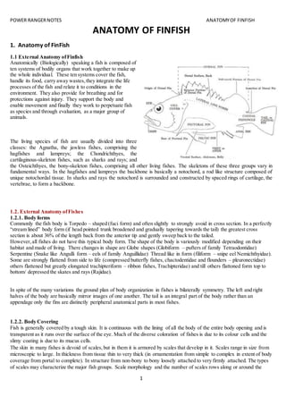
  • 1. POWER RANGERNOTES ANATOMYOF FINFISH 1 ANATOMY OF FINFISH 1. Anatomy of FinFish 1.1 External Anatomy ofFinfish Anatomically (Biologically) speaking a fish is composed of ten systems of bodily organs that work together to make up the whole individual. These ten systems cover the fish, handle its food, carry away wastes,they integrate the life processes of the fish and relate it to conditions in the environment. They also provide for breathing and for protections against injury. They support the body and enable movement and finally they work to perpetuate fish as species and through evaluation, as a major group of animals. The living species of fish are usually divided into three classes: the Agnatha, the jawless fishes, comprising the hagfishes and lampreys; the Chondrichthyes, the cartilaginous-skeleton fishes, such as sharks and rays; and the Osteichthyes, the bony-skeleton fishes, comprising all other living fishes. The skeletons of these three groups vary in fundamental ways. In the hagfishes and lampreys the backbone is basically a notochord, a rod like structure composed of unique notochordal tissue. In sharks and rays the notochord is surrounded and constructed by spaced rings of cartilage, the vertebrae, to form a backbone. 1.2. External Anatomy ofFishes 1.2.1. Body forms Commonly the fish body is Torpedo – shaped (fuci form) and often slightly to strongly avoid in cross section. In a perfectly “stream lined” body form (if head pointed trunk broadened and gradually tapering towards the tail) the greatest cross section is about 36% of the length back from the anterior tip and gently sweep back to the tailed. However,all fishes do not have this typical body form. The shape of the body is variously modified depending on their habitat and made of living. There changes in shape are Globe shapes (Globiform – pufters of family Tetraodontidae) Serpentine (Snake like Angulli form – eels of family Anguillidae) Thread like in form (filiform – snipe eel Nemichthyidae). Some are strongly flattend from side to life (compressed butterfly fishes, chactodentidae and flounders – pleuronectidae) others flattened but greatly elongated trachipteriform – ribbon fishes, Trachipteridae) and till others flattoned form top to bottom/ depressed the skates and rays (Rajidae). In spite of the many variations the ground plan of body organization in fishes is bilaterally symmetry. The left and right halves of the body are basically mirror images of one another. The tail is an integral part of the body rather than an appendage only the fins are distinctly peripheral anatomical parts in most fishes. 1.2.2. Body Covering Fish is generally covered by a tough skin. It is continuous with the lining of all the body of the entire body opening and is transparent as it runs over the surface of the eye. Much of the diverse coloration of fishes is due to its colour cells and the slimy coating is due to its mucus cells. The skin in many fishes is devoid of scales,but in them it is armored by scales that develop in it. Scales range in size from microscopic to large. In thickness from tissue thin to very thick (in ornamentation from simple to complex in extent of body coverage from portal to complete). In structure from non-bony to bony loosely attached to very firmly attached. The types of scales may characterize the major fish groups. Scale morphology and the number of scales rows along or around the
  • 2. POWER RANGERNOTES ANATOMYOF FINFISH 2 body frequently serve as specific and genetic characters. Living Agnathaus are scale less, shark and their relatives have dentinal placoid scales,Bony fishes have various types of bony scales. 1.3. Internal Anatomy of finfish Internal anatomy of a bony fish: finned aquatic vertebrates animal with skin covered with scales. It lives in water and is usually oviparous. Brain: seat of the mental faculties of a fish. Esophagus: part of the digestive tract connecting the mouth to the stomach. Dorsal aorta: vessel in the back that carries blood from the heart to the organs. Stomach: part of the digestive tract between the esophagus and the intestine. Air bladder: pocket in which urine collects. Spinal cord: part of the nervous system that connects the brain to all other parts of a fish. Kidney: blood-purifying organ. Urinary orifice: opening for eliminating urine. Genital Orifice: opening related to the genital organs. Anus: end of the digestive tract. Gonad: hormone-secreting sexual gland of a fish. Intestine: last part of the digestive tract. Pyloric cecum: cul-de-sac related to the intestine. Gall bladder: small sac containing the bile. Liver: bile-producing digestive gland. Heart: blood-pumping organ. Gills: respiratory organ of a fish. Tooth: hard organ of a fish used to shred food. Eye: sight organ of a fish. Olfactory bulb: bulging part of the smell organ of smell of a fish 1.4. Structure of fins Appendages of fishes comprise of fins and cirrti (flaps of flesh) which attain extreme development in the sargassum fish (pterophryne) and the leafy sea dragan (phyllopteryx). The fins are classified as median or paired. Median fins (Unpaired fins) Rayed fins in line with the median axis of a typical fish are these of the back (dorsal fin or fins) the tail (caudal fin) and the lower edge of the body just behind the vent (Anil fin) Although most commonly all of the median fins may be present, but some may be modified or absent in same fishes. Also developed in the median axis may be a rayless fatty adipose fin (as in the trouts – Salmonidae) off fins reduced to a few disconnected spines as in the stickle backs – Gasterosteidae) The anal fin may be modified into an intermittent organ, the gonopodium for use in copulation (as in the live bearers – Poecilidae).
  • 3. POWER RANGERNOTES ANATOMYOF FINFISH 3 Paired fins Paired fins are the Pectorals and Peluics (ventral) the pectorals are supported by the pectoral girdle that joins the skull. Although ordinarily present in fishes, the pectorals are wanting in such kinds as the lamprey (Petromyzonidae) and hag fishes (Myxinidae) They are greatly enlarged in the (Soaring) flying fishes (Exocoetidae) and flying characins (Gastropelecinae). In some cling fishes (Gobiesocidae) and Asiatic suckerbelly loaches (Gastromyzan) the pectorals serve as a part of the ventral hold fast organ. The pelvic fins vary substantially in position and in adoptive modification typically their support is by a pelvic girdle anchored in the bully musculature. In soft rayed fishes (Malacopterygians) the peluics are abdominal in position, Eg. Clupeoids, Salmonids, cypriroids, but they also be situated arterially, just below the pectorals in a thoracic position (as in many spiny rayed species, the Acanthopterygians) or even under the throat in a jugular position (as in blennies). The pelvic are lacking in the lampreys and hag fishes and is lost in various other fishes, rlotably the cels (Angullidae etc) In the sharks and some relatives they are modified into claspers. In cling fishes (Gobiorocidae) sucker belly loaches (Gastromyzon) and certain other fishes, the pelvic from part of a hold fast organ that resembles a ruction cup on the belly. 1.5. Skin and Scales The skin of a fish consists of two layers. The outer layer is epidermis and the inner, the dermis or corium. The epidermis is composed superficially of several layers of flattened, moist epithelial cells. The deepest layers are a zone of active cell growth and multiplication. Here cell multiplication goes on all the time to replace from with in the outermost layers of cells as it is worm off and provide for growth. These epithelial cells from the epidermis are the first to close a surface wound. The dermal layer of the skin contains blood vessels nerves and cutaneous sense organs and connective tissue. The dermis plays the main role in the formation of sales and related integumantory structure. Scattered among the flattened cells of the epidermis are numerous openings of the tubular and flask shaped mucous gland cells that extent into the dermis. These cells secrete the slippery mucus that cover most fishes. The mucus lessons the drag an a fish when it swims through water. As in other vertebrates, the skin of a fish is the envelope for the body and is the first lines of defense against disease. It also affords protection from, and adjustment to environmental factors that influence life, for it contains sensory receptors tunes to the surroundings of a fish. Further more, the skin has respiratory, excretory and osmoregulatory functions.
  • 4. POWER RANGERNOTES ANATOMYOF FINFISH 4 1.6. Scalation Outstanding among the special feature of the skin are the very prominent scales that most fishes have. However some do not have scales. Examples include the lampreys (petromyzonidae) and a number of cat fishes North America (F.W) (Ietaluridae). An intermediate structural category have seals only on a few palces of the body for example: Paddle fish (polydon) three spines stickle bask Gasterosteus aculeatus and mirror carp. Then there are a few fishes that have very small deeply imbedded scales that they look scale less for example Fresh Water Eels (Anguilla) Brook trauts (Salvelius fontinalis) and the barbot (lota) of Cod family (F.W) (Gadidae) In arrangements scales are most often “imbricated” and thus overlap like roof tiles with free margin directed towards the tail in a manner that minimize friction with water. In some fishes such as barbot (lota) and F.W eels (Anguilla) the pattern is mosaic rather than overlapping one another, the scales are minutely separated or meet their neighbors only at their margins Scales shapes Although they comprise only a few basic structural types, Scales exhibit many modifications that are often characteristic of groups or species on the basis of shape one type is plate like (Placoid), with each plate carrying a small cusp, as common among the sharks (Elasmobranchil) a Second type is diamond shaped (rhombic) and found in gars (Lepirosteidae) and the bichirs (pdypterus). A third type of scale is called Cycloid because it is typically smooth disc like and more or less circular mostly found in soft rayed fishes. (Malacopterygi). In fourth type is ctenoid the posterior surface or margin is toothed or comb like and a characteristic of spiny rayed bony fishes (Acunthopterygil) However, some of the soft rayed fishes have ctenoid like contact organs on their scaes, examples characins (characid) Killi fishes (cyprinodontidac). Some spiny rayed fishes have cepeloid scales exclusively. For example the brooke silverside labidestus. Many spiny rayed species exhibit both cycloid and ctenoid scales. Example: Common basses (Micropterus). Structural types Structurally, there are two types of fish scales, placoid and Non-placoid. Non-placoid scales are basically of the three kinds – cosmoid, Ganoid and Bony ridged scales. I) Placoid scale Placoid scales also called dermal denticles have an ectodermal cap or covering of enamel like substances (as an human teeth) termed “Vitrodentine” beneath this is a thicker layer of entine with a pulp cavity and dentinal tubules emanating form it. Each scale has a disc like basal plate in the dermis with a cusp projecting outward from it though the epidermis. Placoid scales occur among the sharks
  • 5. POWER RANGERNOTES ANATOMYOF FINFISH 5 and their relatives (chandrichthyes). II) Non – Placoid scales 1) Cosmoid scales: The cosmoid scales have a thinner, harder outer layer than the placoid ones. It is also termed as vitrodentine. The layer below this hard and non cellular and is called cosmine. The layer beneath is vascularised mid layer of perforated bony substances called “Iropedine” The growth of the scale is at the edge and beneath no ligivng cell layer covers the scale. The cosmoid scales found in (latimeria). 2) Ganoid Scale: In ganoid scale the outer layer is a hard inorganic substances called ganoine which is different from Vitrodentine. The layer below the ganoine is a cosmine like layer. The innermost layer is a bony layer called isopedine. The growth takes place at the edge, beneath and on the surface. Three types of scales are found in bichirs (pdypterus) and gars (Lepisosteidae). 3) Bony Ridges Scales: Bony –ridged scales are typically thin and transclucent lacking both dense enameled and dentinal layers of the three other kinds. Bony ridged scales characterizes the many living species of bony fishes (Osteichthyes) that have either cycloid or ctenoid scales. The outer layer of there scales is marked with bony ridges alternate with depression. The inner part or plate of the scale is made up of layers of criss – crossing fibrous connective tissues. The growth of the scales is both on the outer surface and from beneath. For ridges on the scale two terms are used ridges and circuli changes in the growth pattern of the individuals fish is reflected in the character and distribution of ridges. “Breeding” and “Yearmarks” can be identified on this basis in many species. In both cycloid and ctenoid scales, a nuclear central zone can be recognized. This zone may be called as “focus” the scale. However this central position shifts due to differential growth of fore or after part of the scale, shifting the focus to more posterior and anterior margin. In many species grooves or radii radiate from or near focus towards one or more margins of the scale. The ctenoid scales have teeth on its posterior margins. 2.Cellstructure, tissue and body organization 2.1. Organization: kind of construction of the body A cellular or protoplasmic level of organization: Body is not differentiated into cells. Example: Protozoa (subkingdom: Protozoa); All other animals except Protozoa come under the subkingdom: Metazoa (Body is composed of numerous cells) Cellular level of organization: The cells of the body are more or less loose and Independent. Example: Amoeba Tissue level of organization: In some animals, body cells form tissues. Example: Coelenterata and Ctenophora Organ system level of organization: In most of the animals, tissues combine into organs, which, in turn, combine to form organ systems. Example: Animals like Human being, Fish, Reptiles etc., Germ Layers: The fundamental cell layers laid down in an early embryo of the Metazoa are called germ layers. The Coelenterata have only two layers: Ectoderm and Endoderm. They are termed the diploblastic animals. Most other Metazoa possess three germ layers: Ectoderm, Mesoderm and Endoderm. They are known as the triploblastic animals. Example; Animals except Coelenterata 2.2. Symmetry It referstothe similarityin size,shape andnumberof partsonthe opposite sidesof amedianline.Partsof ananimal bodyare oftensoarrangedthat it ispossible tocutit intotwosimilarhalvesbyone or more planes.Suchanimalsare said to be symmetrical.Some animals,likeAmoebaandSnail,cannotbe dividedintoequal partsbyanyplane.Theyare saidto be asymmetrical.The symmetricalanimalsshow one of the three typesof symmetry:spherical,radial andbilateral.
  • 6. POWER RANGERNOTES ANATOMYOF FINFISH 6 Section– 1 Spherical (universal) symmetry: Bodyis divisible intosimilarhalvesbyanyplane passingthroughthe center.Example:Seaurchin Section– 2 Radial symmetry: Bodyis witha numberof similarpartsradiatingoutfroma central axis.The boycan be splitupintoequal halvesbyany plane passingthroughthe middle fromtoptobottom.The animalswithradial symmetryare calledRadiata.Examples: Spongesandmostcoelenterates. In some animals,some partsof the bodyare single orpairedratherthanradial,so that onlyone or twoplanesthrough the longitudinalaxisdivide the animalintosimilarhalves.Suchasymmetryiscalledbiradial.Examples:Comb-jellies, seanemonesandseastars. Section–3 Bilateral symmetry: In restof the Metazoa,chief organsof the bodyare pairedandare arrangedon the sidesof a central axisconnectingthe headwiththe tail.Withthe result,the bodycan be dividedintotwosimilarhalvesonlybyone plane.Suchare termedas bilateral.The bilaterallysymmetrical animalsare termedthe bilateria. A plane orsectionpassingdorso-ventrally(vertically)throughthe middleof the anterior-posterior(longitudinal) axisis knownas the Sagittal plane orsection.Thisisthe plane thatdividesthe bilateral animal intosimilarandrightand left halves. A plane orsectionrunningantero-posteriorlyatrightanglestothe sagittal plane iscalledthe frontal plane orsection.It dividesthe bodyintodorsal andventral halves. A plane orsectionpassingdorso-ventrallyatrightanglestothe antero-posterioraxisisknownastransverse plane or section.Itdividesthe bodyintoanteriorandposteriorhalves. Section– 4 Digestive tract: The Mesozoa andparazoa lack a digestive tract.The Eumetazoapossessadigestive tract,andare termedthe Enterozoa. Certainenterozoa,viz.,coelenterata,CtenophoraandPlatyhelminthes,have incompletedigestive tract,whichhasasingle external opening,the mouth,thatservesforintake of foodaswell aseliminationof faeces.Allotherenterozoahave complete digestivetract,whichhastwoexternal openings,the mouthforintake of foodandthe anusfor eliminationof faeces. Section– 5 Coelom: Some bilateriahave nocavityintheirbody,exceptthatinthe digestivetract.Suchformsare knownas the acoelomates. All others possessacavityinadditiontothe one in the digestive tract.The space betweenthe twocavitiesiscalledthe bodycavitythe nature andoriginof thiscavity variesindifferentgroups.Whenlinedbyamesodermal epithelium,the peritoneum, itisknown asthe true coelom,andwhenunlinedbyperitoneum, itistermedfalsecoelomorpseudocoel. The animalswithfalse andtrue coelomare respectivelycalledthe pseudocoelomatesendeucoelomates.InMolluscaand Arthropodathe coelomisgreatlyreduced,andthe bloodfillsthe spacesbetweenthe internal organs.Suchabody-cavity iscalledthe haemocoel.The coelomisof greatsignificance inanimal evolution.Itprovidesspace forthe developmentof variousorgansystems. Section– 6 Metamerismor Segmentation: Segmentsoccurinthree phyla:Annelida,ArthropodaandChordata.Eachsegmentiscalledametamere orsomite. Metamerismaffectsexternal andinternal structures.Itissaidto be homonomous,if the somitesare similar,asin earthworm(annelida);andheteronomous,isthe segmentsare dissimilarasinprawn (Arthropoda).Thesegmentationin Chordatesshowsupclearlyinthe embryonicstage inte segmentallyarrangedmesodermalsomites.Inadultchordates segmentationislargelyinternal andisseenintheirvertebrae,ribs,nervesandbloodvessels. 3. Oral region and Associated structures
  • 7. POWER RANGERNOTES ANATOMYOF FINFISH 7 3.1. Adaptations for feeding (The feedinghabitsorfeedingbehaviorof fishesisthe searchforand ingestingof food) The diversityinfeedinghabits that fishesexhibitisthe resultof evaluationleadingtostructural adaptationforgettingfoodfromthe environments.The structural adaptationof feedingorganssuchas 1) positionandshape of the mouth2) Presence orabsence of “Teeth”.3) Gap of the mouthetc.,greatlyhelpinpredictingthe nature of foodandmode of feedingof the fish(inquestion). Section1: Major feedingtypes: On the basis,fishescanbe classified,accordingtotheirfeedinghabitsas1) Predators2) Grazers 3) Foodstrainers4) Food suckersand 5) Parasites. 1) Predators Fishesthatfeedonmacroscopicanimals,theyusuallyhave welldevelopedgraspingandholdingteethasinmanysharks (Elasmobrabch) the Barracuds(Sphyraena),the pikes(Esox) gars(Lepisosteus). 2) Grazers In Grazing,the foodis takenbybitesor continual browsing(Grazingcharacterizemanyfishesthatfeedonplanktonsoron bottomorganisms) (sometimesorganismsare takensingle oratother timesinsmall groups) mayfeedonbottomor column – a bluegill (Lepomismacrochirus),Parrotfishes(scaridae) Butterflyfishes(chaetodontidae) browsingoncoral reef. 3) Strainers(Filterfeeders) Filterthe waterforplankton,here foodsare selectedbysize andnotby kind.Herringclupidae,Gizard,shads(Dorosoma), Paddle fish(polyodon) whaleshark(Rhincodon) alsohave efficientfoodstrainingorfilteringadaptation.The principal adaptationforfilterfeedingorstrainersisthe developemtof numerous,closely –setand elongatedgillrackers. 4) Suckers The suckingintothe mouth of foodor foodcontainingmaterial istakenpracticedbybottomfeedingfishessuchasthe sturgeons(Acipenseridae) andsuckers(catostomidae) 5) Parasites Parasitisumisperhapsthe mostunusual andhighlyevolvedfeedinghabitsamonganimals.Theysuckbodyfluidafter raspinga hole inthe sidesof the body.Parasiticlampreys(Petromyzonidae),Sealamprays(Petromyzonmarinus)Pacific lampreys(Lamptratridentata) 3.3. Oral adaptation for feeding Lips Suctorial feeders have an “inferior mouth” and fleshy modification of lips. Notable among these are the sturgeons (Acipenseridac) and suckeres (catastomidae). The lips of strurgeons and suckers are mobile and described as “Plicate” (having folds) or papillose (having small tufts of skin or papillose). Many sectorial feeders also have well developed barbells bordering the mouth. Sucker mouth : Moxostoma (Redhorse) suctorial lips of free – living fishes may also serve as holdfast organs in fast flowing mountain streams. For example (Glyptorternum). In loach – like Gyriocheilid (Gyrinocheilus) has an extreamly specialized suctorial adaptation, in addition to suctorial lips the opercular structure has separate inhalent and exhalent device for respiration. 3.3.1. Modificationsin the shape of mouth Amongthe grazersand suctorial feeders,thereexistnotonlyspeciallydevelopedlipsbutalsoadaptationsof othermouth parts. The Trumpetfishes(Aulostomidae) the cornetfishes(Fistularidae)andthe pipe fishes(syngnanthidae) aswell as manybutterflyfishes(chaetodontidae) of coral reefs,have mouthsthatresemble elongatedbeaks.Thisadaptationis achievedbya protractionof the hyomandibularbone ratherthanbya lengtheningof the lowerjaw bones(denteries) themselves.
  • 8. POWER RANGERNOTES ANATOMYOF FINFISH 8 Amongthese fishes,the methodof feedingmaybe bysuctionasin the case of trumpetfishes,cornetfishesandpipe fishesoritmay be a selectivegrazingactionwithsharpteethwhenthe longsnoutenablesthe butterflyfishtoreachinto small crevicesof the coral. A peculiarstructure amongmouthmodificationhasariseninthe half beaks(Hemiramphidae)where the lowerjaw projectsintoa beakoftena thirdof the lengthof the fishitself,withthe mouthopeningabove it.Half beaksare usually surface feedingfishes. 3.3.2. Teeth Outstandingamongthe obviousoral adaptationforfeedinginfishesare the teeth.Theyare thoughttohave arisenfrom scalescoveringthe lips,asrepresentedinlivingsharks(squliformies) where the placoidscalesof the skinvisiblygrade intoteethonthe jaws. In bonyfishes(Osteichthyes) Teethare of three kinds,basedonwhere theyare foundJaw,Mouthand Pharyngeal. Jaw Teeth Jaw teethare variouslythose onthe maxillaryandpremaxillarybonesabove andonthe dentariesbelow. In the roof of the oral cavityteethare variouslyborne bythe medianvomerandbythe palatine andectopterygoidbones on eachside.Inthe floorof the mouththe tongue oftenhasteethonit. Pharyngeal teethoccuraspads on variousgill arch eolementsinmanyspecies.Inthe carps(cyprinedae)andsuckers(cjcatastromidae) the onlyteethare those indeep inthe pharynx (gut,mouthandeoesophagous) thatdeveloedmodificationof lowerelementsafterlastgill arch,inclarius and labeothe teethare modifiedforgrasping,tearing,grindingandrazorlike cuttingteethhave developedinpredacious fishes. Kinds ofjaw teeth Basedon theirformmajorkindsof Jaw teethare 1) Cardiform2) Villiform3) Canine 4) Incisorand5) Molariform. Cardiform Teeth Cardiformteethare numerous,shrotfine andpointedsuchdentitionwithvariationsisfoundinmanyfishesthathave multiple rowedteeth.Forexample Americancatfish(Ictaluridae)perches(percidae) andmanyseabases(serranidae) Villiformteeth Villiformteethare more orlesselongatedcardiformteeth.Forexample:Needlefishes(Belonidae) andLionfishesas (Pterois). Canine teeth Caninesare dogtooth like orfange like (longpointedtooth) theyare elongatedandsubconical,straightorcurvedandare adaptedforpiercingandhodlingforexample walleyes(Alskapollock) (Stizostediosn).Incertainfishessuchasmoreys (muraenidae) the caninesare hinged(thehook) yieldotbackwardpressure butlockwhenmovedforwardandadaptation to retainlivingmovingpreyinsidethe mouth. Incisors teeth Incisorsare sharp edgedcuttingteeth.Insome fishesincisorsfuse togetherincutting beakasin parrot fishes(scaridae). Molariformteeth Molariformteethare forcrushingand grindingthusflatwithprodrudingdenticlesonthe surface.These teethare found inbottomdwellingfisheslike skatesandraysand some sciaenidae (drums). Within a single groupthe diversityof dentitiononidentical bonesmayvarylargely.Forexample Incarpsand minnows (cyprinidae)the pharyngealteethrange fromsharpincarnivoressuchas insemotilustomolariforminthe commoncarp (Cyprinuscarpio). In general teethare absentinplanktonfeedersandinsome of the more generalizedomnivore.Theyare presentin increasingnumbersof bonesinmore andmore relative toitspredatorybehaviors.The premaxillarybonesare toothed whenjawboneshave little ornoteeth.Thisistrue for manysoftrayedspeciessuchas bowfin(Amia) the gars (lepisosteus) the salmonsandtrouts(Salmonidae).
  • 9. POWER RANGERNOTES ANATOMYOF FINFISH 9 The maxillae are typicallytoothedinthose softrayedfishesasthatcarry premaxillaryteeth.Howevertoothlessin otherwise tooth –bearingspinyrayedfishes. 3.4. Gill Rakers Besides protecting the tender gill filaments form abrasion (a rubbing off or scrap) by ingested materially that are coarse in texture, gill rakers are specialization in relation to food and feeding habits. They are very stubby (short and thick) and unornamented in most omnivores for example sunfish (Lepomis cynellus). In many plankton feeders, the gill rakers are elongated, numerous are variously lamellate or ornamented to increase efficiency of filtering. Simple but very numerous rakers are possessed by gizzard shads (Dorosoma) and paddlefish (polyodon) Ornamentation of gill rackers are of taxonomic value. In pleuronectdae the gill rakers resembles feather having a main axis with lateral processes these lateral processes are branched. The adjacent gill rakers overlap to form a very fine sieve. 4. Gastro Intestinal Tract OR Digestive system and associated digestive glands 4.1. Gastro Intestinal Tract Esophagus Esophagus forms the beginning of the gastro intestinal tract/ In general the oesophagus is no distensible that it can accommodate anything the fish can get into the mouth and sometimes even accommodate the item if it happens to double on itself two or three times, on its way to the stomesh. The ocsgtagus is a short and narrow to be in a no. of Herbivorus and omnivorus fishes. (Cyprinus corpio, L.rohito,Tor tor etc. A large no. of mucus – secreting cells are scattered in the mucosa at taste buds are also present in some or species. 4.1.2. Stomach Frequently in literature the fishes are classified as stomach or stomach less fishes. 85% telecast have stomach 15% had no stomach. The stomach too shows various adaptations one of which is shape. In carnivore the stomach typically is straight, elongate for example in gars (lepisosteus), bowfins (Amia), Pikes (Esox) and barracudas (sphyraena). In omnivorous species, the stomach is most often sac-shaped. A very special adaptation is the modification of the stomach into a grinding organ as in the sturgeons (Acipenser) gizzard shads (Dorosoma) and mullets (Mugil). Here the stomach is reduced in overall size but its wall greatly thickened and muscularized. The lining too is heavily strengthened with connective tissue and the lumen is very small. The organ is not for storage, mixing and primary digestion but rather a food grinder. Great dispensability is the adaptation of the stomach in the predatory deep sea swallowers (saccopharyngidae) and gulpers (Eurypharyngidae) enabling these fishes to take relatively huge prey. A remarkable modification of the stomach exists in the puffers (Tetradontidae) which can inflate themselves with water or air to assume often on almost globular shape. The adaptive value of this modification of the digestive tract is probably mainly one of defense, for many puffers and porcupine fishes have spines all over the body which can thus be created. Not all fishes have a stomach that is a portion of digestive tube with a typically acid secretion and a distinctive epithelial lining different from that of the intestine. In most of the herbivores and phytoplankton feeders the epithelial tissue of the esophagus grades directly into that of the intestine, thus termed stomach less. Though the primary criterian for being without stomach does not seem to be whether a fish is an herbivore or a carnivore but whether accessory adaptations for trituration and fine grinding of food exist either in the form of teeth or a grinding apparatus such as a gizzard. Where stomach exist
  • 10. POWER RANGERNOTES ANATOMYOF FINFISH 10 most pronouncedly (strongly) in carnivores. They are characterized by a low pH and the prominent presence of pepsin (enzyme) among other digestive juices. 4.1.3. Intestine The intestine too has many variations. It is shortened in carnivores such as in the pike (Erox), perhaps because meaty foods can be digested easily. Where as in herbivores the intestine is highly elongated and several times the body length of the fish and in carps and certain catfishes. In sharks and other elasmobrachs the intestine has a coiled layer of absorptive tissue called spiral valve which increase absorptive surfaces for the relatively in short intestine. 4.1.4. Pyloric caeca (Fishesare onlyvertebratesthathave appendages(Caeca) atthe gastro-intestinaljunctions). Onthe intestine of most bonyfishesatthe pyloricendof the stomach.There maybe fromone to manyblindsacs or pyloniccaecaor intestinal caeca. A fewgroupssuchas cat fishes(Ictaluridae) topminnows(Cyprinidae) andpikes(Esocidae)lackthese structures. In suchgroups as flatfishes(Pleuronectiformies)the pyloriccaecaare few usuallynotmore thanfive (5).Inotherssuchas Mackerels(Scombridae),Salmons(Salmonidae)the numberof these caecamayrange to 200 or more.Generallycaecaof differentspeciesvaryconsiderablyinsize,state of branchingandthe connectionwiththe gut.Insturgeons (Acipenseridae) the manycaecaformalarge mass,but onlya single ductleadstothe intestine.Insalmon,eachcaecum communicatesdirectlywiththe gut,the functionsof pyloriccaecaprobablyinvolve bothdigestionandabsorption. 4.1.5. Rectum Morphologicallythere isnodistinctionbetweenthe rectumadintestine,the ilio-rectal valveispresentinsciana,tetradon and muracnosox. 4.1.6. Anus It isthe posterioropeningof the alimentarycanal ordigestive system.The internalsurface of the regionnearthe rectum iscoveredwithan epitheliumrichmucouscells.The anusismade upof an innercircularand outerlongitudinal muscle layer.The circularmuscle isthickdevelopedformingsphincter.The regionof anusfacingthe anteriorhasthe epithelium continueswiththe skin. 4.2.1. Associated digestive glands Liver Like othervertebrates,liverisanimportantorganinfisheswhichhasboth“secretary”and “storage”function.Thisisa large glandinall fishes,butsharksandraysmay have extremelylarge liverscomprisingabout20% of the bodyweight especiallyinsome pelagicsharks.The liverusuallyliesoverorpartiallysurroundsthe stomach.Itistypicallybilobed,but may have onlyone lobe asinsalmonor three as inmackerel.InHag fishesthe liverisintwodistinctparts,withseparate ducts leadingtothe gallbladder.Adultlampreyhave nobile ductsorgall bladder,gall bladder isalsoabsentinburbot (lota) butinmost otherfishesthe gall bladderispresentandfunctiontostore liversecretions.Ordinarilyone hepatic ducts originatesfromeachlobe of the liverandjoinsthe cysticductsfromthe gall bladderto formthe “bile ducts” Liverfunctionincludesbile secretionandglycogenstorage inadditiontoseveral otherbiochemical processes.Apartfrom theirfunctionfishliveralsostoresfats,vitamin“A”and“D” and the wearyRBCs releasinghemoglobinforrecyclinginto the bodyliveralsohelpsproductionof ureaandothernitrogenouscompounds. 4.2.2. Pancreas Hag fisheshave asmall – pancreaswithseveral ductsthatemptyintothe bile duct.Lampreyshave pancreatictissue locatedthroughoutthe liverandintertinal wall.Amongbonyfishesthe pancreatictissue isusuallydiffusedinoraround the liver.Thisisespeciallytrue of spinyrayedfishes,inwhichthe pancrease andliverincorporate ina“hapatapancrease”, inmany of soft rayedbonyfishesPancreasis adistinctorgan,insharks andrays alsothe pancrease isa compact organ withtwolobes.The pancreaticductmay reach the small intestineseparatelyfromthe bileductasin the sharksor may
  • 11. POWER RANGERNOTES ANATOMYOF FINFISH 11 discharge intothe bile ductas inthe gar (lepisosteus).The pancreassecretsseveral enzymes,thatare active indigestion. In additionthe pancreaticisletshave the endocrinefunctionof producinginsulin. 4.2.3: Spleen The spleenisusuallyrecognizedasadark redstructure lyingon or behindthe stomachto whichit attachesbya bandlike ligament.Althoughitisassociatedwiththe digestive organsithasno digestivefunction,butratherisinstrumentalin bloodcell formation(The functionof redbloodcell distractionisascribedtospleenof higherbony fishes).Inlampreys and Hag fishes,whichdonothave a compact spleen,spleenliketissue isdiffusedalongthe intestine. 4.2.4. Gas bladder The Gas bladderisa thinwalledsactypicallyfoundinthe upperpartof the bodycavity immediatelybelow the kidney.In manyfishesthe shape issimple,usuallysomewhattorpedoshaped,butthere are manyvariations,minnowsandcarps (cyprinidae)have anteriorandposteriorchambers,connectedbyanopeningcontrolledbyasplinter.Featherbacks (Notopteridae) have airbladderdividedlaterally,buttwochamberscommunicate anterioritymostcroackers(scieanidae) have unusual airbladdersinthatvariouslyshapedsacsor branchingcaeca maybe arrangedalongeach side of the organ. In Herrings(chupeidae) the gasbladderhasaposterioropeningtothe exteriornearthe anus. In the embryologyof bonyfishesthe gasbladderoriginatesasan outgrowthof the alimentarycanal andremains attachedto the esophagusorstomach viathe pneumaticductssuch fishesare knownasphysostomes.Some groupsof fisheslose the connectiontothe alimentarytract.These fishesare knownasphysoclistous.Some of suchfishesmay retainthe connectiontill larval orjuvenilestages. Some bottom-dwellingstreamfishessuchasdarter (Etheostoma) andSculpin(cottus) lackthe gasbladder.Variousother bathypelagicfisheshave alsolostgasbladder.Agnathusandcartilaginousfishesalsolackgasbladder. 5. Circulatory System 5.1. Introduction Fishesare cold-bloodedaquaticvertebratesandcanbe foundinbothsaline andfreshwater.The circulatorysystemof fishesisresponsiblefortransportingbloodandnutrientsthroughoutthe body.Ithasa closedcirculatorysystem,i.e. bloodtravelsacrossthe bodythroughthe networkof bloodvessels.Unlike humans,fishesexhibitsingle cycle circulation, where the oxygendeprivedbloodcomestothe heart,fromwhere itispumpedtothe gillsandthencirculatedtothe entire body.Onthe otherhand,inmammals,the deoxygenatedbloodentersthe heart,fromwhere itispumpedintothe lungsforoxygenation.Then,the oxygenatedbloodreturnstothe heartfromthe lungs,tobe transportedthroughoutthe body. 5.1.1. Circulatory System of a Fish The circulatorysystemof fishisquite simple.Like mammals,the circulatorysystemof fishconsistsof aheart,bloodand bloodvessels.The heartof a fishisa simple muscularstructure thatislocatedbetweenthe posteriorgill arches.Itis enclosedbythe pericardial membrane orpericardium.Inmostof the fishes,the heartconsistsof anatrium, a ventricle,a sac-like thinwalledstructure knownassinusvenosusandatube,knownas bulbusarteriosus.Inspite of containingfour parts,the heartof a fishisconsideredtwo-chambered. The bloodcontainsplasma(the fluidportionof blood) andthe bloodcells.The redbloodcellsorthe erythrocytescontain hemoglobin,aproteinthatfacilitatesthe transportof oxygentothe entire body,whilethe white bloodcellsare an indispensable partof the immune system.The thrombocytesperformthe functionsthatisequivalenttothe role executedbythe plateletsinthe humanbody,i.e.theyhelpinbloodclotting.Bloodiscirculatedthroughoutthe bodywith the helpof bloodvessels.The blood vesselsare of twotypes,arteriesandveins.The arteriesare responsibleforcarrying oxygenatedbloodfromthe hearttothe restof the body,while the veinsreturndeoxygenatedbloodfromthe different parts of the bodyto the heart.
  • 12. POWER RANGERNOTES ANATOMYOF FINFISH 12 5.1.2. Heart The heart ismodifiedbloodvessel exhibitingthe three layerscharacteristicof arteriesaninnerlining(endocardium) of endotheliumandelastictissue onmuscularlayer(myocardium) thatisverythick,especiallyinthe ventricularregionand an outerfibroustunic(epicardium) onthe surface of whichisthe visceral pericardium.The heartpulsatesasaresultof the response of the muscle cellstothe electrolytes(Bloodelectrolytes) thatinfuseit.The rhythmicityof the pulsationsis regulatedbyflexlybythe autonomicnervoussystemexceptinhagfishes,inwhichnonerve fiberssupplythe muscle.The heartoccupiesthe pericardial cavity(asubdivisionof the coelomanteriortothe septumtransversem).The heartof all vertebratesisbuiltinaccordance withabasic architectural pattern.Itisdemonstratedinitssimplestforminhagfishes where itsexhibitsaseriesof fourchamberssinusvenous,atrium, ventricle andconusarteriousthroughwhichfloodflow inthat sequences. Chondrychthyeshascontractile,muscularandvalvedbase the conusarteriosustothe ventral aortawhere itleavesthe ventricle.Inhigherbonyfishesthe planislike thatof the sharkand itsrelatives,butthe firstsectionof the ventral aortais typicallythin-walledandvalvedbulbusarteriosusandnotcontractile butelastic,alternatelyenlargingandshrinkingin response tochangesinbloodpressure fromalternate ventricularcontraction,systole(contraction) anddiastole (relaxation) The ventral aorta ina fishismedianinposition,beneaththe gillsfromitsbranchthe afferentbronchial arteriestoeach gill pouchor arch. Within the gills,afferentbronchial breakdownintocapillariesandcollectagaininto efferentvessel that formthe dorsal aorta mainvessel fordistributionof bloodtothe body. 5.2.2. Arterial Systemin Elasmobranchs Afferentbranchial arteries The ventral aorta arisesformthe conus arteriosus andveinsforwardsalongthe ventral surface of the pharynx rightupto the posteriorborderof the hyoidarch where itbifurcatesintotwobrancheseachbranchdivide intofirstandsecond afferentbranchial arteries.The firstafferentbranchial arteryrunsalongthe posteriorborderof the firstbranchial arch and suppliedarterialbranchestoanteriorandposteriorgill lamellacof the firstbranchial archof the third,fourthand fifthafferentbranchialarteriesarise formthe ventral aortaalmostequidistantfromone anotherandrunsalongthe outer borderof the secondthirdandfourthbranchial arches.Thissupplythe bloodtothe 3rd 4th and5th gill arches
  • 13. POWER RANGERNOTES ANATOMYOF FINFISH 13 respectively. Efferentbranchial arteries The bloodfromthe capillariesof gilllamellacis collectedbyaseriousof bloodvessel calledthe efferentbranchial arteries.There are nine efferentbranchialvesselsoneachside (maybe lessdependinguponspecies)of these the first eightjoininpairsto formfour complete toopsaroundthe 1stfour gill clefts.The 9thrunsalongthe anteriorborderof the fifthgill cleft. The four loopand5th efferentbranchialarteriesare joinedbyshortlongitudinalconnectivesrunningacrossthe inter branchial septa.The efferentbranchial subsequentlyjointoformthe mediumdorsal aorta. Arteriesof the head The firstefferentbranchial andasmall part fromthe dorsal aorta supplybloodtothe head.The firstefferentbranchial vessel givesrisetothree branchesthe external carotidafferentspiracular andhyoidean. The afferentspiracularrunsforwardandgivesrise toophthalmicartery.Thisarterymovesforwardjoinswithexternal carotidto form cerebral artery. - 5.2.3. Dorsal aorta and its branches The dorsal aorta runsbackwardsalongthe whole lengthof the bodylyingbeneaththe vertebral column.Inthe tail region it continueswithinthe haemal canal ascaudal artery.Anteriorlyitgivesoff several small branchestothe roof of buccal cavity.The followingarteriesthatarise fromdorsal aorta,the celiacsuppliesbloodtostomachand liver,the anterior mesentericsuppliesthe pancreasthe intestineandthe rectumthe posteriormesenterestothe gonad.The parietal tothe bodywall the renel tothe kidney(andthe femoral tothe griddle region). 5.2.4. Arterial systemin Bony fishes Heart issimilartothat of shark.But there isno “conusarteriosis”.However,the base of the ventral aorta isenlargedto forma non-contractile “bulbusarteriosis”. Arterial system The ventral aorta iscomparativelyshorter.Itgivesoff 4pairsof afferentbrachial arteriesthroughwhichvenousbloodis suppliedtothe gills( 4 pairs) forpurification.The bloodfromthe gillsiscollectedby4 pairsof “efferentbranchial arteries”Inthe bonyfisheseach gill archhas a single efferentarteryunlike the conditioninshark.The fourefferent branchial arteriesunite toformthe lateral aortaon each side.The twolateral aortaare joinedtogetherbothanteriorly and posteriorlyformingaringshapedvessel knownasthe “circularcephalicus”.Inthe anteriorend,the external and internal carotidsarise oneachside formthe circular cephalicusposteriorlythe twolaterial aortafuse togethertoproduce the dorsal aorta whichsuppliesall the organsof the abdomenandendsinthe tail. 5.2.5. Venous system The blood distributed to the different parts of the body by the arteries and their braches through capillaries and returned to the heart by the veins. The veins differ in structure from the arteries in possessing thin walls and in forming wide irregular spaces called sinuses (during then curve) The Venus system can be devided into the following systems.  Anterior cardinal system.  Posterior cardinal system  Hepatic portal system  Ventral sinus  Cutaneous system. Anterior cardinal system: the anterior cardinal system consists of the following viens, the internal juglar veins it has the alfactory sinus, orbital sinus, post orbital sinus and anterior cardinal sinus. The orbital sinus finally opens into large anterior cardinal sinus. It opens into ductus cuaveri. The posterior cardinal system: This system consists of medium caudal vein two renal veins and two large posterior cardinal sinuses. The renal portal vein gives of branches to kidney recollect into renal veins to form the posterior cardinal sinuses.
  • 14. POWER RANGERNOTES ANATOMYOF FINFISH 14 Hepatic portal system: This system has the hepatic portal vein anterior and posterior intestinal veins, gastric vein and the anterior and posterior gastric. These subsequently form hepatic sinuses which open into sinus venosus. Ventral veins: The ventral vein comprises two groups. 1) the anterior veins which discharges the blood into ductus curvier through the inferior jugular sinuses and the posterior veins which empty their blood through the subclavians. Cutaneous systems: The cutaneous system includes a dorsal, a ventral and two paired lateral veins. The ventral joins the abdominal veins. The laterals empties into the branchial vein. 6. Respiratory system in fishes 6.1.1. Gills Gaseous Respiration in Fish Just like you and me fish need a constant supply oxygen in the form of O2 in order to run their metabolism. Without oxygen they can't turn their food into energy or make any new fish body. All the free oxygen on this planet on this planet, was, or is being, released into the air by plants, the atmosphere at the moment is about 21% O2. However oxygen will dissolve in water, in a similar sort of way that the bubbles in your Coca Cola are dissolved into the liquid, which is mostly water, that makes up the drink. Fish could of course breathe air like Seals and Whales, and some do, but if they wish to stay safe under the water for longer periods of time it would be much easier if they could get the oxygen they need from the water, and this is exactly what they do. In fact they were doing this long before any vertebrate animals learned how to breath the air. So fish live in the water and they breathe the water, to do this they have special organs called gills. Gills are wonderfully well designed, and they have to be because although the water does hold some oxygen it never holds any where near as much as the air, and to make things more difficult the amount of oxygen a body of water can hold decreases the warmer it becomes, and also, salt water holds less oxygen than fresh water. If the air above a body of water is 21% O2, this means that 210 parts per thousand are O2, but if we do the math for one of the figures below, such as cold salt water we see that it contains only 7.58 parts per thousand O2. What this means to the fish is that their gills need to be a lot more efficient at extracting O2 from the water than our lungs need to be at extracting it from the air. Fish solve the problems of extracting the O2 they need from the water they live in in a variety of ways. Firstly they have different life styles, obviously a fish that spends most of its life resting on the bottom of the ocean waiting for its dinner to swim by needs less O2 than a fish which actively chases smaller fish for its dinner. However most of the problem is solved in the design of the gills. A fish's gills are situated one set on either side of the body and near the back of the head They are open to the gullet at the front, and open to the external environment behind. They are designed so that water can flow continually passed them, coming in through the mouth, and/or the spiracle in sharks and their allies, and passing out through the single external gill opening in fish or through one of the 5 to 7 gill clefts in sharks and rays. In fish there is a bony plate protecting the gills, this is called the operculum, and it is hinged and has muscles attached to it so it can be regularly opened and closed. This ability to have water continually passing over the gills is one of the major factors making gills more efficient than lungs. With lungs the air comes in, fills the space, and then has to be expelled before any more O2
  • 15. POWER RANGERNOTES ANATOMYOF FINFISH 15 rich air can be brought in. With gills there is no time wasted getting rid of the old air/water and no energy wasted reversing the direction of the flow. In sharks and rays the number of gills is usually 5, but there are some species with 6 or 7 sets, in fish the number of gills is 4 on either side of the body. Each gill is supported by a gill arch and protected by gill rakers. Each gill arch supports one set of paired gill filaments. The gill rakers help make sure that no extraneous material gets into the gill filaments to clog them up. Each paired gill filament in turn supports numerous lamellae (sing. lamella), extending out from both sides of the filament body. It's here in the lamellae that the uptake of O2 actually occurs. The lamellae are very fine structures, however there exact dimensions depend on the normal activity levels of the fish in question. The more active the fish the thinner they are and the less distance there is between them. Also the absolute thickness of the individual lamellae walls varies, this is important in considering to facility with which O2can diffuse from the water to the fish's blood, the thinner the membrane the more quickly. and easily the O2 can pass across it. Thus in sluggish fish like the American Brown Bullhead (Amerius nebulosus) the lamellae are 25 µ thick, 45 µ apart and the lamellae walls are 10 µ thick giving you 14 lamellae per mm, whereas in a highly active fish such as the Atlantic Herring (Clupea harengus) the lamellae are 7µ thick, 20 µ apart and the lamellae walls are >1µ in width, giving you 32 lamellae per mm. The presence of all these lamellae greatly increases the surface area of the gills, meaning that a large amount of water is available for gaseous exchange at any particular moment of time. In active fish, such as the Atlantic Mackerel (Scomber scombrus), which has nearly the same gill dimensions as the Atlantic Herring, there may be as much as 1,000 square mm of lamellae surface for every gram of body weight. Such a fish weighing 1 kg will have approximately 1 square metre of gill surface area. Having such a large amount of gill area obviously helps the fish in its battle to extract enough O2 from the water it lives in. 6.2.1. Structure ofgills 6.2.2. Gill slits There are six or sevenpairsof gillsincartilaginousfisheswhile fourpairsinbonyfishesdue tothe lossof spiracle.Gill slits of bonyfishesare coveredbyoperculumwhileoperculumisabsentincartilaginousfishes.Insharksgill slitsare situated while inraystheyare ventrallyplaced.A pairof spiracle ispresentinElasmobranchiianteriortofirstgill which
  • 16. POWER RANGERNOTES ANATOMYOF FINFISH 16 correspondstoa vestigial primitivefirstgillslit.Althoughspiracle isabsentinbonyfishes,inActinopterygiiitisreplaced by a pseudobranchwhichisfree insome fishesbutskincoveredinothers. 6.2.3. Pseudobranch In carp and rainbowtroutthe pseudobranchisembeddedinsubmucosal connective tissue of pharyngealwall andshows a glandularappearance due tocomplete conglutinationof branchial filaments.Insome species,apseudobranchwith hemibranchsstructure islocatedinsidethe operculum.However,ineelthe pseudobranchisnotpresent.itisalsoabsent incat fishes(Siluroidae) andfeatherback(Notopteridae). In glandularpseudobranch,abundantdistributionof bloodcapillariesisfoundinthe parenchymaenclosedbyconnective tissue.Itcontainsacidophiliccellsinmitochondriaandendoplasmicreticulumandisrichin enzyme carbonicanhydrase. The pseudobranchregulatesthe flow of the arterial bloodtothe ophthalmicarterytoincrease the amountof blood carbon dioxide.Inrainbowtroutextirpationof pseudobranchindusedmelanophore expansionandbodycolourchange, suggestingthe secretionof amelanophore-aggregatinghormone frome tissue.Italsohelpsinmetabolicgasexchange of retinaand fillingof gasbladder.Because of itsdirectvascularconnectionwithchoroidglandonthe eyeball,the pseudobranchhasbeenimplicatedinthe regulationof intercellularpressure.The structure of gillshasbeenstudied extensivelyinIndianfishesbylight,transmissionandscanningelectronmicroscopy.The gill comprisesgillrakers,gill filaments(primarygill lamellae) andlamellae (Secondarygill lamellae). A complete gill isknownasholobranch.Itconsistsof abonyor cartilaginousarch.The anteriorandposteriorpartof each gill arch possessesplatelikegill filaments.Eachholobranchconsistsof ananterior(oral) anda posterior(aboral) hemibrnch.The architectural planof teleosteangillsshowshetergenityintheirfunctional unitwhichisdue tovaried osmoregulatory,feedingandrespiratorybehaviourandtothe physcochemical statusof theirenvironment. In teleostfishes,five pairsof branchial archesare presentof whichfirstfourbeargill lamellae butthe fifthisdevoid of gill lamellae andtransformedintothe pharyngeal boneformasticationof food.Itdoesnotplayanyrole in respiration.The gill arch isan importantunitand bearsprimary(gill filament) andsecondarylamellae.The branchial archtypicallyconsists of pairedpharyngobranchials,epibranchials,ceratobranchials,hypobranchialsandamedianunpairedbasibranchial.The epibranchial andthe ceratobranchial elementsof eachbranchial arch bearstworows of gill filamentsof the two hemibranchsof the holobranch.,whichare the seatgaseousexchange.Itenclosesafferentandefferentbranchialarteries and veins.Itisalsoprovidedbynerves.The branchesof 9th(glossopharyngeal)cranial nerve innervatethe firstgill,while II,III and IV archesare suppliedbythe branchesof vages(10thcranial nerve).Italsocontainsabductorandabductor muscles.Inside itcontainesgill rakers,taste buds,mucousglandcellsandsensorypapillae. 6.2.4. Gill raker It occurs intwo rowson the innermarginof each gill arch.Each gill arch isshort stumpystructure supportedbybony elements(fig3,a& b).The gill archprojectsacross the pharyngeal opening.Theyare modifiedinrelationtofoodand feedinghabits.The mucouscellsof the epitheliumhelptoremove sedimentsfromthe coveringepitheliuminorderto enable the taste budstofunctioneffectivelyandtosense the chemical nature of foodpassingthroughthe gill sieve. 6.2.5. Gill Filaments(Primarygill lamellae) Each hemibranch consistsof bothprimaryandsecondarylamellae.The primarygill filamentsremainseparatedfromthe branchial septumattheirdistal endmakingtwohemibranchinoppositionwhichdirectthe waterflow betweenthegill filaments.Amongstduel breathersthe heterogeneityinthe gill systemismore pronouncedparticularlyinthe swampeel, Monopterus,Amphipnouscuchiaandclimbingperch,Anabastestudineus.InMonopterusgill filamentsare stumpyand are presentonlyinsecondpairof gill andlack gill lamellae.AccordingtoMunshi andSingh(1968,a) and Mnshi et al,(1990), the remainingthree pairsare withoutfunctional lamellae.Itisthe modificationforanotherwayof exchange of gases. The gill filamentsare blad-like structuressupportedbygill rays.The headsof the gill raysof boththe hemibranchsare connectedbyligaments(Yadavetal.1993). Theyprovidedwithtwotypesof adductormuscle unitsinteleosts.The gill filamentsare alsolinedbyepitheliumreferredtoasprimaryepithelium.The epitheliumhasglandularandnonglandular part.
  • 17. POWER RANGERNOTES ANATOMYOF FINFISH 17 6.2.6. Lamellae (secondary lamellae) The each gill filamentsismade upof secondarylamellaewhichare actual seatof exchange of gases.Theyare generally semicircularandlinedupalongbothsidesof the gill filaments.The lamellae frequencyisdirectlyproportional tothe dimensionandresistance of the gill sieve.The secondarylamellaeare havingtwosheetsof epitheliumwhichare separatedbyspace and throughthese spacesbloodcirculates.The epithelial sheetsare separatedbyaseriesof pillar cells.Eachcell consistsof a central bodyand is providedwithextensionateachend. 6.3.1.Functions of Air bladder or Gas bladder As a Hydrostatic organ: It isimportantfunctionof airbladderishydrostatic.Thisisbecause of the fact that the bonyfish can fill upor emptiesthe bladderatwill.Thishelpthemtomaintaintheirbodydensitycloselytothatof the surrounding environmentthusthe airbladderactuallyactas “float” inthe bodycavity.Whenthe gas contentinthe bladderisincreased,the fishbecomeslighteranditrisestohigher levelsandconverselywhendiminishedthe fishsinksdeeperinwater. As a respiratoryorgan: fisheswhichhave tolive indraughtconditionorfoul water, the bladdersometimesactsas a subsidiaryoraccessoryrespiratoryorgan.Insome deepwatermarine fishesthe airbladderactsasa store house of oxygen.Insome gill breatheralsostore or intheirair bladderforemergencypurpose. As a sound,producing organ: A large numberof fishspeciesusesthe airbladderfor soundproduction.Byproducingcharacteristicssound,the fishprobablycommunicates withthe apart sex.Soundisalsoproducedwhenfish“apprehendsany danger”. As a sensoryorgan: Itis viewedthatthe wall of the airbladderishighlysensitiveto waterpressure aswell astemperature fluctuations.Thusairbladderactsas a barometerormonometerora Hydrophone. 7. Nervous system in fishes 7.1. Introduction As inall vertebrates,the nervoussystemof fishesisthe primarymechanismcoordinatingbodyactivities,aswell as integratingthese activitiesinthe appropriate mannerwithstimuli fromthe environment. The central nervoussystem,the brain, andspinal cord,are the primaryintegratingmechanisms.The peripheral nervous system,consistingof nervesthatconnectthe brainand spinal cordto variousbodyorgans,carriessensoryinformation fromspecial receptororganssuchas the eyes,internal ears,nares(senseof smell),taste glands,andotherstothe integratingcentresof the brainandspinal cord.The peripheral nervoussystemalsocarriesinformationviadifferent nerve cellsfromthe integratingcentresof the brainandspinal cord.This codedinformationiscarriedtothe various organs andbodysystems,suchas the skeletal muscularsystem, forappropriate actioninresponsetothe original external or internal stimulus.Anotherbranchof the nervoussystem, the autonomicsystem, helps tocoordinate the activitiesof manyglandsand organs andis itself closelyconnectedtothe integratingcentresof the brain. The nervoussystemconsistsof Brain,Spinal Cordandthe nervoustoco-ordinate variousactivities(Peripheral nervesand Autonomicnervous) of the body.Fishbrainisanenlargedanteriorendof the spinal cord.Itisdividedintoseveral anatomical andfunctional parts,all closelyinterconnectedbuteachservingasthe primarycentre of integratingparticular kindsof responsesandactivities.Several of these centresorpartsare primarilyassociatedwithone type of sensory perceptionsuchassight,hearing,orsmell (olfaction).Itspartsprogresslinearlyfroma“forebrain”region(enlarged cerebral hemisphere andthe connectingtweenbrain),throughthe “Midbrain”withitsswellings(the opticlobes) tothe “hindbrain”(cerebellumandmedulla) andcontinue towardthe caudal finwiththe spinal card(Inembryonic) the forebrainiscalled“Prosencephalon”the midbrain“mesencephalon”andhindbrain – Rhombencephalon,the brainand spinal cordare whitishandsoft.(The brainishousedincraniumof the skull).The spinal cordrunslengthwise of the fish inneural canal of the vertebral column.The cerebral hemisphere (forebrain) andcerebellumare more prominentin sharksand relatives(Chondrichthyes) andbonyfishes(Osteichthyes) thaninthe lampreysandhagfishes(Cyclostomes). The “mid brain”prominentinthe cyclostomesisalsoprominentinthe Chondrichthyens.However,thatof the higher bonyfishes(Actinopterygi)isoftenverylarge.The cavitiesof the brainare continuouswiththatof the spinal cord. Beinghighlycomplexlifeformsfishneedabrainand a nervoussystemtocontrol theirbody'sactions.The nervous systemof fish,muchlike ours,iscomposedof a central co-ordinatingbrain,aspinal cordandmany,many nerves.
  • 18. POWER RANGERNOTES ANATOMYOF FINFISH 18 7.1.1. The Brain Generallyspeakingfishhave smallbrainsinrelationshiptotheiroverall bodyweight.Elasmobranchs(SharksandRays) in general have aslightlylargerbrainforthe same bodymass as Teleosts(BonyFish),howeverthere isgreatvarietywithin the teleostsscientistshave learnedsomethingquitesurprisingaboutthe Elephantnose Fish(Gnathonemuspetersii). The brainsof cyclostomes(HagfishandLampreys) are simple butspecificallyevolvedtosuittheirlifestyles.Forinstance the opticlobe iswell developedinthe visuallyorientedLampreysbutindiscernibleinthe blindHagfish.Inbothhowever the medullaislarge andthe cerebellumsmall.Togetherthe cerebellumandthe medullamake upthe hindbrain. The medullacontrolsthe operationsof the innerorganssuch as heartrate,bloodpressure,digestionandwaste disposal. It isalso a relaycentre formany nervessendingmessagestoandfromthe midandor forebrain. The cerebellumcontrolsmotorco-ordination(butitdoesnotinitiatemotoractivities).Thismeansitcontrolsthe timing and interactionof musclesonce amuscularactionhas beeninitiated.The cerebellumisalsoimportantinmaintaining equilibrium. The mid-brain of a fishconsistsmostlyof the opticlobes,whichvarygreatlyinsize betweenspeciesinaccordance with theirdependance onsight,andinsome speciesthe opticlobesmaybe solarge theycompletelycoverthe forebrain.In fishthe mid-brainisimportantinsortingoutincominginformationanditisalsothe maincentre of learning(whereasin mammalsitis the forebrainthatisthe maincentre of learning). The forebrain of fishisdominatedbythe olfactorylobeswhichextendforwardsandmaybe placedatthe endof stalks. These olfactorylobesare large inthe cyclostomesandverylarge inthe elasmobranchsreflectingthe importance of smell to these togroupsof fish.The teleosts, forwhomsightisoftenthe mostimportantsense have smallerolfactorylobes. In manyelasmobranchsandsome teleoststhere existsacerebrumorpairof cerebral hemispheresThese alsoseemtobe predominantlyinvolvedwiththe senseof smell (inmammalsthe cerebrumismuchlargerandinvolvedinplanningand learning).The pituitaryalsoarisesoutof the forebrain,itplaysandimportantrole inthe regulationof metabolism. A fish'sbrainnevercompletelyfillsthe cranium,the cavityinthe skull where itliesprotected.The remainingspace is filledupwithagelatinousmaterial.Finallyasinall vertebratesthe brain,plusthe gel,are surroundedbyamembrane that helpskeepforeignmatterandmicro-organismsfromcontactingthismostimportant organ. The forebraininfishesdenotedtothe receptionelaborationandconductionof smell impulses.The midbrainconsistsof the optictectumandall visionsensesare receivedhere.Thisoptictediumisgreatfunctionalimportance inthe central nervoussystem(CNS) andhasseveral layersof nerve cells. Hindbraincontrolsthe swimmingequilibrium,maintenance of musculartomesandorientationinspace inthe anterior mostregions.The braindivisionshave developedasthe sensoryintegrations.Itis the centre to whichleadthe sensory nervesexceptthose of smell(I) andsight(II) 7.2. Nerves Apart fromthe brainand the spinal cord the fishbodyissuppliedwithavastnetworkof nerves,the electricwiresof the bodyalongwhichmessagestravel.Nervesare builtof of numerousneuronsandneuronsare aone-waysystem, messageseithertravel toor fromthe brain or the spinal cordalonga particularneuronal path,butneverbothways. Those nervesthatarise fromthe spinal cord are calledspinal nervesandthose whicharise fromthe brainare called cranial nerves. Normallythere isone pairof spinal nerves(leftandright) foreachvertebrae,thuslongthinfishwithmanyvertebrae such as eelswill have manymore pairsof spinal nervesthan amuchshorterfishsuch as a gobi.In fishthere are 10 pairsof cranial nervesall withwell definedroles. 7.2.1. Peripheral nervoussystem There are twokindof nerves,spinal andcranial nerves.The formertake theiroriginfromthe spinal cordandare metamericallyarranged,thatisto lay,theirnumberissame as thatof the vertebrae. Spinal Cord: The spinal cord,or nerve cordis similarinall fish.Itisa thicksheathof nervousmaterial thatrunsfrom the base of the brainback alongthe fish'sbodythrough,and protectedby,the neural canal of the spinal column.Normallyitextendsthe full lengthof the fish'sbody,buta notable exceptiontothisisthe giantSunfish(Molamola) whereinthe spinal cordis actuallyshorterthanthe brain.It servesas the basisof many simple responsesandasthe majorlinkto the brainfor
  • 19. POWER RANGERNOTES ANATOMYOF FINFISH 19 sensoryinputandbrain-mediatedresponses. Cranial nerves The cranial nervesarise fromthe brainandTen (10) pairsof themare typicallypresentinateleost(twelve i nhigher vertebrates). Theyare as follows: 1) Olfactory2) Optic3) Oculomotor4) Trochlear5) Trigeminal 6) Abducens7) Facial 8) Acousticor Auditory9) Glorrophoryngeal 10) Vagus. Olfactory nerve:It is purelysensoryone connectingnasal organwitholfactorylobe.Itisaspecial sensorynerves conveyingsmellimpulsestothe brain. Optic nerve:Likewise sensoryandsuppliestothe eyes.Inbonyfishes,the twocrosseseachotherbelow the brain immediatelyafterleavingthe opticlobes.The nerve fromthe leftlobejoinstothe righteye andvice versa.Itsuppliesthe visual impulses. Oculomotor nerve:Arisesfromthe lowersurface of the brainand innervatesfourof the six striatedmusclesof the eye ball. Trochlear nerve:Arisesformthe dorsolateral side of the brainbetweenthe opticlobesandthe cerebellumandsupplies the superioroblique muscle of the eye ball. Trigeminal nerve:Arisesfromthe lateral side of the medullaoblongataandsuppliesthe snoutandupperandlowerjaws. It isa mixednerve andisdividedintothree importantbranches –the ophthalmic,maxillaryandmandibullar. Abducensnerve:Arise fromthe ventral side of the medullaoblongata,alittle behindthe trigeminal nerve.Italsoenters the orbitand suppliesthe posteriorrectusmuscle thatmovesthe eye ball. The Facial nerve:has independentoriginfromthe side of medullaoblongatabehindthe trigeminal,butsoonjoinsthe laterto formthe trigemino –facial complex whichdividesintothree branches,the supra–orbital,infraorbital and hyomandibular. The Auditory nerve or acoustic nerve: Arisesformthe side of the medullaoblongatabehindthe facial andsupplies nervestothe “innerear”. The Glassopharyngeal nerve:Arisesfromthe ventro –lateral aspectof the medullaoblongatabehindthe auditoryand entersthe firstgill slit.Itisa mixednerve andsuppliesapartof the lateral line system, taste budsinthe pharynx and the musclesof the firstgill slit. The Vagus: The vagus nerve arisesbehindthe glasso-pharyngeal andhasanextensive distribution.Itdividesintobranchia – visceral trunkanda lateralisbranch.The branchiovisceral trunkdividesintothree branchiolisnervesanda visceralis branch.Each branchial branchgivesoff a slenderbranchtosupplythe musclesof the gills.The visceralisbranchsupplies variousorgansof the viscera.The lateraliesisastoutnerve thatruns upto the endof the tail alongthe lateral line canal and innervatesitbya several branches. 8. Urino-Genital System 8.1. Introduction The fish,like mostanimals,beginslife asanegg andas in otherinvertebrates,the single cell eggcannot developunlessit isfertilizedbyaspermproducedby a male.Fishspermismostcommonlyreferredtoasmilt. Eggs may be fertilizedeitherexternallyorinternally.External fertilizationtakesplace whenthe eggispenetratedbythe spermafterthe egg leavesthe female’sbody.Mostfishare reproducedbythissystem.Internal fertilizationoccurswhen the male introducesthe spermintothe female’sbody,where itmakescontactwithandfertilizesthe egg.Some sharks are ovoviviparous;thatis,the eggisfertilizedinternallyandheldwithinthe female withoutattachmenttoheruntil itis readyto be extrudedalive.Inotherspecies,suchassome of the sharksand the sculpin,andthe skate,the egg is penetratedbythe sperminside the female’sbody,butitdoesnothatchuntil some time afterbeingreleasedfromthe female.Reproductionandassociatedactivitiesinfishare generallyreferredtoasspawning.The spawningseason,or
  • 20. POWER RANGERNOTES ANATOMYOF FINFISH 20 breedingperiod,isthattime whenthe eggsof the female andthe milt,orsperm, of the male are ripe.Thisperiodmay lastonlya fewdays or itmay extendintoweeksandevenmonths.Fishthatlive intropical watersof fairlyconstant temperature mayspawnyearround. 8.2. Reproduction The reproductioninfishesisbisexual,hermaphrodite orparthenogenicthe mostpredominate processisbisexual reproduction.Insuchfishesthe sexesare separate.Eg:- The individualsare dioecious.Infew fishesbothsexesare presentinsame individualsi.e.,suchfishesare hermaphrodite.Eg.Perca,Stizortadian,Micropterus.Insome fishes, juvenilehermaphroditismhasbeennoticed.InPoecilliafermosaparthenogenesisoccurs,actuallythe correctprocessis gynogenesisi.e.,the developmentof youngwithoutfertilization. MembersbelongingtoclassPiscesshowavarietyof sexualityfromsynchronoushermaphroditism, protogynousand protandroushermaphroditism(consecutivehermophroditism) togonochorism(Dioecious). Types ofReproduction: Synchronus hermaphroditism: In synchronushemophroditism, bothtestisandovarymature at the same time witha possibilityof self fertilization.Eg. Scrranus subligirus Family – Serranidae,Cyprinadontidae,Maenidae,Labridae,Ipoopidae etc. Consecutive hermaphroditism: In Consecutivehermaphroditismthe fishmaybe firstafemale withfunctional ovariesandconsequentlyafunctional male,suchhermaphroditismiscalled“Protogynoushermaphroditism”andwhenthe fishfirstafunctional male andthen a functional femalesuchhermaphroditismis term“Protandroushermaphroditism”. Protandrous - Gonostamatidae,Sparidae,Labridae. Protogynous –Synbranchidae,Serranidae,Maenidae. Gonochorism(Dioecious): Gonochorismmeanswhenboththe sexes,maleandfemale are twoseparate functional individuals (unisexual the male and female reproductiveorgansbornondifferentindividuals). 8.3.Sexual differences The characteristicsof sexual differencesorsexual dimorphismthatunable identificationof the sexesisclassifiedas primaryand secondary.Primarysexual charactersare those thatare concernedactuallywiththe reproductive processes. Testisandtheirducts inthe male and ovariesandtheirductin the female constituteprimarysexual characters. The secondarysexual charactersthemselvesare reallyof twokindsthose whichhave noprimaryrelationshipwiththe reproductive actat all,andthose whichare definitelyaccessorytoreproduction.A genital papillaispresentinthe male fishesof lampreys(Petromyzonidae) darters(Etheostomanigrum) white bass(Moronechrysops). “Tubercles”appearonvariousareasof the bodysurface in manymalesduringsexual maturity.Forexample,smelts (Ormerus),some minnows(Cyprinidae).The “finsoften”providecharacteristicdistinctiveof males:onanaverage they are largerthan infemale.Insome fishes,the “caudal fin”mayshow sexual dimorphism,forexample the lowerlobe as greatlyextendedinthe malesof the swordtail (Xiphopherushelleri) andsomewhatenlargedinwhitesucker(Catastomus commersoni). Obviously“colouration”infishesoftenservesasamark of sexual distinctionandrecognition;itistermedsexualdichromatism.In general,the malesare brighterormore intense incolourthanthe females.Forexample,inorange spottedsunfish(Lepomishumilis) Bowfin(Amia calva),the male hasa darkereye spoton the tail region.Otherexampleare Wrasses(Labridae)andparrotfishes (Scaridae).Several differentheadcharacteristicsalsoserve to distinguishthe sexesamongfishesforexample,InSalmonsand Trouts (Salmonidae) the breedingmakestypicallydevelopaknobby hookor kype nearthe tipsof both the upperand lowerjaw.
  • 21. POWER RANGERNOTES ANATOMYOF FINFISH 21 An accessorysexual charactersmarksthe malesof several speciesinwhichthe anal finbecomesenlargedintoan “intermittentcopulatoryorgan”.Thisorgandesignatedasthe “gonopodium”,occursinsuch fishesasthe mosquitofish (Gambusia affinis) the guppy(Lebistes) andothermalesof live bearingtopminnows(Poecilidae). Pelvisfinsare variouslymodifiedinthe sharksandtheirrelatives(Elasmobranchi)asintermittentstructures,the “myxopterygia”(Claspers) thathelptoensure internalfertilization,whichiswidespreadinthisgroupof fishes.A few accessoryreproductive structuresamongfemalesserve assexual characteristics.Anoutstandingexampleisthe egglayingtube or ovipositorinthe female of the Europeanbetterlings –Rhodeusamarus. 8.4. Reproductive system Gonads The gonads of fishesare usuallyelongatedstructuressuspendedby mesentriesfromthe dorsal aspectof the abdominal cavity.Their relationshiptothe kidneyandtheirductsdifferswidelyamonggroups. 8.4.1. Lampreys The gonads are single,suspendedfromthe midlineandreachmostof the lengthof the bodycavity. Thoughno spermductsor oviductsare present at spawningbutboththe eggsandspermare shedintothe bodycavityfrom whichtheyexitthroughpairedabdominal poresto the urogenital sinus.A prominenturogenital papillaisdevelopedinmature specimen. 8.4.2. Hag fishes As inlampreys,asingle elongategonadispresentinbothsexes;the testisisirregularandlobate,withoutaspermduct and spermreleasedintothe bodycavity.Unlikethe lampreys,hagfishesproducelarge eggswithtoughshells.There isno oviduct.Botheggsand spermreachthe exteriorthroughanabdominal pore justbehindthe anus.Inhagfishesthe gonadsare suspendedbyamesenteryfromthe gut. 8.4.3. Sharks The testes are paired and usually placed anteriorly in the body cavity suspended dorsally by means of a “mesorchium”. Often right testis is larger than the left, sperm discharges into a central canal network that communicates with the anterior part of the kidney through efferent ducts tranversing the mesorchium. The front part of the kidney is modified into a glandular “epididimis” where the archinephric ducts receives the efferent ductles and runs down posteriorly. Just behind the testis the kidney is modified into “Leydig gland” in which the tubules secrete a “seminal fluid” into the archinephric duct. As the archinephric ducts runs down along the kidney as the vas deferens, it enlarges into a seminal vesicle from which a sperm sac opens dorsally. The vesicles and sperm sacs open into the urinogenital sinus, which in turn empties into the cloaca. From the cloaca sperm enter the grooves of the claspers, through which they can be transferred to the female. Ovaries are paired, but the left one may be greatly reduced in size in some species. Like the testes, they are placed well anteriorly in the body cavity; each is suspended by a mesovarium. The oviducts open anteriorly to the ovaries and usually have a common mouth or funnel. Eggs are released into the coelom, proceed into this funnel and then traveled down the oviduct to the region of the shellgland (Nidamental gland) where fertilization
  • 22. POWER RANGERNOTES ANATOMYOF FINFISH 22 occurs and a horney shell or membrane as secreted. In oviparous species, the shell is tough and protects the developing embryo. In viviparous species, the shell is slight or vestigial and the young develop in the posterior, uterine portion of the oviducts. 8.4.4. Bony Fishes In mostof the bonyfishestestisare whittish,lobulateorgans,lyingalongthe gasbladder,althoughinsome groupssuch as Salmonids,the organsare smoothand entire withoutlobules.Inmostof the forms,there isnoconnectionatall betweenthe reproductiveopeningstothe exteriorforthe twosystemswiththe urinarypore posteriortothe genital pore. In some the spermductsconnectswiththe urinarysystem ina urogenital sinuslocatedatthe posteriorendof the body cavity.Ovariesof bonyfishesare typicallysaccularandcontinue withthe oviduct. Interestingexceptionstothissysteminvolvesthe more primitive formsviz., Acipenser,PolydonandAmia all shedeggs fromthe incompletelycoveredovariesintothe bodycavity.Eggsare caught bya coelomicfunnelopeningpartiallyfrom the back of the ovaryand conveyedthroughthe oviduct. Otherexceptionsare seeninthe Smelts,Salmonids,Eelsandfew others,none of whichhave saccular(A halloworbagor a pouch and a flexiblestructure inthe body).Ovariescontinue withoviducts.Smeltshave coelomicoviductwithfunnels openingbehindthe ovaries.Salmonidsshowincompletelyenclosedovariesandextrude eggsthoughaveryshortfunnel leadingtoa pore justanteriorto the urinarypore.Eelshave no funnels;the eggssimplypassoutthrougha pore.The ovariesof fishesare usuallywell separated,butfusionof the rightandleftorganscan be seenin some Percoids(fishesof the familyParciformes).Inlarge mouthbars,the ovariesjoinposteriorlytoproduce a“V” shapedstructure.Ovariesof the yellowperch,Percaflavaseensare socompletelyfusedsoasto give the appearance of a single organ.Thisovaryisfused to the bodywall justposteriortothe anus and eggsare extrudedwhenthisarearuptures,sothose oviductsare not functional.The rupture of the bodywall healssoonafteroviposition. 8.5. Excretory system The primaryexcretoryorgan in fishes,asinothervertebrates,isthe kidney.Infishessome excretionalsotakesplace in the digestive tract,skin,andespeciallythe gills(where ammoniaisgivenoff).Comparedwithlandvertebrates,fishes have a special probleminmaintaining theirinternalenvironmentata constantconcentrationof wateranddissolved substances,suchas salts.Properbalance of the internal environment(homeostasis) of afishisina greatpart maintained by the excretorysystem,especiallythe kidney. The kidney,gills,andskinplayanimportantrole inmaintainingafish'sinternal environmentandcheckingthe effectsof osmosis.Marine fisheslive inanenvironmentinwhichthe wateraroundthemhasa greaterconcentrationof saltsthan theycan have inside theirbodyandstill maintainlife.Freshwaterfishes,onthe otherhand,live inwaterwithamuch lowerconcentrationof saltsthantheyrequire inside theirbodies. Osmosistendstopromote the lossof waterfrom the bodyof a marine fishandabsorptionof waterbythat of a freshwaterfish.Mucusinthe skintendstoslowthe processbut isnot a sufficientbarriertopreventthe movementof fluidsthroughthe permeable skin.Whensolutionsontwosidesof apermeable membrane have differentconcentrations of dissolvedsubstances,waterwill passthroughthe membrane intothe more concentratedsolution,whilethe dissolved chemicalsmove intothe areaof lowerconcentration(diffusion). The kidneyof freshwaterfishesisoftenlargerinrelationtobodyweightthanthatof marine fishes.Inbothgroupsthe
  • 23. POWER RANGERNOTES ANATOMYOF FINFISH 23 kidneyexcreteswastesfromthe body,butthatof freshwaterfishesalsoexcreteslarge amountsof water,counteracting the waterabsorbedthroughthe skin.Freshwaterfishestendtolose salttothe environmentandmustreplace it.Theyget some saltfrom theirfood,butthe gillsandskininside the mouthactivelyabsorbsaltfromwaterpassedthroughthe mouth.Thisabsorptionisperformedbyspecial cellscapable (likethose of the kidney) of movingsaltsagainstthe diffusiongradient.Freshwaterfishesdrinkverylittle waterandtake inlittle waterintheirfood. Marine fishesmustconserve water,therefore theirkidneysexcrete littlewater.Tomaintaintheirwaterbalance marine fishesdrinklarge quantitiesof seawater,retainingmostof the waterandexcretingthe salt.Byreabsorptionof needed waterin the kidneytubules,theydischargeamore concentratedurine thandofreshwaterfishes.Mostnitrogenouswaste inmarine fishesappearstobe secretedbythe gillsasammonia.Some marine fishes,atleast,canexcrete saltbyclusters of special cellsinthe gillsandintestine. There are several teleosts-forexample,the salmon-thattravel betweenfreshwaterandseawaterandmustadjustto the reversal of osmoticgradients.Theyadjusttheirphysiological processesbyspendingtime (oftensurprisinglylittle time) in the intermediatebrackishenvironment. Marine lampreys,hagfishes,sharks,andrayshave osmoticconcentrationsintheirblood aboutequal tothatof seawater so do nothave to drinkwaternor performmuchphysiologicalworktomaintaintheirosmoticbalance.Insharksandrays the osmoticconcentrationiskepthighbyretentionof ureainthe blood.Freshwatersharkshave aloweredconcentration of ureainthe blood. 8.5.1. Structure of Kidney “Kidney is the organ through which most of the metabolic wastes are excreted”. The kidneys of fishes are reddish brown, soft elongated paired structures lying ventrally to the vertebral column. Though paired, the two kidneys show multiple shapes in different fishes due to fusion at various positions. Ventrally the kidneys are covered by coelomic epithelium. The teleostean kidney may be divided into a “head kidney” and a “trunk kidney” but this differentiation sometimes is not possible by external observations. Anatomically fish kidney is of two types – Pronephric and Mesonephric. In fishes with kidney distinguishable to a head and trunk kidney show the pronephric part in the head region (generally non functional in terms of excretion and mesonephric part in the trunk region. Generally there are no conspicuous differences in shape between the two sexes. “Ogawa” (1961a) has classified the marine teleostean kidney into five (5) configurational classes.  Type I: The two sides of the kidney are completely fused throughout. No clear distinction between trunk and head kidney. Ex. Clupeidae (Herrings).  Type 2: The middle and posterior portions only are fused. Clear distinction between head and trunk kidney. Ex. Plotosidae (Marine catfishes), Angulidae (Eels).  Type 3: Posterior portion only is fused, anterior portion represented by two slender branches, clear distinction between head and trunk kidney. Most marine fishes have this type of kidney. Ex. Bellonidae, Mugilidae, Scombridae, Carringidae and Pleuronectidae.  Type 4: Extreme posterior portion only is fused and head kidney is not recognizable. Ex. Syngnathidae (Pike fishes).  Types 5 : The two kidneys are completely separate. Eg: Lophidae (Angler fishes) Ogawa has observed that all the freshwater teleost species that he examined can be grouped into the first 3 or the 5 groups described above. Examples are as follows. Type 1 – Salmonidae – Salmons and trouts. Type 2 – Cyprinidae – Carps and minnows Type 3 – Cyprinodontidae (Killi fishes), Gastrostidae and Cottidae. Generally the anterior part of the kidney (Head kidney) is pronephric and possesses lymphoid, haematopoitic internal and chromofin tissue. The nephron is generally absent in this region but when present it is devoid of renal corpusele and glomerulis. In most fishes the pronephrons is traditional and it is taken over by the mesonephros in the later period of the life (Adult stage). In other words the head kidney in majority of fishes is not functional and in some other it is functional only in the early stage of the individual (life). Some teleosts, however, show the extension of posterior kidney (trunk kidney) towards the anterior kidney. In this part of the kidney typical nephrons are present with glomerulli, renal corpuscle, renal tubule and collecting ducts. The variable amount of haematopoitic and pigment cells are distributed among the tubules and vascular spaces in the
  • 24. POWER RANGERNOTES ANATOMYOF FINFISH 24 trunkate region. The two archineptric ducts are always fused. This fusion may occur at the posterior end of the kidney or at some point between the kidney and the urinary papilla. Dilation of the orchinephric duct may found a bladder like enlargement (Urinary bladder) in many bony fishes. 9. Endocrine system 9.1. Introduction The vertebrate endocrine system is present in it essence already in Lampreys and Hag fishes,further similarities with higher vertebrates appear in both the shark and bony fishes. Fishes possess a well developed endocrine system comparable to that of other vertebrate .The recent demonstration of tissue with parathyroid like function (Rasquin and Rosenbloom 1954) has filled a gap formerly thought to exist in the list of fish endocrine organs. Major contributions in the fish endocrine have also been made in studies of physiology of migration (Fontain,1954,Oliverean 1954)and reproduction (Bretsch-neider,Wit 1947,Docid 1955,Hoar,1957). Important Endocrine Glands in Fishes. Following are important endocrine glands found in fishes  Pituitary gland.  Thyroid gland.  Chromaffin tissue (Supra renal body, medullary tissue represent the adrenal).  Interrenal tissue (Adrenocortical tissue).  Gonads.  Ultimobranchial tissue.  Intestinal mucosa. Following are main tissue with possible endocrine function:  Carpuscles of stanius.  Pineal organ.  Thymus  Urohypophysis  Pseudobranches. 9.2. Pituitary gland The arrangement and organizations of the component parts of pituitary gland varies greatly in different species.Olivereace,1954,has recently discussed the terminology and the homologus of the different area.the pituitary gland or the hypophysis develops from two parts as follows:  Neurohypophysis derived from the brain.  Adenohypophysis which develops as an ectodermal up-growth the roof of the buccal cavity (Rathke’s pouch). The Hypophysis of the ventral side of the brain just behind the optic chaisma and is attached to the infundibulam by means of stalk.In some fishes the stalk is absent and the gland is pressed closely against the floor of the brain. Adenohyphophysis
  • 25. POWER RANGERNOTES ANATOMYOF FINFISH 25 The adenohypophysis is divided into pars distallis and pars intermedia. Of these the parsis further divided into rostral pars distalis (Pro-adenohypophysis)and posterior distalis (meso adenohyphophysis).the pars intermedia corresponds to the meta adenohypophysis of the older terminology. Neurohypophysis The region consists of a large number of nerve fibres that arises from cells bodies in the hypothalamus and enter the hyphophysis,where they branch repeatedly and ramify in the pairs intermedia. Pituitary in Elasmobranch The neurohypophysis of this group is diffused and intermingled with the pars intermedia.the two parts are often been collectively referred to as neurointermediate lobe.Neurosecretory axons are arose from the paraoptic nuclei and lateral hypothalamus terminate in the neurohypophysis but,these are absent from the anterior infaundibulam floor.a few such fibres termed the saccus vasculosus are folded and highly vascularised structure laying posterior to the neurohypophysis. The pars distalis lies below the infandibulam and is divisible in the procentral and rostral zone.the ventral lobe is a peculiar feature of the Elasmobranches adenohypophysis and varies greatly in size and shape among different species. 9.2.1. Pituitary in teleosts In teleosts the adenohypophysis consists of a pars intermedia and an intermediate lobe which is sometimes divisible topographically into a rostral and procentral region.This region of pars destalis appears to contain cell and the cell types characteristic of the mammalian anterior lobe .some workers have homologues the rostral pars distalis with the tetradopods pars tuberalis,but it is not certain that this is true. the neurohypophysis of teleost as in Elasmobranch is diffused and interdigites with the cells of pars intermedia and to a lesser extentwith the cells of pars distalis.Neurosecretory fibrous axons from the paraoptic nucleus and the lateral tubular nucleus terminate in all parts of neurohypophysis but their secretions have also been found in both regions of pars distalis. The Saccus vasculosus is well developed in many teleosts but it does not appear to be supplied by neurosecretory tract. 9.2.2. Thyroid gland In elasmobranchs,thyroidisaneucapsulatedorganusuallylocatednearthe pointwhere the afferentbranchial arteries leave the ventral aorta.the thyroidistypicallyanunpairedorganlyingbelow the pharynx.Inteleoststhe thyroidfollicles are scatterdalongthe ventral aorta.thyroidfolliclesare oftenfoundinthe headkidneysof the platyfi sh (platypolcitus).tyhe numberincreasingwiththe age.inafew teleostssuchas‘Bermuda’Parrotfish,the folliclesare arrangedto form a compact unpairedgland. 9.2.3. Chromaffintissue Adrenal tissue are presentinall vertebratesfromcyclostomatatomammalsbutprofounddifferencesare encountedin the arrangementof the functional componentsi.e.,steroidproducingcellsandcatecholamine producingcells.in Elasmobranchesthe stereogenetictissueiscomposedintoseveral well formbodieslyingbetweenthe caudal endsare kidneys.theseare interregnal glands.pairedaggregationof chromaffinpairsare presentbetweenkidneys.the two componentsof the adrenal are typicallyseparatedthroughsmallcellsof chromaffincellsandhave beendescribedinthe interregnal glandof the ray.Greatvariationisfoundamongactinopterygianfishes(teleost) withrespecttothe condensationanddispersionof adrenal tissue.these are generallylocatedwithinorjustanteriortothe cephalickidney and occur aroundthe postcordinal veinsandtheirbranches. 9.2.4. Interrenal tissue ‘Baecker’-1928 and ‘Dettus’-1940 have described the interregnal tissue of many fishes.Abion-1940 has made an extensive review of the literature.In Elasmobranchs interregnal is located posteriorly in one or more compact
  • 26. POWER RANGERNOTES ANATOMYOF FINFISH 26 masses between the opisthonephric and is discreate enough to permit surgical removal.In teleosts the interregnal gland is located historically.here it is confined to the cranial regions of the head kidney. Inter-renal tissue (a) 9.2.5. Gonads In fishes, the maturation and possibly also the elaboration of gametes requires the presence of sex harmones in addition to secondary sex characteristics such as coloration,breeding tubercells and the maturation of gonopodium depends upon the pressure of endocrine substances from the sex glands. Testis Testis is made up of lobular units.in teleost lobular unit may be short and long tubules and internally divided space with their apex at the center of the organ and their broader ends directed towards the periphery.In most of fishes however the com-plex network of elongated and intergreatly divided lobules, and lobularspaces are bound together forming a compact organ. Septum and covering contain much elastic but no muscle.the lobules open into a spermatic duct which may be long and tortuous with a lining of secretory epitheliumor may be straight and simple. Interstitial tissue and serotoli cells.Seasonal variation and the amount of interstitial and connective tissue cells of gonads have been described in details.in this species the development of interstitial tissue probably the source of testicular androgen considers with the appearance of secondary sexual characters and complex breeding behavior. Sertoli cells have been described in both Elasmobranch and teleost and show cyclical changes in relation to spermatogenesis.this is noted sperms are often discharged at the stage. Ovary The ovary is made up of numerous ovarian follicles embedded in connective tissue.the arrangement of ovarian follicles within the supporting tissue varies greatly in different groups.The ovary in most teleost fishes is a hollow sac-like organ into which extend numerous ovigerous folds lined by germinal epithelium. The germ cells, the endodermally derived oogonia, multiply mitotically and get transformed into nonyolky primary oocytes whose, nuclei are arrested at the prophase of the first meiotic division until maturation. These processes can take place even in the absence of the pituitary (Barr, 1968; Hoar, 1969). Primary oocytes, covered generally by two layers of follicle cells, an outer thecal layer and an inner granulosa layer, undergo vitellogenesis when yolk is deposited in the. ooplasm. During maturation, the first polar body is given cut and the second meiotic division is arrested at metaphase. The eggs are spawned by the fish at this stage and the second polar body is released only after fertilization. In some fishes, ovulation and- spawning occur almost at the same time, whereas in ethers
  • 27. POWER RANGERNOTES ANATOMYOF FINFISH 27 (rainbow trout and milkfish) ovulated oocytes are retained in the ovarian or peritoneal cavity and spawning takes place much, later. 9.2.6. Isletsof Langerhans Isletsof Langerhans,alsocalledislandsof Langerhans,irregularly shapedpatchesof endocrine tissue locatedwithinthe pancreas of mostvertebrates.The isletsconsistof fourdistinctcell types, of whichthree (alpha,beta,anddeltacells)produce important hormones;the fourthcomponent(Ccells) hasnoknown function.Withinthe pancreasof higherformsof sharks,isletsof Langerhansdevelopfromthe tubulesof the otherwise called digestive gland.isletstissue inthe actinopterygianfishestendto be concentrated.There isusuallyone isletorsometimes twolarge isletsinthe regionof the bile duct.inagnathanfishespancreaticisletshave beenobservedinthe lampreybut not inthe Hag fish.The endocrine pancreasispresentinmostfishasisletof Langerhansandisassociatedwiththe exocrine pancreas.Insome speciesthe isletsare verylarge andmay be grosslyvisible(Brockmanbodies).Duringthe spawningseasonthe size andnumberof isletwill increaseinsome fish. 9.2.7. Thymus In the actinopterygian fishes a rather massive thymus is observed in the medial wall of the branchial cavity.this thymns apparently arises from contributions of several of the branchial pouches.the thymus of Latimeria is a comparable to that of the actinopterygian but is lobular.in the cyclostomes all of the gill pouches give rise to thymus tissue dorsally.the anterior pouches also have ventral anlagen. In the adult only the dorsal parts remain. 9.2.8. Ultimobranchial Body An ultimobranchial bodyissaidto be presentinthe sharks,holocephalans,andsome ray-finnedfishes.insome of the teleosts,thereisflatdiscoidalmasslyinginthe connectivetissuebetweenthe floorof the oesophagousandthe sinus venosus.Thisorgansecretescalcitonin(lowersserum calciumlevels)thatactswithhypocalcin(secretedbythe corpusclesof Stannius) toregulate calciummetabolism. 9.2.9. Intestinal mucosa The intestinal mucosabeingproducingharmoneswhichjoinwithnervouscontrolstoregulate the pancreatic secretions.the intestinal harmonesare secretinandpancreozymin.secretinproducesflowof the enzyme carryingliquids fromthe pancreasand pancreozyminenhancesflowof the zymogens.bothharmonesare foundinthe anteriorportionof the small intestineandothertissue withpossibleendocrine function. 9.2.10. CorpusclesofStannius The Corpusclesof Stannius,whichoccurinthe kidneyof manyactinopterygianfishes,have oftenbeenviewedas potentiallyendocrine.these arise asmany(40 or) more)diverticulate fromthe nephricductsinthe anteriorpartof the opisthonephros(segments9to 12) of Amiaand Lepisosteusor,inteleosts,asafew or evena single pairof diverticulaein the posteriorregionof the kidney.Theseare islandsof eosinophilicgranularcellslocatedinpairedorgansonthe ventral surface of the kidney.Thisorgansecretesaproteincalledhypocalcin(teleocalcin) thatactswithcalcitonintoregulate calciummetabolism.The parathyroidglandsare absentinthe sharksandin the actinopterygianfishes;theirfunctionis takenoverby otherendocrine organs(Corpusclesof Stannius).
  • 28. POWER RANGERNOTES ANATOMYOF FINFISH 28 9.2.11. Pineal Gland The pineal organ,outgrowthfromthe roof of the brain,isrecognizedasa photoreceptorinfishes(itisvestigial inthe hag fish),butitsglandularstructure suggestsendocrine activity.removal of the pineal organof the guppy(Lebistes) is followedbyreducedgrowthrate,skeletal abnormalities,are markedstimulationof bothpituitaryandthyroidglands.The pineal glandisa lightsensitive neuroendocrine structure that liesinthe anteriorbrainandis a well-vascularizedorgan.This glandsecretesmelatoninthatmayplaya role incontrolling reproduction,growth,andmigration. 10. Skeletal system 10.1. Introduction The skeletal systemof vertebratesiscomposedof bone and/orcartilage.Bone tissueisfoundonlyinthe Subphylum Vertebrata.Some of the lowervertebratesdonotpossessbone,butall the highervertebratesdo.Assuch,bone isoften thoughtof as beingtypical of vertebrates.Invertebrates,bone functionsasa supportingtissue,acalciumreserve andas a hemopoietic(bloodforming) tissue. The skeletonisthe basisof formand supportof the vertebrate body.Musclesattachtothe skeletonandvital organsare surroundedandprotectedbyskeletal elements.Asyouexamine the skeletonsof the perchandthe rat youshouldnote a numberof basicchangesthat have occurred inthe evolutionof the vertebrate skeleton.Some of the changesinvolve a reduction inthe numberof bonesinthe skull anda reductioninthe numberof ribs.Correlatedwiththe move froman aquaticto a terrestrial environmentare the increase inthe complexityof the limbsandlimbjoints,the developmentof the pectoral and pelvicgirdlesandthe strengtheningof individual bonestosupportthe weightof the organismonland. The central structure of supportinthe lowervertebrates,the notochord,isprogressivelyreplacedfunctionallyby elementsof the vertebrae.Althoughthe notochordrunsthe lengthof the vertebral columninfish,inmanyithasbeen greatlyrestrictedbythe vertebrae.Inadulttetrapods,the onlyremnantof the notochordisthe gelatinousmaterial found inthe intervertebral discsbetweensuccessivevertebrae of the vertebral column. In the studyof the skeletal systeminthe perchanda more advancedvertebrate,the rat,youshouldtryto determine whichskeletal featuresare signsof typical evolutionaryadvancementandwhichmaybe specializationsdue tothe animal’swayof life. The skeletonof vertebratesisbroadlydividedintotwoparts:the axial skeletonconsistingof the skull,vertebrae and ribs; and the appendicularskeletonconsistingof the pectoral andpelvicgirdlesand the bonesof the appendages. 10.2.1. The Axial Skeleton Skull The skull of the perch isactuallya double structure consistingof two“boxes”of bone,one enclosedbythe other.The
  • 29. POWER RANGERNOTES ANATOMYOF FINFISH 29 outerskull isan armourof dermal bone.Primitive extinctbonyfishhaddermal bonyarmourcovering mostof theirbodies.Inthe modernfish,the outer skull isvirtuallyall thatremainsof thisarmour.Dermal bone forms,as itsname implies,inthe dermisof the skinand isnot proceededbyacartilage structure.The innerskull iscomposedof endochondral bone. Endochondral bone developsunderthe dermisand replacesexistingcartilaginousstructures.Hence the name “endochondral”denotesthe bonytissue develops“within”existingcartilage structures. Elementsof the innerskull formthe craniumorbrain case. The perch skull consistsof manysmall bones.Youare not responsible forknowingthe identityof these bones,butlookcloselyatthe skull tosee the inner endochondral skull encasedbythe outerdermal skull. 10.2.2. Vertebral Column A seriesof endochondral bonescalledvertebrae formthe vertebral column.Vertebraehave several commonfeatures. The large spool-shapedcentral portionof eachisthe centrum.Extendingthroughthe middleof eachcentrumisa canal for the passage of the notochord.As mentioned previously,manyfishretainanotochordthroughout life. Above the centrum,anarch of bone surroundsand protectsthe spinal cord.A dorsal projection,the neural spine,extendsoutwardfromthe vertebral column.The fishvertebral columnisdividedintotwosubdivisions: the trunk andthe tail (caudal). Althoughfishdonothave a neck,the firsttwo trunk vertebrae are modified.Thesevertebrae lackribs.The restof the trunk vertebrae possessribs.Caudal (tail) vertebrae possessaventral portionwhichformsahemal archwhichsurroundsbloodvessels. 10.3. Appendicular Skeleton and Fins Using the preserved perch and the skeleton, locate the following structures: Median Fins The dorsal fins of the perch have fin rays for support. The anterior dorsal fin has ossified fin rays which provide
  • 30. POWER RANGERNOTES ANATOMYOF FINFISH 30 stiff support, while the fin rays of the posterior dorsal fin are not ossified and are flexible. Only the first two fin rays of the anal fin are ossified. The caudal fin is composed entirely of soft, unossified fin rays. Pectoral Girdle and Fins The pectoral fins are attached to a bony girdle, the pectoral girdle. The pectoral girdle is composed of a number of fused elements. The girdle is also fused to the skull (the head and trunk of the perch move as a unit). The fins are supported by soft fin rays. Pelvic Girdle and Fins The pelvic fins are attached to the pelvic girdle, which is composed of two bony pelvic plates. The plates may be fused along the midline. The pelvic girdle is not attached to the vertebral column nor to the pectoral girdle, but is free-floating (embedded in muscle only). Only the medial fin rays are bony, the rest are the typical soft unossified type. 11. Sensory System in fishes 11.1. Introduction A fish’s eyes are adapted or modified for underwater vision, but they are not very different from human eyes. Fish do not have true eyelids. Human eyelids prevent the eyes from becoming dry and also protect against dirt. A fish’s eyes are always covered by water; therefore, they require no lids. The metallic-looking ring, called the iris, encircling the dark center, or lens, of the fish’s eye cannot move as it does in the human eye. The human iris can expand or contract, depending upon light conditions. Because light never attains great intensity underwater, a fish needs no such adaptation. The big difference between a human eye and the eye of a fish occurs in the lens. In humans it is fairly flat or disc like; in fish, it is spherical or globular. Human eyes are capable of changing the curvature of the lens to focus at varying distances—flatter for long-range focusing and more curved for shorter range. Although the eye of a fish has a rigid lens and its curvature is incapable of change, it can be moved toward or away from the retina (like the focusing action of a camera). Fish can distinguish colors. There are indications that some kinds of fish prefer one color to another and also that water conditions may make one color more easily distinguished than others. Many kinds of fish have excellent vision at close range. Fish that live in the dusky or dimly lit regions of the sea commonly have eyes that are comparatively larger than the eyes of any other animal with backbones. Fish that live in the perpetual darkness of caves or other subterranean waters usually have no eyes, but those inhabiting the deep sea, far below the depth to which light rays can penetrate, may or may not have eyes. The reason that most deep-sea fish have well-developed eyes is the prevalence of bioluminescence. Deep-sea squid, shrimp, and other creatures, as well as fish, are equipped with light-producing organs. The light they produce is used to recognize enemies or to capture prey. Many fish with poor vision have well-developed senses of smell, taste, and touch. Improbable as it may seem, a fish does possess nostrils. Four nostrils are located close to the top of the snout, one pair on each side. Each pair opens into a small blind sac immediately below the skin. Water, carrying odors, passes through the sacs, which