SlideShare ist ein Scribd-Unternehmen logo
1 von 137
MANDIBULAR OSTEOTOMIES
Dept Of Oral And Maxillofacial Surgery
VSPM’S Dental College, Nagpur
Presented by: Guided by:
Dr. Sapna K Vadera Dr. S.R.Shenoi
(P.G. Student) (Prof, Guide and H.O.D)
CONTENTS
• Introduction
• History
• Aims of mandibular osteotomies
• Principals in treatment of mandibular deformities
• Surgical anatomy- Vessels, Nerves, Muscles
• Classification
• Sagittal split osteotomy
• IVRO
• Body osteotomy- Anterior & Posterior
• Subapical Osteotomies- Anterior,Posterior, Total
• Genioplasty
• Conclusion
INTRODUCTION
Orthognathic in Greek
Orthos- straight ; Gnathos- jaw
Orthognathic surgery refers to surgical procedures designed to correct
jaw deformities.
Orthognathic surgery is the art and science of diagnosis, treatment
planning, and execution of treatment by combining orthodontics and
oral and maxillofacial surgery to correct musculoskeletal, dento-
osseous, and soft tissue deformities of the jaws and associated
structures
Fonseca RJ, Marciani RD. Oral and Maxillofacial Surgery: Orthognathic surgery, esthetic surgery, cleft and
craniofacial surgery. Turvey TA, editor. Saunders; 2009.
Basic Therapeutic Goals For Orthognathic
Surgery
• To establish proper function ( normal mastication, speech, respiratory
function)
• To establish aesthetics ( Establishment of facial harmony)
• Provide stability (Prevention of short and long term relapse)
• Minimizing of treatment time
Fonseca RJ, Marciani RD. Oral and Maxillofacial Surgery: Orthognathic surgery, esthetic surgery, cleft and
craniofacial surgery. Turvey TA, editor. Saunders; 2009.
Once growth has ceased, the combination of orthognathic surgery
with orthodontics, usually becomes one of the common means of
correcting severe dentofacial deformities
In severe malocclusion there are three possibilities for correction:
• Growth modification
• Orthodontic treatment
• Orthognathic surgery in conjunction with orthodontics to establish
proper jaw relationship
Orthognathic procedures are divided into three categories:
Maxillary surgery
Mandibular surgery
Bimaxillary procedures
Fonseca RJ, Marciani RD. Oral and Maxillofacial Surgery: Orthognathic surgery, esthetic surgery, cleft and
craniofacial surgery. Turvey TA, editor. Saunders; 2009.
INTRODUCTION
• Orthognathic surgery was originally developed in the United States
of America (Steinhäuser ).
• The first mandibular osteotomy is
considered to be Hullihen´s procedure
in 1846 to correct anterior open bite &
mandibular dento-alveolar protrusion
with an intraoral osteotomy.
• 50 years later Osteotomy of the
mandibular body for the correction of
mandibular horizontal excess was
performed by Vilray Blair. 1906
HISTORY
Fonseca RJ, Marciani RD. Oral and Maxillofacial Surgery: Orthognathic surgery, esthetic surgery, cleft and
craniofacial surgery. Turvey TA, editor. Saunders; 2009.
1907 - Blair – Horizontal
Subcondylar osteotomy of the
ramus, external approach
1932 Intraoral Subcondylar
osteotomy was given by Earnst
HISTORY
Fonseca RJ, Marciani RD. Oral and Maxillofacial Surgery: Orthognathic surgery, esthetic surgery, cleft and
craniofacial surgery. Turvey TA, editor. Saunders; 2009.
1925 - Limberg
Posterior oblique vertical ramal osteotomy,
external approach
Wassmund 1927
Vertical subcondylar osteotomy
inverted –L-osteotomy.
HISTORY
Fonseca RJ, Marciani RD. Oral and Maxillofacial Surgery: Orthognathic surgery, esthetic surgery, cleft and
craniofacial surgery. Turvey TA, editor. Saunders; 2009.
Hofer in 1936 demonstrated
an anterior mandibular
alveolar osteotomy to
advance anterior teeth in
correction of a mandibular
dentoalveolar retrusion.
HISTORY
Fonseca RJ, Marciani RD. Oral and Maxillofacial Surgery: Orthognathic surgery, esthetic surgery, cleft and
craniofacial surgery. Turvey TA, editor. Saunders; 2009.
Horizontal sliding osteotomy
first described by Hofner in
1942
Hofer O. Operation der prognathic and mikrogenie. Dtsch Zahn Mund
Kieferheilkd. 1942;0:121-32.
HISTORY
In 1954, Caldwell and Letterman developed a vertical ramus
osteotomy technique, which had the advantage of minimizing
trauma to the inferior alveolar neurovascular bundle.
HISTORY
Fonseca RJ, Marciani RD. Oral and Maxillofacial Surgery: Orthognathic surgery, esthetic surgery, cleft and
craniofacial surgery. Turvey TA, editor. Saunders; 2009.
The greatest development in osteotomies of the vertical ramus is
the sagittal split osteotomy credited to obwegeser in 1955. The
major modifications in the osteotomies design were first made by
Dalpont in 1961.This was further discussed by Hunsuck in 1968 in
order to decrease the trauma to overlying soft tissues.
HISTORY
Kent & Hinds in 1971 initially presented the use of single
tooth osteotomies of the mandible.
Macintosh closely followed with his description of the
total mandibular alveolar osteotomy in 1974.
HISTORY
Fonseca RJ, Marciani RD. Oral and Maxillofacial Surgery: Orthognathic surgery, esthetic surgery, cleft and
craniofacial surgery. Turvey TA, editor. Saunders; 2009.
Aesthetics
Function
Stability
AIMS OF MANDIBULAR OSTEOTOMIES
Fonseca RJ, Marciani RD. Oral and Maxillofacial Surgery: Orthognathic surgery, esthetic surgery, cleft and
craniofacial surgery. Turvey TA, editor. Saunders; 2009.
• Patient’s perception of the deformity and expectations
• Surgeon’s recognition of the deformity
• Complete physical examination, model surgery, cephalometric
analysis
• Optimal treatment plan
• Counseling of the patient
• Informed consent
PRINCIPLES IN TREATING MANDIBULAR
DEFORMITIES
Fonseca RJ, Marciani RD. Oral and Maxillofacial Surgery: Orthognathic surgery, esthetic surgery, cleft and
craniofacial surgery. Turvey TA, editor. Saunders; 2009.
Surgical Anatomy
Vascular
structures
NervesMuscles
ANATOMICAL & PHYSIOLOGICAL
CONSIDERATIONS OF MANDIBULAR
OSTEOTOMIES
Fonseca RJ, Marciani RD. Oral and Maxillofacial Surgery: Orthognathic surgery, esthetic surgery, cleft and
craniofacial surgery. Turvey TA, editor. Saunders; 2009.
• Bell and Levy’s work {1970} demonstrated that blood flow
through the mandibular periosteum could easily maintain a
sufficient blood supply to the teeth of a mobile segment, even
when the labial periosteum was degloved.
VASCULAR STRUCTURES
Fonseca RJ, Marciani RD. Oral and Maxillofacial Surgery: Orthognathic surgery, esthetic surgery, cleft and
craniofacial surgery. Turvey TA, editor. Saunders; 2009.
VASCULAR STRUCTURES
Fonseca RJ, Marciani RD. Oral and Maxillofacial Surgery: Orthognathic surgery, esthetic surgery, cleft and
craniofacial surgery. Turvey TA, editor. Saunders; 2009.
• The proximal segment of VRO maintains its blood supply
through TMJ & capsule and attachment of lateral
pterygoid muscle.
• But inferior tip of this segment undergoes avascular
necrosis.
VASCULAR STRUCTURES
Fonseca RJ, Marciani RD. Oral and Maxillofacial Surgery: Orthognathic surgery, esthetic surgery, cleft and
craniofacial surgery. Turvey TA, editor. Saunders; 2009.
• Determination of safe distance away from the apex of
teeth is vital factor to be considered
• If the vascularity of the segments and teeth are to be
preserved. The safer distance is 5 mm but studies have
shown even 10 mm distance shows pulpal changes.
• A cut of 10 mm below apex shows greater safety.
VASCULAR STRUCTURES
Fonseca RJ, Marciani RD. Oral and Maxillofacial Surgery: Orthognathic surgery, esthetic surgery, cleft and
craniofacial surgery. Turvey TA, editor. Saunders; 2009.
NERVE SUPPLY
• The position of the lingula is
posterior-inferior relative to the
position of the antilingula
• Any osteotomies performed at a
measurement of 5 mm posterior to
the antilingula (at the level of the
antilingula)- no risk of damaging the
neurovascular bundle
Aziz SR, Dorfman BJ, Ziccardi VB, Janal M. Accuracy of using the antilingula as a sole determinant of
vertical ramus osteotomy position. Journal of oral and maxillofacial surgery. 2007 May 31;65(5):859-62.
NERVE SUPPLY
• A- S to lingula - 14.8 +/- 2.90 mm
• C- S to mandibular foramen –
21.6 +/- 3.31 mm
• B- Horizontal distance from linguala to
anterior border of ramus –
17.7 +/- 2.89 mm
• D- Mandibular foramen to ramus –
18.6 +/- 2.49 mm
Aziz SR, Dorfman BJ, Ziccardi VB, Janal M. Accuracy of using the antilingula as a sole determinant of
vertical ramus osteotomy position. Journal of oral and maxillofacial surgery. 2007 May 31;65(5):859-62.
NERVE SUPPLY
At a distance between 7.5 to 13.3 mm above
the lingula Buccal and lingual cortex fusion
occurs at a rate of
• 20% in the anterior ramus
• 39% in the posterior ramus
• The medial horizontal cut be at or
just above the tip of the lingula
because a higher cut may be
associated with an increased
difficulty in splitting or incidence of
unfavorable fracture.
Aziz SR, Dorfman BJ, Ziccardi VB, Janal M. Accuracy of using the antilingula as a sole determinant of
vertical ramus osteotomy position. Journal of oral and maxillofacial surgery. 2007 May 31;65(5):859-62.
NERVE SUPPLY
V1- 9.15 mm
H1-0.57mm
Dias GJ, de Silva RK, Shah T, Sim E, Song N, Colombage S, Cornwall J. Multivariate assessment of site
of lingual nerve. British Journal of Oral and Maxillofacial Surgery. 2015 Apr 30;53(4):347-51.
NERVE SUPPLY
MUSCLES
Orthognathic surgery affects muscles in primarily two
ways:
• It changes the length of a muscle or it changes the direction of
muscle function.
The muscles commonly discussed in orthognathic surgery of the
mandible have been the muscles of mastication and the
suprahyoid group of muscles .
Fonseca RJ, Marciani RD. Oral and Maxillofacial Surgery: Orthognathic surgery, esthetic surgery, cleft and
craniofacial surgery. Turvey TA, editor. Saunders; 2009.
Removal of masseter & medial pterygoid attachment
Condylar luxation
(lateral pterygoid muscle pulling the condyle forward)
2 postulates
• Medial and forward displacement of the mandibular disk- by
the upper head of the lateral pterygoid muscle.
• After sectioning - the mandibular condyle is displaced in the
same direction as the disk - by the pull of the lower head of
the lateral pterygoid muscle.
Fonseca RJ, Marciani RD. Oral and Maxillofacial Surgery: Orthognathic surgery, esthetic surgery, cleft and
craniofacial surgery. Turvey TA, editor. Saunders; 2009.
REVASCULARISATION &
HEALING
Immediate post-operatively
• Intermedullary circulation between the proximal and distal
segments
• Margins of osteotomy- avascular
One week post-op
• Level of hypervascularity around surgical site
• No soft tissue re-attachment
• Isolated areas of sub- periosteal bone formation
Fonseca RJ, Marciani RD. Oral and Maxillofacial Surgery: Orthognathic surgery, esthetic surgery, cleft and
craniofacial surgery. Turvey TA, editor. Saunders; 2009.
2 weeks post-op
• Avascular zone at the proximal osteotomy site
• Necrotic zone at the distal osteotomy site
• No soft tissue attachment at the distal necrotic zone
3 weeks post-op
• Soft tissue re-attachment
• Vascular anastamoses between proximal and distal segments
• Osteoid formation through out marrow formation
REVASCULARISATION &
HEALING
Fonseca RJ, Marciani RD. Oral and Maxillofacial Surgery: Orthognathic surgery, esthetic surgery, cleft and
craniofacial surgery. Turvey TA, editor. Saunders; 2009.
6 weeks post-op
• Circulation reconstituted across the osteotomy site
• Soft tissue re- attachment established
12 weeks post- op
• Circulation between the segments is continuous
REVASCULARISATION &
HEALING
Fonseca RJ, Marciani RD. Oral and Maxillofacial Surgery: Orthognathic surgery, esthetic surgery, cleft and
craniofacial surgery. Turvey TA, editor. Saunders; 2009.
CLASSIFICATION
MANDUBULAR ORTHOGNATHIC PROCEDURES
BODY
OSTEOTOMIES
SUB APICAL
OSTEOTOMIES
RAMUS
OSTEOTOMIES
HORIZONTAL
OSTEOTOMY OF
CHIN
Sagital Split Osteotomy
Vertical Ramus
Osteotomy
Inverted “L” & “C”
Osteotomy
Anterior Sub Apical
Osteotomies
Posterior Sub Apical
Osteotomies
Total Sub Apical
Osteotomies
Condylotomy/
Condylectomy Anterior To Mental
Foramen
Step osteotomy/ostectomy
Posterior To Mental Foramen
Y Ostectomy
Rectangular ostectomy
Trapizoid ostectomy
Inverted V ostectomy
SAGITTAL SPLIT OSTEOTOMY
• A surgical procedure resembling the saggital split osteotomy was
described in 1942 in the German literature by Schuchardt.
• Lane evidently described a similar procedure earlier, which was
done extraorally.
Schuchardt G: Ein Beitrag zur chirurgischen Kieferorthopadie unter Berucksichtigung ihrer fur die Behandlung angeborener
und erworbener Kieferdeformitaten bei Soldaten. Dtsch Zahn Mund Kieferheilkd 1942;9;73.
Parallel horizontal cuts through the cortex of
the vertical ramus, the medial cut being
placed above the lingula and a lateral cut
being made about 1 cm below that.
HUGO OBWEGESER & TRAUNER 1957
• The first to discuss its use in English literature
• Satisfactory for prognathism but very little bone contact in
mandibular retrusion.
Traunar R, Obwegeser H: Operative Oral Surgery: The surgical correction of mandibular prognathism and
retrognathia with consideration of genioplasty. Oral Surg Oral Med Oral Pathol 1957;10;677
DALPONT (1961)
Advanced the oblique cut towards molar region and made it vertical
through the lateral cortex.
Dal Pont G: Retromolar osteotomy for correction of prognathism. J Oral Surg 1961:19:42
HUNSUCK (1968)
Shortened the cut through the medial cortex taking it only as far
as the mandibular foramen.
Hunsuk E: A modified intraoral saggital splitting technique for correction of mandibular prognathism. J Oral
Surg 1968;26;249
BELL SCHENDEL (1977) & EPKER (1978)
• Hunsuck technique is adopted but on the lateral aspect the
vertical cut is taken downwards from an oblique line through
outer cortex to lower border where the lower border is
sectioned.
• Minimal detachment of the pterygomassetric sling there is
decreased intra- osseous ischemia, and necrosis of the
proximal segment
Epker BN: Modifications in the saggital split osteotomy of the mandible. J Oral Surg 1977;35;157.
Bell W, Schendel S: Biological basis for the saggital ramus split operation J Oral Surg 1977;35;362
Bell schendel (1977) and Epker (1978) :
Hunsuck technique is adopted but on the lateral
aspect the vertical cut is taen downwards from
an oblique line through outer cortex to lower
border where the lower border is sectioned.
Bell schendel (1977) & Epker (1978)
SAGITTAL SPLIT OSTEOTOMY
Bell W, Schendel S: Biological basis for the saggital ramus split operation J Oral Surg 1977;35;362
Epker BN: Modifications in the saggital split osteotomy of the mandible. J Oral Surg 1977;35;157.
FIXATION- ADVANCEMENT
• Development of rigid internal fixation by Spiessl in 1974 replaced
osteosynthesis by wire fixation or IMF.
• Jeter described technique of placing 3 bicortical 2.0 mm position
screws to fix the proximal and distal segments.
• Blomqvist and others showed no significant difference in terms of
relapse between monocortical screws with miniplates and
bicortical screws for mandibular advancement.
• Recently good stability after BSSO is also shown by polylactate
bone plates and screws
Beukes J, Reyneke JP, Damstra J. Unilateral sagittal split mandibular ramus osteotomy: indications and
geometry. British Journal of Oral and Maxillofacial Surgery. 2016 Feb 29;54(2):219-23.
INDICATIONS
1.Mandibular deficiency-with normal or short face,
-with long face- increase maxillary vertical dimension
- excessive chin height
- for correction of sleep apnea
Limitation-
• Advancements beyond 10- 12 mm, extra oral approach should be considered
• Additional surgery for most dentofacial deformity
2.Mandibular prognathism- short face
- long face
• Limitation -Large setbacks of more than 7 -8 mm, IVRO/ inverted L osteotomy should be
considered
3.Mandibular asymmetry- Hemi mandibular hypertrophy
- Hemi mandibular elongation
4. Open bite
5. Cross bite
Beukes J, Reyneke JP, Damstra J. Unilateral sagittal split mandibular ramus osteotomy: indications and
geometry. British Journal of Oral and Maxillofacial Surgery. 2016 Feb 29;54(2):219-23.
CONTRAINDICATIONS
• Severe decreased posterior mandibular body height
• Extremely thin medial –lateral width of ramus
• Severe ramus hypoplasia and
• Severe asymmetries
Beukes J, Reyneke JP, Damstra J. Unilateral sagittal split mandibular ramus osteotomy: indications and
geometry. British Journal of Oral and Maxillofacial Surgery. 2016 Feb 29;54(2):219-23.
Incision & Dissection
Beukes J, Reyneke JP, Damstra J. Unilateral sagittal split mandibular ramus osteotomy: indications and
geometry. British Journal of Oral and Maxillofacial Surgery. 2016 Feb 29;54(2):219-23.
• Medial dissection done after ascertaining the position of lingula
• Sigmoid notch identified
• Minimal traction on medial side to avoid injury to the neurovascular
bundle.
Incision & Dissection
Beukes J, Reyneke JP, Damstra J. Unilateral sagittal split mandibular ramus osteotomy: indications and
geometry. British Journal of Oral and Maxillofacial Surgery. 2016 Feb 29;54(2):219-23.
Sagittal Bone Cut 5mm Above The MandibularForamen With Long Lindemann Burr
Osteotomy cut
Beukes J, Reyneke JP, Damstra J. Unilateral sagittal split mandibular ramus osteotomy: indications and
geometry. British Journal of Oral and Maxillofacial Surgery. 2016 Feb 29;54(2):219-23.
Smith & colleagues noted that in 2% of cases cortical plates were
fused inferior to the lingula at the anterior portion of the ramus.
Osteotomy cut
Smith B, Rajchel J, Waite D, et al: Mandibular ramus anatomy as it relates to the medial osteotomy of the
saggital split osteotomy J Oral Maxillofac Surg 1991;49;112
• Vertical cut in 2nd molar region
• Depth of cut should be just enough to reach the cancellous bone
• Rotary instrument or reciprocating saw
Osteotomy cut
• Small spatula osteotome is
malleted and directed laterally
• Smith spreader used to separate
the segments
Beukes J, Reyneke JP, Damstra J. Unilateral sagittal split mandibular ramus osteotomy: indications and
geometry. British Journal of Oral and Maxillofacial Surgery. 2016 Feb 29;54(2):219-23.
Great care should be undertaken to avoid fracturing the buccal plate or the proximal
extension of the distal segment, especially if the Smith spreader instrument is used
,either of those fractures may preclude the use of rigid internal fixation. Any prying or
torquing of these segments should be minimized.
Care Of The Neurovascular Bundle
• Neurovascular bundle visualized
• It should be on the medial fragment
• If mandible is to be advanced,
medial pterygoid is separated from
the inferior border
• If mandible is to set back, medial
pterygoid and masseter needs to be
stripped off to prevent posterior
displacement of the condylar
segment
Beukes J, Reyneke JP, Damstra J. Unilateral sagittal split mandibular ramus osteotomy: indications and
geometry. British Journal of Oral and Maxillofacial Surgery. 2016 Feb 29;54(2):219-23.
Mandibular Advancement Mandibular Setback
Beukes J, Reyneke JP, Damstra J. Unilateral sagittal split mandibular ramus osteotomy: indications and
geometry. British Journal of Oral and Maxillofacial Surgery. 2016 Feb 29;54(2):219-23.
Fixation techniques
Fujioka M, Fujii T, Hirano A. Comparative study of mandibular stability after sagittal split osteotomies:
bicortical versus monocortical osteosynthesis. Cleft palate craniofacial journal 2000; 37:551.
• With wire at upper and lower border
• Lag screws
• Bicortical screws – 2 or 3 screws are used
• Mini plates
• Bioresorbable plates and screws
Fujioka M, Fujii T, Hirano A. Comparative study of mandibular stability after sagittal split osteotomies:
bicortical versus monocortical osteosynthesis. Cleft palate craniofacial journal 2000; 37:551.
Fixation techniques
ADVANTAGES
• Healing is quick because of a good bony interface
• Three dimensional flexibility in repositioning the distal fragments.
• Broad bony overlap of osteotomized segments
• Minimal alteration of the position of muscles of mastication – prevents
relapse from muscular traction
• The surgery can advance or set back the mandible, correct most
asymmetries
• Rigid fixation can be used, eliminating the need for MMF. Rigid fixation
significantly improves the stability and predictability of results.
• Minimal alteration of the position of TMJ – prevents Post operative TMJ
dysfunction
Beukes J, Reyneke JP, Damstra J. Unilateral sagittal split mandibular ramus osteotomy: indications and
geometry. British Journal of Oral and Maxillofacial Surgery. 2016 Feb 29;54(2):219-23.
COMPLICATIONS
• Relapse
• Nerve injury
• TMJ Dysfunction and hypomobility
• Haemorrhage
Beukes J, Reyneke JP, Damstra J. Unilateral sagittal split mandibular ramus osteotomy: indications and
geometry. British Journal of Oral and Maxillofacial Surgery. 2016 Feb 29;54(2):219-23.
CONDYLAR POSITION
• Rotation of the proximal segment
• Condylar sag
• Condylar torque
These malpositions can lead to
• Skeletal relapse
• Malocclusion
• Hypomobility
• Remodeling of the condylar head
Condylar Sag
Central
Unilateral Bilateral
Peripheral
Type I Type II
TYPES
Johan. P. Reyneke, C. Ferretti, Intraoperative diagnosis of condylar sag after bilateral sagittal split ramus
osteotomy ,British Journal of Oral and Maxillofacial Surgery (2002) 40, 285–292
CENTRAL CONDYLAR SAG
Johan. P. Reyneke, C. Ferretti, Intraoperative diagnosis of condylar sag after bilateral sagittal split ramus
osteotomy ,British Journal of Oral and Maxillofacial Surgery (2002) 40, 285–292
PERIPHERAL CONDYLAR SAG - I
Johan. P. Reyneke, C. Ferretti, Intraoperative diagnosis of condylar sag after bilateral sagittal split ramus
osteotomy ,British Journal of Oral and Maxillofacial Surgery (2002) 40, 285–292
PERIPHERAL CONDYLAR SAG - II
Johan. P. Reyneke, C. Ferretti, Intraoperative diagnosis of condylar sag after bilateral sagittal split ramus
osteotomy ,British Journal of Oral and Maxillofacial Surgery (2002) 40, 285–292
MALOCCLUSION
Open Bite
• Inadequate Fixation
• Posterior open bite during fixation
Lateral shift
• Inadequate advancement on 1 side
• Equal bilateral advancement with midline shift
• Torquing of the proximal segment
Beukes J, Reyneke JP, Damstra J. Unilateral sagittal split mandibular ramus osteotomy: indications and
geometry. British Journal of Oral and Maxillofacial Surgery. 2016 Feb 29;54(2):219-23.
• Elastic traction when there is no anterioposterior discrepancies for
2-3 weeks
• When there is AP discrepencies reenter, explore and advance the
descrepant site
MALOCCLUSION
Beukes J, Reyneke JP, Damstra J. Unilateral sagittal split mandibular ramus osteotomy: indications and
geometry. British Journal of Oral and Maxillofacial Surgery. 2016 Feb 29;54(2):219-23.
Bad split
Abort the procedure
& perform after healing
Correct the split &
Complete the procedure
Incidence-18%
Beukes J, Reyneke JP, Damstra J. Unilateral sagittal split mandibular ramus osteotomy: indications and
geometry. British Journal of Oral and Maxillofacial Surgery. 2016 Feb 29;54(2):219-23.
BAD SPLIT
BAD SPLIT
• Fracture of buccal plate
• Fracture of lingual flange
• Inferior border left in the proximal fragment
• Condylar split
• Impacted 3rd molars
Beukes J, Reyneke JP, Damstra J. Unilateral sagittal split mandibular ramus osteotomy: indications and
geometry. British Journal of Oral and Maxillofacial Surgery. 2016 Feb 29;54(2):219-23.
FRACTURES ON THE PROXIMAL PORTION
• Also called “Buccal plate fracture”
• Small Proximal Fragment
• Bicortical Screws
• Free Fragment is checked for
compatibility
• Secure the segment with plate
Beukes J, Reyneke JP, Damstra J. Unilateral sagittal split mandibular ramus osteotomy: indications and
geometry. British Journal of Oral and Maxillofacial Surgery. 2016 Feb 29;54(2):219-23.
Most frequent
• Presence of impacted 3rd molar
• Recent removal of 3rd molar
• Age of the patient
• Incomplete transection of the
inferior border
• Surgeon’s experience
FRACTURES ON THE PROXIMAL PORTION
Role of impacted 3rd molars in unfavorable # is debatable
Advocated removal 6months prior surgery
Beukes J, Reyneke JP, Damstra J. Unilateral sagittal split mandibular ramus osteotomy: indications and
geometry. British Journal of Oral and Maxillofacial Surgery. 2016 Feb 29;54(2):219-23.
Medial Splits Up The Condyle
• Medial cut more superior to the lingula
• Angling the cut in an oblique fashion towards condylar neck
• Chiesel should not be used
Fracture of coronoid process
• Occurs when the horizontal cut is placed too high where the ramus
is thin
Fracture of distal segments
• Inferior border remains attached to distal segment
Beukes J, Reyneke JP, Damstra J. Unilateral sagittal split mandibular ramus osteotomy: indications and
geometry. British Journal of Oral and Maxillofacial Surgery. 2016 Feb 29;54(2):219-23.
RELAPSE
• Mandibular advancements greater than 7 mm
• Suprahyoid myotomies and orthodontic overcorrection
• Prevention
• Proximal segment control
• Proper condylar positioning
• Avoidance of condylar rotation
• Decreased when 1-2 week skeletal fixation used
• Suprahyoid myotomies and orthodontic overcorrection
Van Sickels JE: a comparative study of bicortical screws and suspension wires versus bicortical
screws in large mandibular advancements. J Oral & Maxillofac Surg 1991;49;1970
TMJ DYSFUNCTION & HYPOMOBILITY
• 20 – 25%
• Prolonged immobility
• Intraarticular haemorrhage
• Fibrosis
• Preexisting TMJ disorder
Beukes J, Reyneke JP, Damstra J. Unilateral sagittal split mandibular ramus osteotomy: indications and
geometry. British Journal of Oral and Maxillofacial Surgery. 2016 Feb 29;54(2):219-23.
NERVE DAMAGE
-Injury to the Inferior Alveolar Nerve (IAN)
-White et al pointed out that damage to the inferior alveolar nerve
most likely occurs either during the medial retraction of the soft
tissues and the nerve as it enters the canal or during the vertical
bone cut
-Guernsey et al felt that damage occurred during the splitting of
the mandible and reported the problem of parts of the nerve
staying in the proximal fragment after the split.
Beukes J, Reyneke JP, Damstra J. Unilateral sagittal split mandibular ramus osteotomy: indications and
geometry. British Journal of Oral and Maxillofacial Surgery. 2016 Feb 29;54(2):219-23.
Injury to the Lingual Nerve
• Less common
• Higher incidence of neurosensory disturbance with bicortical
screws than monocortical screws
NERVE DAMAGE
Beukes J, Reyneke JP, Damstra J. Unilateral sagittal split mandibular ramus osteotomy: indications and
geometry. British Journal of Oral and Maxillofacial Surgery. 2016 Feb 29;54(2):219-23.
Reasons for Tear or Cut Inferior Alveolar Nerve (IAN)
• Application of local anesthesia
• Stretching of the nerve from the medial protecting retractors
• Forced osteotomy with reciprocating saw or chisel during splitting
• Abnormal anatomic position of the IAN canal
• Presence of an Impacted third molar
NERVE DAMAGE
Beukes J, Reyneke JP, Damstra J. Unilateral sagittal split mandibular ramus osteotomy: indications and
geometry. British Journal of Oral and Maxillofacial Surgery. 2016 Feb 29;54(2):219-23.
Measures to Prevent Injury to the IAN During BSSO
• Preparation subperiosteal at the medial side of the ascending ramus
• Prevent Stretching of the nerve from the medial protecting
retractors
• Rotating instruments and saws may be used only to open the
mandibular cortex
• Osteotome may penetrate only very superficially into the mandible
during splitting
• Removal of impacted third molars at least 6 months before surgery
NERVE DAMAGE
Beukes J, Reyneke JP, Damstra J. Unilateral sagittal split mandibular ramus osteotomy: indications and
geometry. British Journal of Oral and Maxillofacial Surgery. 2016 Feb 29;54(2):219-23.
Measures to Prevent Lingual Nerve
• Measurement of the depth of the drilling hole when screws longer
than 15mm are used
• Drainage of larger hematomas to allow quick recovery from
pressure on lingual nerve
NERVE DAMAGE
Beukes J, Reyneke JP, Damstra J. Unilateral sagittal split mandibular ramus osteotomy: indications and
geometry. British Journal of Oral and Maxillofacial Surgery. 2016 Feb 29;54(2):219-23.
HAEMORRHAGE
Incidence decreased from 38% in 1972 to 1% in 2005
Most common sources
• Maxillary artery and its branches (massetric and inferior alveolar
artery)
• Retromandibular vein
• Facial artery and vein
Beukes J, Reyneke JP, Damstra J. Unilateral sagittal split mandibular ramus osteotomy: indications and
geometry. British Journal of Oral and Maxillofacial Surgery. 2016 Feb 29;54(2):219-23.
VERTICAL RAMUS
OSTEOTOMIES
• 1st described by Caldwell and
Letterman in 1954- extra oral
• Introduced by Moose in 1964- intra-
oral technique performed from lingual
aspect
• Wistanley, 1968- performing the
technique from the lateral aspect of
the mandible
• Modified by herbert in 1970. used at
present
INTRA-ORAL VERTICAL RAMUS
OSTEOTOMIES
McKenna SJ, King EE. Intraoral Vertical Ramus Osteotomy Procedure and Technique. Atlas of the oral and
maxillofacial surgery clinics of North America. 2016 Mar 1;24(1):37-43.
INDICATIONS
• Horizontal mandibular excess
• Mandibular asymmetry
• Minor occlusal discrepancy after isolated Le Fort I osteotomy
• Asymmetric lateral open bite
• Failure to achieve passive rotation of the mandible after the release of
MMF
• Patients with significant TMJ complaints
McKenna SJ, King EE. Intraoral Vertical Ramus Osteotomy Procedure and Technique. Atlas of the oral and
maxillofacial surgery clinics of North America. 2016 Mar 1;24(1):37-43.
• Advancement of the distal segment
• Aesthetic assessment of the soft tissues of the neck is the integral
factor in planning mandibular set back by ramus surgery
• Recent condylar fractures
• A-p decrepany more than 6-7mm
• Prexisting heavy neck
CONTRAINDICATIONS
McKenna SJ, King EE. Intraoral Vertical Ramus Osteotomy Procedure and Technique. Atlas of the oral and
maxillofacial surgery clinics of North America. 2016 Mar 1;24(1):37-43.
ADVANTAGES
• Can be performed on OPD basis
• Inherent anatomic architecture of the mandible poses little
interference to place the cuts
• Less chance of damaging the IAN bundle
• Found to have curable effects in pts with pre-op TMD
• Excellent post op stability
DISADVANTAGES
• Need for MMF 7-10 days
• Post op physiotherapy- for 1-2 week
McKenna SJ, King EE. Intraoral Vertical Ramus Osteotomy Procedure and Technique. Atlas of the oral and
maxillofacial surgery clinics of North America. 2016 Mar 1;24(1):37-43.
SURGICAL PROCEDURE
Intra orally the incision is made in the mucosa
from midway up the anterior border
of the ramus to the first molar area
McKenna SJ, King EE. Intraoral Vertical Ramus Osteotomy Procedure and Technique. Atlas of the oral and
maxillofacial surgery clinics of North America. 2016 Mar 1;24(1):37-43.
SURGICAL PROCEDURE
McKenna SJ, King EE. Intraoral Vertical Ramus Osteotomy Procedure and Technique. Atlas of the oral and
maxillofacial surgery clinics of North America. 2016 Mar 1;24(1):37-43.
SURGICAL PROCEDURE
McKenna SJ, King EE. Intraoral Vertical Ramus Osteotomy Procedure and Technique. Atlas of the oral and
maxillofacial surgery clinics of North America. 2016 Mar 1;24(1):37-43.
McKenna SJ, King EE. Intraoral Vertical Ramus Osteotomy Procedure and Technique. Atlas of the oral and
maxillofacial surgery clinics of North America. 2016 Mar 1;24(1):37-43.
The effect of the temporalis on relapse has led to other
recommendations that include either stripping the temporalis
attachment completely off the coronoid or cutting off the coronoid.
Hamid Mahmood . Evaluation of intraoral verticosagittal ramus osteotomy for correction
of mandibular prognathism : A 10 yr study . J Oral Maxillofac Surg 2008: 66:509
COMPARISON BETWEEN SSRO AND VRO
SSRO VRO
OSTEOTOMY PA Saggital split Latero medial cut
Open procedure Blind procedure
Along IAN Rear to IAN
Frequent exposure of IAN No exposure of IAN
BONE HEALING Contact on marrow to
marrow
Contact on cortex to
cortex
BONE FIXATION Rigid internal fixation No fixation
CONDYLAR HEAD Original position New equilibrated
position
POST OP IMF
prognosis
None or shorter period
Weakly dependent on pt
Required 7-10 day
Strongly dependent on pt
McKenna SJ, King EE. Intraoral Vertical Ramus Osteotomy Procedure and Technique. Atlas of the oral and
maxillofacial surgery clinics of North America. 2016 Mar 1;24(1):37-43.
COMPLICATIONS
• UNFAVOURABLE OSTEOTOMY
Inadvertent subcondylar osteotomy
More likely in
• Prognathic mandible with high mandibular plane angle and ill-
defined gonial angle
McKenna SJ, King EE. Intraoral Vertical Ramus Osteotomy Procedure and Technique. Atlas of the oral and
maxillofacial surgery clinics of North America. 2016 Mar 1;24(1):37-43.
NERVE INJURY
Incidence ranges from 0%- 14%
Less incidence when compared to SSO
Can occur in 2 phases
• If osteotomy is close to mandibular foramen
• Medial displacement of the proximal segment compressing and
tearing the nerve
McKenna SJ, King EE. Intraoral Vertical Ramus Osteotomy Procedure and Technique. Atlas of the oral and
maxillofacial surgery clinics of North America. 2016 Mar 1;24(1):37-43.
Bleeding
Common source- maxillary artery and its branches
Proximal Segment Malpositioning
Control of proximal segment- major disadvantage
• May be displaced antero- medially, anteriorly towards articular
eminence or can be displaced medially and inferiorly
McKenna SJ, King EE. Intraoral Vertical Ramus Osteotomy Procedure and Technique. Atlas of the oral and
maxillofacial surgery clinics of North America. 2016 Mar 1;24(1):37-43.
CONDYLOTOMY
Mild mandibular prognathism
TMJ internal derrangements
Approaches:
Extra oral
Intraoral
Blind :gigli saw
Fonseca RJ, Marciani RD. Oral and Maxillofacial Surgery: Orthognathic surgery, esthetic surgery, cleft and
craniofacial surgery. Turvey TA, editor. Saunders; 2009.
Fonseca RJ, Marciani RD. Oral and Maxillofacial Surgery: Orthognathic surgery, esthetic surgery, cleft and
craniofacial surgery. Turvey TA, editor. Saunders; 2009.
CONDYLECTOMY
Indications:
• Ankylosis
• Tumors
• Condylar hyperplasia
• Mandibular asymmetry:
1.Hemifacial microsomia
2. Unilateral: Hypertrophy
Elongation
Can be combined with joint reconstruction Or with other osteotomy
procedures
Fonseca RJ, Marciani RD. Oral and Maxillofacial Surgery: Orthognathic surgery, esthetic surgery, cleft and
craniofacial surgery. Turvey TA, editor. Saunders; 2009.
Blair -1907-as an extra oral procedure
Dingman –combination of extra-oral and intra oral access with
preservation of IAN and bon grafting-assist bony union.
Now contemplated only as an intraoral procedure.
Fonseca RJ, Marciani RD. Oral and Maxillofacial Surgery: Orthognathic surgery, esthetic surgery, cleft and
craniofacial surgery. Turvey TA, editor. Saunders; 2009.
INDICATIONS
Mandibular setback
• In Mandibular prognathism with ramus procedure.
• In Mandibular prognathism where long body in relation to
ramus
Anterior open bite closure-superior repositioning with sub apical
will make increase ant teeth show
Curve of spee reduction
Progenia jaw correction
• In class III-anterior body osteotomy –wedge of bone
removed and set back
Mandibular advancement- less used
Fonseca RJ, Marciani RD. Oral and Maxillofacial Surgery: Orthognathic surgery, esthetic surgery, cleft and
craniofacial surgery. Turvey TA, editor. Saunders; 2009.
Potential for sensory problem
CONTRAINDICATIONS
PITFALLS
• Anatomic discrepancies leading to reduction in bone to bone contact-
distal segment is set back into wider proximal segment intra-bony
width.
• Segment control
• Torqueing of the proximal segments is the classic problem.
• Intermediate wafers , cast cap splints can be used- some control over
displacing muscle pull.
• Neurovascular bundle position- lingual orientation in 2nd molar region
to more buccal position at mental foramen. Lowest point of canal is
distal half of 1st molar
• Root anatomy is variable
• Difficult to perform osteotomy in the premolar region when trying to
protect the mental nerve and root of the 1st premolar
• Root torquing may help
Anterior body osteotomy
( Straight Vertical
osteotomy)
Bilaterally small vestibular incisions
are taken leaving attached gingiva intact,
into first or second premolar regions,
depending on the extraction.
Extract the tooth in the segment
which is going to be resected
before performing the
osteotomies.
The inferior alveolar nerve can be identified
and mobilized after removing the lateral
cortical bone overlying the nerve.
After the alveolar nerve is identified and mobilized,
two parallel vertical osteotomy lines are marked with
a pen or drill on the bone surface. The lingual
mucoperiosteal layer is detached from the bone with a
periosteal elevator. The osteotomy is then performed with
either a saw, drill .
Internal fixation is usually performed with two straight miniplates one above and one
below the inferior alveolar nerve. The plate placement and drilling is usually performed
from the transoral route
After completion of both osteotomies
the segment of bone is removed.
After bilateral resection, the anterior segment of the mandible
is moved posteriorly into the preplanned position.
Mandibulo-maxillary fixation is performed to position
the mandibular segments to the desired relationship
with the maxilla. A prefabricated surgical splint
(or wafer) may be used to facilitate this.
Fonseca RJ, Marciani RD. Oral and Maxillofacial Surgery: Orthognathic surgery, esthetic surgery, cleft and
craniofacial surgery. Turvey TA, editor. Saunders; 2009.
• The step osteotomy may be indicated in cases of mandibular
prognathism, retrognathism, asymmetry, and apertognathia.
•
Fonseca RJ, Marciani RD. Oral and Maxillofacial Surgery: Orthognathic surgery, esthetic surgery, cleft and
craniofacial surgery. Turvey TA, editor. Saunders; 2009.
Planned osteotomies in 1st molar region
Diverging vertical incisions in buccal vestibule adjacent to
area of planned osteotomy; horizontal osteotomy is made
superior to level of inferior alveolar nerve to intersect
with vertical bone incisions.
Posterior Body
Osteotomy
Fonseca RJ, Marciani RD. Oral and Maxillofacial Surgery: Orthognathic surgery, esthetic surgery, cleft and
craniofacial surgery. Turvey TA, editor. Saunders; 2009.
Sequential excision of buccal,
lingual and inferior cortical plates
Exposure of inferior alveolar bundle
by careful excision of cancellous bone
Excision of residual inferior lingual
cortical bone facilitated by retraction
of the inferior alveolar nerve
Interosseous wire placed through margins of
osteotomised bone before body osteotomy is
completed
Apposition of segments fixed with interosseous
wires
ANTERIOR MADIBULAR SUB-APICAL
OSTEOTOMY
• Earliest referenced description of symphyseal osteotomies was by Trauner
in 1952
• Aids in correction of dentofacial deformities.
• When combined with AMO non skeletal open bite or bimaxillary
protrusion can be corrected
• Useful to level the plane of occlusion with out decreasing the vertical
facial height
Fonseca RJ, Marciani RD. Oral and Maxillofacial Surgery: Orthognathic surgery, esthetic surgery, cleft and
craniofacial surgery. Turvey TA, editor. Saunders; 2009.
Fonseca RJ, Marciani RD. Oral and Maxillofacial Surgery: Orthognathic surgery, esthetic surgery, cleft and
craniofacial surgery. Turvey TA, editor. Saunders; 2009.
Modification by KOLE
Bone gaps caused by movement of the
segment,especially by vertical movement
necessary for the closure of an anterior
open bite, should be grafted. The use of
cortical bone from the symphysis, as
advocated by Kole
Kole H. Surgical operations on the alveolar ridge to correct occlusal abnormalities. Oral
Surg Oral Med Oral Pathol 1959; 12:277.
COMPLICATIONS
• Loss of bone or teeth in osteotomised segment.(lingual tissues
not protected-decrease in blood supply)
• Bone cuts placed close to the teeth-loss of vitality and
periodontal defects
• Mental nerve paresthesia-directly related to the amount of
trauma
Fonseca RJ, Marciani RD. Oral and Maxillofacial Surgery: Orthognathic surgery, esthetic surgery, cleft and
craniofacial surgery. Turvey TA, editor. Saunders; 2009.
POSTERIOR SUBAPICAL
OSTEOTOMY
First described by- Peterson
Indications
• Correction of super eruption of posterior mandibular teeth
• Ankylosis of one or more posterior teeth
• Abnormal buccal or lingual position of these teeth especially if
orthodontics is not feasible
Fonseca RJ, Marciani RD. Oral and Maxillofacial Surgery: Orthognathic surgery, esthetic surgery, cleft and
craniofacial surgery. Turvey TA, editor. Saunders; 2009.
TOTAL MANDIBULAR
SUBAPICAL OSTEOTOMY
• Oldest procedures used to correct Jaw Deformity.
• Described by HULLIHEN in 1849.
• Popularised by Hofer and Kole.
Fonseca RJ, Marciani RD. Oral and Maxillofacial Surgery: Orthognathic surgery, esthetic surgery, cleft and
craniofacial surgery. Turvey TA, editor. Saunders; 2009.
Primary indication
Malocclusion caused by Mandibular
Dentoalveolar deformity with normally
positioned Maxilla and Mandibular skeletal
bases
• To increase the height of the mandible
• To level the occlusal plane
Fonseca RJ, Marciani RD. Oral and Maxillofacial Surgery: Orthognathic surgery, esthetic surgery, cleft and
craniofacial surgery. Turvey TA, editor. Saunders; 2009.
Modification by Booth et al
Booth et al suggested a variation of the total mandibular subapical osteotomy
that combines the sagittal split osteotomy of the vertical ramus with the
total mandibular alveolar osteotomy.
Advantages :osteotomy is made below the inferior alveolar nerve, thereby
decreasing the risk of damaging the inferior alveolar nerve and the apices of the
teeth, at the same time preserving much of the vascular supply to the mobile
segment. Also the sagittal part of the osteotomy allows a larger bone contact area
to assist in healing
Booth DF, Dietz V, Gianelly AA. Correction of class III malocclusion by combined sagittal ramus and subapical body osteotomy.
J Oral Surg 1976; 34:630
• Facial features often form a basis for stereotyping of personality
characteristics
• Chin is most prominent facial feature
• Chin deformities can manifest in 3 dimensions but majority are in
the horizontal direction
Fonseca RJ, Marciani RD. Oral and Maxillofacial Surgery: Orthognathic surgery, esthetic surgery, cleft and
craniofacial surgery. Turvey TA, editor. Saunders; 2009.
Horizontal sliding osteotomy-first described by Hofer in 1942-
through extra oral approach.
Converse 1950- fesibility of bone graft by intra oral approach
Trauner and Obwegeser-1957- horizontal osteotomy through an
intra oral incision.
Reichenbach-1965-wedge osteotomy and vertical shortening of the
chin.
Fonseca RJ, Marciani RD. Oral and Maxillofacial Surgery: Orthognathic surgery, esthetic surgery, cleft and
craniofacial surgery. Turvey TA, editor. Saunders; 2009.
HORIZONTAL OSTEOTOMY
WITH ADVANCEMENT
Incision halfway between the depth of the vestibule extended upto canine
B/L
Reflection of periosteum by keeping periosteum intact in inferior border
Maintain 5-10 mm of periosteum in the midpoint of symphysis region so
that soft tissue support and blood supply are maintained
Osteotomy cut should be 5 mm below the canine tooth root and 10-15 mm
above the inferior border.and 4-5 mm below lowest mental foramen
The more parallel the osteotomy cut with the occlusal plane and the
mandibular plane , more easy AP movement
Buccal and lingual cortical cuts be complete on proximal region.-
reciprocating saw
Fonseca RJ, Marciani RD. Oral and Maxillofacial Surgery: Orthognathic surgery, esthetic surgery, cleft and
craniofacial surgery. Turvey TA, editor. Saunders; 2009.
HORIZONTAL
OSTEOTOMY
WITH
ADVANCEMENT
Same as advancement
Reduced proximal tips of mobilised segments to ensure smooth
transition along inferior border and avoid palpable wings
Plus take ant vertical height of mandible into consideration
HORIZONTAL OSTEOTOMY
WITH A-P REDUCTION
TENON TECHNIQUE
Michelet 1974
Adv-
Symmetry is ensured by tenon
and visual inspection of
proximal extention
Single lag screw required
Disadvantage- amount of
advancement is limited by
oerall thickness of ant
mandible.
DOUBLE SLIDING HORIZONTAL OSTEOTOMY
Wedge vertical reduction osteotomy allows for anteroposterior repositioning
in addition to vertical shortening
Fonseca RJ, Marciani RD. Oral and Maxillofacial Surgery: Orthognathic surgery, esthetic surgery, cleft and
craniofacial surgery. Turvey TA, editor. Saunders; 2009.
...
Downward movements are associated with gap formation.
This gap needs to be filled with either autogenous bone or a bone substitute,
to ensure a predictable contour
Fonseca RJ, Marciani RD. Oral and Maxillofacial Surgery: Orthognathic surgery, esthetic surgery, cleft and
craniofacial surgery. Turvey TA, editor. Saunders; 2009.
For transverse genial deformities, vertical osteotomies/ostectomies can also be
performed.
This allows widening or narrowing of the chin. In case of widening,
additional bone grafts or alloplastic implants (eg, ceramic blocks) are needed
vertical osteotomies/ostectomies
PROPELLAR GENIOPLASTY
COMPLICATIONS
UNFAVOURABLE OSTEOTOMY
• Inadvertent # of body and ramus
• Damage to teeth roots
NERVE INJURY
Mental nerve is commonly injured
• incision, reflection and retraction, osteotomies, plating or closure
Fonseca RJ, Marciani RD. Oral and Maxillofacial Surgery: Orthognathic surgery, esthetic surgery, cleft and
craniofacial surgery. Turvey TA, editor. Saunders; 2009.
BLEEDING
Damage to lingual soft tissues
• Injury to genioglossus, geniohyoid muscles
• Laceration of sublingual and submental arteries
Usually not life threatening
Managed by local measures
Fonseca RJ, Marciani RD. Oral and Maxillofacial Surgery: Orthognathic surgery, esthetic surgery, cleft and
craniofacial surgery. Turvey TA, editor. Saunders; 2009.
POST-OPERATIVE PHASE
EARLY POST OPERATIVE
COMPLICATION
Excessive swelling
Haemorrhage & Haematoma
PONV
Neurological dysfunction
Mandibular dysfunction
• Hypomobility, reduction in bite
force, TMJ dysfunction
Relapse
• Genioplasty
• Neurological dysfunction
• Chin asymmetry
• Uneven mentalis muscle
contraction
• Chin ptosis
Fonseca RJ, Marciani RD. Oral and Maxillofacial Surgery: Orthognathic surgery, esthetic surgery, cleft and
craniofacial surgery. Turvey TA, editor. Saunders; 2009.
LATE POST-OPERATIVE COMPLICATIONS
• Long term neurological dysfunction
• TMJ dysfunction
• Dental and periodontal problems
Fonseca RJ, Marciani RD. Oral and Maxillofacial Surgery: Orthognathic surgery, esthetic surgery, cleft and
craniofacial surgery. Turvey TA, editor. Saunders; 2009.
CONCLUSION
Over the past three decades our knowledge and
understanding of all aspects of orthognathic surgery has
increased greatly. Not only has there been an evolution in
the sophistication of diagnostic skills and treatment
planning, but through experience, surgical techniques
have attained a level enabling surgeons to treat the most
complex jaw deformities with confidence.
It is preferable that the surgeon develops a familiarity with a
small selected group of instruments that will ultimately
achieve the same goal. No matter how accurate and
meticulous the surgeon, complications may and will occur
during and after orthognathic surgery.
The surgeon should therefore have a routine and an
understanding of the step-by-step sequence of the
procedure. For each step, there are relevant tips to improve
the outcome. The surgeon should also be aware of specific
traps that may lead to consequences or complications. This
will enable him or her to recognize and manage a
complication before it occurs
THANK YOU….

Weitere ähnliche Inhalte

Was ist angesagt? (20)

Costochondral graft in maxillofacial surgery
Costochondral graft in maxillofacial surgeryCostochondral graft in maxillofacial surgery
Costochondral graft in maxillofacial surgery
 
Condylar sag
Condylar sagCondylar sag
Condylar sag
 
Condylar fractures
Condylar fracturesCondylar fractures
Condylar fractures
 
Bsso
BssoBsso
Bsso
 
Bsso
BssoBsso
Bsso
 
Orthognathic surgery
Orthognathic surgeryOrthognathic surgery
Orthognathic surgery
 
8. mandibular orthognathic procedures(113) Dr. RAHUL TIWARI
8. mandibular orthognathic procedures(113) Dr. RAHUL TIWARI8. mandibular orthognathic procedures(113) Dr. RAHUL TIWARI
8. mandibular orthognathic procedures(113) Dr. RAHUL TIWARI
 
Arthrocentesis of Temporomandibular Joint
Arthrocentesis of Temporomandibular JointArthrocentesis of Temporomandibular Joint
Arthrocentesis of Temporomandibular Joint
 
Le fort i maxillary osteotomy
Le fort i maxillary osteotomyLe fort i maxillary osteotomy
Le fort i maxillary osteotomy
 
Mandibular osteotomy and genioplasty
Mandibular osteotomy and genioplastyMandibular osteotomy and genioplasty
Mandibular osteotomy and genioplasty
 
Genioplasty
GenioplastyGenioplasty
Genioplasty
 
3 approaches to the tmj
3 approaches to the tmj3 approaches to the tmj
3 approaches to the tmj
 
Bone grafts in oral surgery
Bone grafts in oral surgeryBone grafts in oral surgery
Bone grafts in oral surgery
 
Kaban protocol tmj ankylosis treatment new 2009
Kaban protocol tmj ankylosis treatment new 2009Kaban protocol tmj ankylosis treatment new 2009
Kaban protocol tmj ankylosis treatment new 2009
 
Zygomatic Complex Fracture- ZMC
Zygomatic Complex Fracture- ZMCZygomatic Complex Fracture- ZMC
Zygomatic Complex Fracture- ZMC
 
diagnosis and treatment planning for orthognathic surgery
diagnosis and treatment planning for orthognathic surgerydiagnosis and treatment planning for orthognathic surgery
diagnosis and treatment planning for orthognathic surgery
 
Ramus osteotomy
Ramus osteotomy Ramus osteotomy
Ramus osteotomy
 
Surgical approaches to tmj
Surgical approaches to tmjSurgical approaches to tmj
Surgical approaches to tmj
 
NOE FRACTURE PPT
NOE FRACTURE PPTNOE FRACTURE PPT
NOE FRACTURE PPT
 
Tmj ankylosis
Tmj ankylosisTmj ankylosis
Tmj ankylosis
 

Ähnlich wie Mandibular osteotomies in orthognathic surgery of Face

MANDIBULAR-OSTEOTOMIES.pptx
MANDIBULAR-OSTEOTOMIES.pptxMANDIBULAR-OSTEOTOMIES.pptx
MANDIBULAR-OSTEOTOMIES.pptxDrHarjeetYadav
 
Mandibular orthognathic procedures 1- ih
Mandibular orthognathic procedures 1- ihMandibular orthognathic procedures 1- ih
Mandibular orthognathic procedures 1- ihitrat hussain
 
PREPROSTHETIC SURGERY 2.pptx
PREPROSTHETIC SURGERY 2.pptxPREPROSTHETIC SURGERY 2.pptx
PREPROSTHETIC SURGERY 2.pptxMalaM67
 
MAXILLRY OSTEOTOMY.pptx
MAXILLRY OSTEOTOMY.pptxMAXILLRY OSTEOTOMY.pptx
MAXILLRY OSTEOTOMY.pptxDentalYoutube
 
Diagnosis in surgical orthodonics
Diagnosis in surgical orthodonics Diagnosis in surgical orthodonics
Diagnosis in surgical orthodonics harshavinod
 
11. Mandibular osteotomies psk.pptx
11. Mandibular osteotomies psk.pptx11. Mandibular osteotomies psk.pptx
11. Mandibular osteotomies psk.pptxishwaryar19
 
Vertical jaw relation kalpana new
Vertical jaw relation kalpana newVertical jaw relation kalpana new
Vertical jaw relation kalpana newKumari Kalpana
 
Rapid Maxillary Expansion : An Update
Rapid Maxillary Expansion : An UpdateRapid Maxillary Expansion : An Update
Rapid Maxillary Expansion : An UpdateNalaka Jayaratne
 
Over dentures and its attachment
Over dentures and its attachmentOver dentures and its attachment
Over dentures and its attachmentChaithraPrabhu3
 
Corticotomy for Rapid Acceleration
Corticotomy for Rapid AccelerationCorticotomy for Rapid Acceleration
Corticotomy for Rapid AccelerationRohan Bhagat
 
ORTHOGNATHIC SURGERY.pptx
ORTHOGNATHIC SURGERY.pptxORTHOGNATHIC SURGERY.pptx
ORTHOGNATHIC SURGERY.pptxDerrickOM
 
orthodontic correction of canted occlusal plane part 2
orthodontic correction of canted occlusal plane part 2orthodontic correction of canted occlusal plane part 2
orthodontic correction of canted occlusal plane part 2Maher Fouda
 
Lingualised occlusion revisited
Lingualised occlusion revisitedLingualised occlusion revisited
Lingualised occlusion revisitedAarti Ahuja
 
Surgical Orthodontics O.ppt
Surgical Orthodontics O.pptSurgical Orthodontics O.ppt
Surgical Orthodontics O.pptDentalYoutube
 
Orthognathic surgery ...
Orthognathic surgery ...Orthognathic surgery ...
Orthognathic surgery ...Aiman Niaz
 
Surgery first orthognathic approach
Surgery first orthognathic approach Surgery first orthognathic approach
Surgery first orthognathic approach Dr.Lekshmi Vijayan
 
distraction osteogenesis
 distraction  osteogenesis  distraction  osteogenesis
distraction osteogenesis MIKYJOLY
 
calcium and it's metabolism.pptx calcium and it's metabolism in orthodonic field
calcium and it's metabolism.pptx calcium and it's metabolism in orthodonic fieldcalcium and it's metabolism.pptx calcium and it's metabolism in orthodonic field
calcium and it's metabolism.pptx calcium and it's metabolism in orthodonic fieldShahVidhi10
 

Ähnlich wie Mandibular osteotomies in orthognathic surgery of Face (20)

MANDIBULAR-OSTEOTOMIES.pptx
MANDIBULAR-OSTEOTOMIES.pptxMANDIBULAR-OSTEOTOMIES.pptx
MANDIBULAR-OSTEOTOMIES.pptx
 
Mandibular orthognathic procedures 1- ih
Mandibular orthognathic procedures 1- ihMandibular orthognathic procedures 1- ih
Mandibular orthognathic procedures 1- ih
 
PREPROSTHETIC SURGERY 2.pptx
PREPROSTHETIC SURGERY 2.pptxPREPROSTHETIC SURGERY 2.pptx
PREPROSTHETIC SURGERY 2.pptx
 
Biologic width 2
Biologic width 2Biologic width 2
Biologic width 2
 
MAXILLRY OSTEOTOMY.pptx
MAXILLRY OSTEOTOMY.pptxMAXILLRY OSTEOTOMY.pptx
MAXILLRY OSTEOTOMY.pptx
 
Diagnosis in surgical orthodonics
Diagnosis in surgical orthodonics Diagnosis in surgical orthodonics
Diagnosis in surgical orthodonics
 
11. Mandibular osteotomies psk.pptx
11. Mandibular osteotomies psk.pptx11. Mandibular osteotomies psk.pptx
11. Mandibular osteotomies psk.pptx
 
Vertical jaw relation kalpana new
Vertical jaw relation kalpana newVertical jaw relation kalpana new
Vertical jaw relation kalpana new
 
Rapid Maxillary Expansion : An Update
Rapid Maxillary Expansion : An UpdateRapid Maxillary Expansion : An Update
Rapid Maxillary Expansion : An Update
 
Over dentures and its attachment
Over dentures and its attachmentOver dentures and its attachment
Over dentures and its attachment
 
Corticotomy for Rapid Acceleration
Corticotomy for Rapid AccelerationCorticotomy for Rapid Acceleration
Corticotomy for Rapid Acceleration
 
ORTHOGNATHIC SURGERY.pptx
ORTHOGNATHIC SURGERY.pptxORTHOGNATHIC SURGERY.pptx
ORTHOGNATHIC SURGERY.pptx
 
orthodontic correction of canted occlusal plane part 2
orthodontic correction of canted occlusal plane part 2orthodontic correction of canted occlusal plane part 2
orthodontic correction of canted occlusal plane part 2
 
Lingualised occlusion revisited
Lingualised occlusion revisitedLingualised occlusion revisited
Lingualised occlusion revisited
 
Maxillary Orthognathic surgery
Maxillary Orthognathic surgeryMaxillary Orthognathic surgery
Maxillary Orthognathic surgery
 
Surgical Orthodontics O.ppt
Surgical Orthodontics O.pptSurgical Orthodontics O.ppt
Surgical Orthodontics O.ppt
 
Orthognathic surgery ...
Orthognathic surgery ...Orthognathic surgery ...
Orthognathic surgery ...
 
Surgery first orthognathic approach
Surgery first orthognathic approach Surgery first orthognathic approach
Surgery first orthognathic approach
 
distraction osteogenesis
 distraction  osteogenesis  distraction  osteogenesis
distraction osteogenesis
 
calcium and it's metabolism.pptx calcium and it's metabolism in orthodonic field
calcium and it's metabolism.pptx calcium and it's metabolism in orthodonic fieldcalcium and it's metabolism.pptx calcium and it's metabolism in orthodonic field
calcium and it's metabolism.pptx calcium and it's metabolism in orthodonic field
 

Mehr von Sapna Vadera

Dr. Sapnna Vaderra.pdf
Dr. Sapnna Vaderra.pdfDr. Sapnna Vaderra.pdf
Dr. Sapnna Vaderra.pdfSapna Vadera
 
Fibro-osseous lesions of jaw
Fibro-osseous lesions of jawFibro-osseous lesions of jaw
Fibro-osseous lesions of jawSapna Vadera
 
General anesthesia & oral and Maxillofacial Surgery
General anesthesia & oral and Maxillofacial SurgeryGeneral anesthesia & oral and Maxillofacial Surgery
General anesthesia & oral and Maxillofacial SurgerySapna Vadera
 
Role of Social Media in Oral and Maxillofacial Surgery
Role of Social Media in Oral and Maxillofacial SurgeryRole of Social Media in Oral and Maxillofacial Surgery
Role of Social Media in Oral and Maxillofacial SurgerySapna Vadera
 
Deep space infections of Head and Neck
Deep space infections of Head and NeckDeep space infections of Head and Neck
Deep space infections of Head and NeckSapna Vadera
 
Osteology of facial bones
Osteology of facial bonesOsteology of facial bones
Osteology of facial bonesSapna Vadera
 
Local Anesthesia in Oral and Maxillofacial Surgery
Local Anesthesia in Oral and Maxillofacial SurgeryLocal Anesthesia in Oral and Maxillofacial Surgery
Local Anesthesia in Oral and Maxillofacial SurgerySapna Vadera
 
Tumors of salivary glands
Tumors of salivary glandsTumors of salivary glands
Tumors of salivary glandsSapna Vadera
 
Calcium metabolism
Calcium metabolism Calcium metabolism
Calcium metabolism Sapna Vadera
 
Autonomic nervous system
Autonomic nervous systemAutonomic nervous system
Autonomic nervous systemSapna Vadera
 

Mehr von Sapna Vadera (11)

Dr. Sapnna Vaderra.pdf
Dr. Sapnna Vaderra.pdfDr. Sapnna Vaderra.pdf
Dr. Sapnna Vaderra.pdf
 
Fibro-osseous lesions of jaw
Fibro-osseous lesions of jawFibro-osseous lesions of jaw
Fibro-osseous lesions of jaw
 
General anesthesia & oral and Maxillofacial Surgery
General anesthesia & oral and Maxillofacial SurgeryGeneral anesthesia & oral and Maxillofacial Surgery
General anesthesia & oral and Maxillofacial Surgery
 
Role of Social Media in Oral and Maxillofacial Surgery
Role of Social Media in Oral and Maxillofacial SurgeryRole of Social Media in Oral and Maxillofacial Surgery
Role of Social Media in Oral and Maxillofacial Surgery
 
Deep space infections of Head and Neck
Deep space infections of Head and NeckDeep space infections of Head and Neck
Deep space infections of Head and Neck
 
Osteology of facial bones
Osteology of facial bonesOsteology of facial bones
Osteology of facial bones
 
Local Anesthesia in Oral and Maxillofacial Surgery
Local Anesthesia in Oral and Maxillofacial SurgeryLocal Anesthesia in Oral and Maxillofacial Surgery
Local Anesthesia in Oral and Maxillofacial Surgery
 
Bone grafts
Bone graftsBone grafts
Bone grafts
 
Tumors of salivary glands
Tumors of salivary glandsTumors of salivary glands
Tumors of salivary glands
 
Calcium metabolism
Calcium metabolism Calcium metabolism
Calcium metabolism
 
Autonomic nervous system
Autonomic nervous systemAutonomic nervous system
Autonomic nervous system
 

Kürzlich hochgeladen

Saket * Call Girls in Delhi - Phone 9711199012 Escorts Service at 6k to 50k a...
Saket * Call Girls in Delhi - Phone 9711199012 Escorts Service at 6k to 50k a...Saket * Call Girls in Delhi - Phone 9711199012 Escorts Service at 6k to 50k a...
Saket * Call Girls in Delhi - Phone 9711199012 Escorts Service at 6k to 50k a...BhumiSaxena1
 
Top Rated Hyderabad Call Girls Chintal ⟟ 9332606886 ⟟ Call Me For Genuine Se...
Top Rated  Hyderabad Call Girls Chintal ⟟ 9332606886 ⟟ Call Me For Genuine Se...Top Rated  Hyderabad Call Girls Chintal ⟟ 9332606886 ⟟ Call Me For Genuine Se...
Top Rated Hyderabad Call Girls Chintal ⟟ 9332606886 ⟟ Call Me For Genuine Se...chandars293
 
Call Girls Madurai Just Call 9630942363 Top Class Call Girl Service Available
Call Girls Madurai Just Call 9630942363 Top Class Call Girl Service AvailableCall Girls Madurai Just Call 9630942363 Top Class Call Girl Service Available
Call Girls Madurai Just Call 9630942363 Top Class Call Girl Service AvailableGENUINE ESCORT AGENCY
 
Call Girls Mysore Just Call 8250077686 Top Class Call Girl Service Available
Call Girls Mysore Just Call 8250077686 Top Class Call Girl Service AvailableCall Girls Mysore Just Call 8250077686 Top Class Call Girl Service Available
Call Girls Mysore Just Call 8250077686 Top Class Call Girl Service AvailableDipal Arora
 
Call Girls Jaipur Just Call 9521753030 Top Class Call Girl Service Available
Call Girls Jaipur Just Call 9521753030 Top Class Call Girl Service AvailableCall Girls Jaipur Just Call 9521753030 Top Class Call Girl Service Available
Call Girls Jaipur Just Call 9521753030 Top Class Call Girl Service AvailableJanvi Singh
 
Dehradun Call Girls Service {8854095900} ❤️VVIP ROCKY Call Girl in Dehradun U...
Dehradun Call Girls Service {8854095900} ❤️VVIP ROCKY Call Girl in Dehradun U...Dehradun Call Girls Service {8854095900} ❤️VVIP ROCKY Call Girl in Dehradun U...
Dehradun Call Girls Service {8854095900} ❤️VVIP ROCKY Call Girl in Dehradun U...Sheetaleventcompany
 
Call Girl in Indore 8827247818 {LowPrice} ❤️ (ahana) Indore Call Girls * UPA...
Call Girl in Indore 8827247818 {LowPrice} ❤️ (ahana) Indore Call Girls  * UPA...Call Girl in Indore 8827247818 {LowPrice} ❤️ (ahana) Indore Call Girls  * UPA...
Call Girl in Indore 8827247818 {LowPrice} ❤️ (ahana) Indore Call Girls * UPA...mahaiklolahd
 
💕SONAM KUMAR💕Premium Call Girls Jaipur ↘️9257276172 ↙️One Night Stand With Lo...
💕SONAM KUMAR💕Premium Call Girls Jaipur ↘️9257276172 ↙️One Night Stand With Lo...💕SONAM KUMAR💕Premium Call Girls Jaipur ↘️9257276172 ↙️One Night Stand With Lo...
💕SONAM KUMAR💕Premium Call Girls Jaipur ↘️9257276172 ↙️One Night Stand With Lo...khalifaescort01
 
Most Beautiful Call Girl in Bangalore Contact on Whatsapp
Most Beautiful Call Girl in Bangalore Contact on WhatsappMost Beautiful Call Girl in Bangalore Contact on Whatsapp
Most Beautiful Call Girl in Bangalore Contact on WhatsappInaaya Sharma
 
Models Call Girls In Hyderabad 9630942363 Hyderabad Call Girl & Hyderabad Esc...
Models Call Girls In Hyderabad 9630942363 Hyderabad Call Girl & Hyderabad Esc...Models Call Girls In Hyderabad 9630942363 Hyderabad Call Girl & Hyderabad Esc...
Models Call Girls In Hyderabad 9630942363 Hyderabad Call Girl & Hyderabad Esc...GENUINE ESCORT AGENCY
 
9630942363 Genuine Call Girls In Ahmedabad Gujarat Call Girls Service
9630942363 Genuine Call Girls In Ahmedabad Gujarat Call Girls Service9630942363 Genuine Call Girls In Ahmedabad Gujarat Call Girls Service
9630942363 Genuine Call Girls In Ahmedabad Gujarat Call Girls ServiceGENUINE ESCORT AGENCY
 
Russian Call Girls Lucknow Just Call 👉👉7877925207 Top Class Call Girl Service...
Russian Call Girls Lucknow Just Call 👉👉7877925207 Top Class Call Girl Service...Russian Call Girls Lucknow Just Call 👉👉7877925207 Top Class Call Girl Service...
Russian Call Girls Lucknow Just Call 👉👉7877925207 Top Class Call Girl Service...adilkhan87451
 
Coimbatore Call Girls in Thudiyalur : 7427069034 High Profile Model Escorts |...
Coimbatore Call Girls in Thudiyalur : 7427069034 High Profile Model Escorts |...Coimbatore Call Girls in Thudiyalur : 7427069034 High Profile Model Escorts |...
Coimbatore Call Girls in Thudiyalur : 7427069034 High Profile Model Escorts |...chennailover
 
Russian Call Girls Service Jaipur {8445551418} ❤️PALLAVI VIP Jaipur Call Gir...
Russian Call Girls Service  Jaipur {8445551418} ❤️PALLAVI VIP Jaipur Call Gir...Russian Call Girls Service  Jaipur {8445551418} ❤️PALLAVI VIP Jaipur Call Gir...
Russian Call Girls Service Jaipur {8445551418} ❤️PALLAVI VIP Jaipur Call Gir...parulsinha
 
Premium Bangalore Call Girls Jigani Dail 6378878445 Escort Service For Hot Ma...
Premium Bangalore Call Girls Jigani Dail 6378878445 Escort Service For Hot Ma...Premium Bangalore Call Girls Jigani Dail 6378878445 Escort Service For Hot Ma...
Premium Bangalore Call Girls Jigani Dail 6378878445 Escort Service For Hot Ma...tanya dube
 
Independent Call Girls Service Mohali Sector 116 | 6367187148 | Call Girl Ser...
Independent Call Girls Service Mohali Sector 116 | 6367187148 | Call Girl Ser...Independent Call Girls Service Mohali Sector 116 | 6367187148 | Call Girl Ser...
Independent Call Girls Service Mohali Sector 116 | 6367187148 | Call Girl Ser...karishmasinghjnh
 
8980367676 Call Girls In Ahmedabad Escort Service Available 24×7 In Ahmedabad
8980367676 Call Girls In Ahmedabad Escort Service Available 24×7 In Ahmedabad8980367676 Call Girls In Ahmedabad Escort Service Available 24×7 In Ahmedabad
8980367676 Call Girls In Ahmedabad Escort Service Available 24×7 In AhmedabadGENUINE ESCORT AGENCY
 
Call Girls in Delhi Triveni Complex Escort Service(🔝))/WhatsApp 97111⇛47426
Call Girls in Delhi Triveni Complex Escort Service(🔝))/WhatsApp 97111⇛47426Call Girls in Delhi Triveni Complex Escort Service(🔝))/WhatsApp 97111⇛47426
Call Girls in Delhi Triveni Complex Escort Service(🔝))/WhatsApp 97111⇛47426jennyeacort
 

Kürzlich hochgeladen (20)

Saket * Call Girls in Delhi - Phone 9711199012 Escorts Service at 6k to 50k a...
Saket * Call Girls in Delhi - Phone 9711199012 Escorts Service at 6k to 50k a...Saket * Call Girls in Delhi - Phone 9711199012 Escorts Service at 6k to 50k a...
Saket * Call Girls in Delhi - Phone 9711199012 Escorts Service at 6k to 50k a...
 
Call Girls in Gagan Vihar (delhi) call me [🔝 9953056974 🔝] escort service 24X7
Call Girls in Gagan Vihar (delhi) call me [🔝  9953056974 🔝] escort service 24X7Call Girls in Gagan Vihar (delhi) call me [🔝  9953056974 🔝] escort service 24X7
Call Girls in Gagan Vihar (delhi) call me [🔝 9953056974 🔝] escort service 24X7
 
Top Rated Hyderabad Call Girls Chintal ⟟ 9332606886 ⟟ Call Me For Genuine Se...
Top Rated  Hyderabad Call Girls Chintal ⟟ 9332606886 ⟟ Call Me For Genuine Se...Top Rated  Hyderabad Call Girls Chintal ⟟ 9332606886 ⟟ Call Me For Genuine Se...
Top Rated Hyderabad Call Girls Chintal ⟟ 9332606886 ⟟ Call Me For Genuine Se...
 
Call Girls Madurai Just Call 9630942363 Top Class Call Girl Service Available
Call Girls Madurai Just Call 9630942363 Top Class Call Girl Service AvailableCall Girls Madurai Just Call 9630942363 Top Class Call Girl Service Available
Call Girls Madurai Just Call 9630942363 Top Class Call Girl Service Available
 
Call Girls Mysore Just Call 8250077686 Top Class Call Girl Service Available
Call Girls Mysore Just Call 8250077686 Top Class Call Girl Service AvailableCall Girls Mysore Just Call 8250077686 Top Class Call Girl Service Available
Call Girls Mysore Just Call 8250077686 Top Class Call Girl Service Available
 
Call Girls Jaipur Just Call 9521753030 Top Class Call Girl Service Available
Call Girls Jaipur Just Call 9521753030 Top Class Call Girl Service AvailableCall Girls Jaipur Just Call 9521753030 Top Class Call Girl Service Available
Call Girls Jaipur Just Call 9521753030 Top Class Call Girl Service Available
 
Dehradun Call Girls Service {8854095900} ❤️VVIP ROCKY Call Girl in Dehradun U...
Dehradun Call Girls Service {8854095900} ❤️VVIP ROCKY Call Girl in Dehradun U...Dehradun Call Girls Service {8854095900} ❤️VVIP ROCKY Call Girl in Dehradun U...
Dehradun Call Girls Service {8854095900} ❤️VVIP ROCKY Call Girl in Dehradun U...
 
Call Girl in Indore 8827247818 {LowPrice} ❤️ (ahana) Indore Call Girls * UPA...
Call Girl in Indore 8827247818 {LowPrice} ❤️ (ahana) Indore Call Girls  * UPA...Call Girl in Indore 8827247818 {LowPrice} ❤️ (ahana) Indore Call Girls  * UPA...
Call Girl in Indore 8827247818 {LowPrice} ❤️ (ahana) Indore Call Girls * UPA...
 
💕SONAM KUMAR💕Premium Call Girls Jaipur ↘️9257276172 ↙️One Night Stand With Lo...
💕SONAM KUMAR💕Premium Call Girls Jaipur ↘️9257276172 ↙️One Night Stand With Lo...💕SONAM KUMAR💕Premium Call Girls Jaipur ↘️9257276172 ↙️One Night Stand With Lo...
💕SONAM KUMAR💕Premium Call Girls Jaipur ↘️9257276172 ↙️One Night Stand With Lo...
 
Most Beautiful Call Girl in Bangalore Contact on Whatsapp
Most Beautiful Call Girl in Bangalore Contact on WhatsappMost Beautiful Call Girl in Bangalore Contact on Whatsapp
Most Beautiful Call Girl in Bangalore Contact on Whatsapp
 
Models Call Girls In Hyderabad 9630942363 Hyderabad Call Girl & Hyderabad Esc...
Models Call Girls In Hyderabad 9630942363 Hyderabad Call Girl & Hyderabad Esc...Models Call Girls In Hyderabad 9630942363 Hyderabad Call Girl & Hyderabad Esc...
Models Call Girls In Hyderabad 9630942363 Hyderabad Call Girl & Hyderabad Esc...
 
9630942363 Genuine Call Girls In Ahmedabad Gujarat Call Girls Service
9630942363 Genuine Call Girls In Ahmedabad Gujarat Call Girls Service9630942363 Genuine Call Girls In Ahmedabad Gujarat Call Girls Service
9630942363 Genuine Call Girls In Ahmedabad Gujarat Call Girls Service
 
Russian Call Girls Lucknow Just Call 👉👉7877925207 Top Class Call Girl Service...
Russian Call Girls Lucknow Just Call 👉👉7877925207 Top Class Call Girl Service...Russian Call Girls Lucknow Just Call 👉👉7877925207 Top Class Call Girl Service...
Russian Call Girls Lucknow Just Call 👉👉7877925207 Top Class Call Girl Service...
 
Coimbatore Call Girls in Thudiyalur : 7427069034 High Profile Model Escorts |...
Coimbatore Call Girls in Thudiyalur : 7427069034 High Profile Model Escorts |...Coimbatore Call Girls in Thudiyalur : 7427069034 High Profile Model Escorts |...
Coimbatore Call Girls in Thudiyalur : 7427069034 High Profile Model Escorts |...
 
🌹Attapur⬅️ Vip Call Girls Hyderabad 📱9352852248 Book Well Trand Call Girls In...
🌹Attapur⬅️ Vip Call Girls Hyderabad 📱9352852248 Book Well Trand Call Girls In...🌹Attapur⬅️ Vip Call Girls Hyderabad 📱9352852248 Book Well Trand Call Girls In...
🌹Attapur⬅️ Vip Call Girls Hyderabad 📱9352852248 Book Well Trand Call Girls In...
 
Russian Call Girls Service Jaipur {8445551418} ❤️PALLAVI VIP Jaipur Call Gir...
Russian Call Girls Service  Jaipur {8445551418} ❤️PALLAVI VIP Jaipur Call Gir...Russian Call Girls Service  Jaipur {8445551418} ❤️PALLAVI VIP Jaipur Call Gir...
Russian Call Girls Service Jaipur {8445551418} ❤️PALLAVI VIP Jaipur Call Gir...
 
Premium Bangalore Call Girls Jigani Dail 6378878445 Escort Service For Hot Ma...
Premium Bangalore Call Girls Jigani Dail 6378878445 Escort Service For Hot Ma...Premium Bangalore Call Girls Jigani Dail 6378878445 Escort Service For Hot Ma...
Premium Bangalore Call Girls Jigani Dail 6378878445 Escort Service For Hot Ma...
 
Independent Call Girls Service Mohali Sector 116 | 6367187148 | Call Girl Ser...
Independent Call Girls Service Mohali Sector 116 | 6367187148 | Call Girl Ser...Independent Call Girls Service Mohali Sector 116 | 6367187148 | Call Girl Ser...
Independent Call Girls Service Mohali Sector 116 | 6367187148 | Call Girl Ser...
 
8980367676 Call Girls In Ahmedabad Escort Service Available 24×7 In Ahmedabad
8980367676 Call Girls In Ahmedabad Escort Service Available 24×7 In Ahmedabad8980367676 Call Girls In Ahmedabad Escort Service Available 24×7 In Ahmedabad
8980367676 Call Girls In Ahmedabad Escort Service Available 24×7 In Ahmedabad
 
Call Girls in Delhi Triveni Complex Escort Service(🔝))/WhatsApp 97111⇛47426
Call Girls in Delhi Triveni Complex Escort Service(🔝))/WhatsApp 97111⇛47426Call Girls in Delhi Triveni Complex Escort Service(🔝))/WhatsApp 97111⇛47426
Call Girls in Delhi Triveni Complex Escort Service(🔝))/WhatsApp 97111⇛47426
 

Mandibular osteotomies in orthognathic surgery of Face

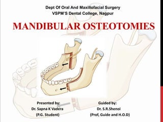
  • 1. MANDIBULAR OSTEOTOMIES Dept Of Oral And Maxillofacial Surgery VSPM’S Dental College, Nagpur Presented by: Guided by: Dr. Sapna K Vadera Dr. S.R.Shenoi (P.G. Student) (Prof, Guide and H.O.D)
  • 2. CONTENTS • Introduction • History • Aims of mandibular osteotomies • Principals in treatment of mandibular deformities • Surgical anatomy- Vessels, Nerves, Muscles • Classification • Sagittal split osteotomy • IVRO • Body osteotomy- Anterior & Posterior • Subapical Osteotomies- Anterior,Posterior, Total • Genioplasty • Conclusion
  • 3. INTRODUCTION Orthognathic in Greek Orthos- straight ; Gnathos- jaw Orthognathic surgery refers to surgical procedures designed to correct jaw deformities. Orthognathic surgery is the art and science of diagnosis, treatment planning, and execution of treatment by combining orthodontics and oral and maxillofacial surgery to correct musculoskeletal, dento- osseous, and soft tissue deformities of the jaws and associated structures Fonseca RJ, Marciani RD. Oral and Maxillofacial Surgery: Orthognathic surgery, esthetic surgery, cleft and craniofacial surgery. Turvey TA, editor. Saunders; 2009.
  • 4. Basic Therapeutic Goals For Orthognathic Surgery • To establish proper function ( normal mastication, speech, respiratory function) • To establish aesthetics ( Establishment of facial harmony) • Provide stability (Prevention of short and long term relapse) • Minimizing of treatment time Fonseca RJ, Marciani RD. Oral and Maxillofacial Surgery: Orthognathic surgery, esthetic surgery, cleft and craniofacial surgery. Turvey TA, editor. Saunders; 2009.
  • 5. Once growth has ceased, the combination of orthognathic surgery with orthodontics, usually becomes one of the common means of correcting severe dentofacial deformities In severe malocclusion there are three possibilities for correction: • Growth modification • Orthodontic treatment • Orthognathic surgery in conjunction with orthodontics to establish proper jaw relationship Orthognathic procedures are divided into three categories: Maxillary surgery Mandibular surgery Bimaxillary procedures Fonseca RJ, Marciani RD. Oral and Maxillofacial Surgery: Orthognathic surgery, esthetic surgery, cleft and craniofacial surgery. Turvey TA, editor. Saunders; 2009. INTRODUCTION
  • 6. • Orthognathic surgery was originally developed in the United States of America (Steinhäuser ). • The first mandibular osteotomy is considered to be Hullihen´s procedure in 1846 to correct anterior open bite & mandibular dento-alveolar protrusion with an intraoral osteotomy. • 50 years later Osteotomy of the mandibular body for the correction of mandibular horizontal excess was performed by Vilray Blair. 1906 HISTORY Fonseca RJ, Marciani RD. Oral and Maxillofacial Surgery: Orthognathic surgery, esthetic surgery, cleft and craniofacial surgery. Turvey TA, editor. Saunders; 2009.
  • 7. 1907 - Blair – Horizontal Subcondylar osteotomy of the ramus, external approach 1932 Intraoral Subcondylar osteotomy was given by Earnst HISTORY Fonseca RJ, Marciani RD. Oral and Maxillofacial Surgery: Orthognathic surgery, esthetic surgery, cleft and craniofacial surgery. Turvey TA, editor. Saunders; 2009.
  • 8. 1925 - Limberg Posterior oblique vertical ramal osteotomy, external approach Wassmund 1927 Vertical subcondylar osteotomy inverted –L-osteotomy. HISTORY Fonseca RJ, Marciani RD. Oral and Maxillofacial Surgery: Orthognathic surgery, esthetic surgery, cleft and craniofacial surgery. Turvey TA, editor. Saunders; 2009.
  • 9. Hofer in 1936 demonstrated an anterior mandibular alveolar osteotomy to advance anterior teeth in correction of a mandibular dentoalveolar retrusion. HISTORY Fonseca RJ, Marciani RD. Oral and Maxillofacial Surgery: Orthognathic surgery, esthetic surgery, cleft and craniofacial surgery. Turvey TA, editor. Saunders; 2009.
  • 10. Horizontal sliding osteotomy first described by Hofner in 1942 Hofer O. Operation der prognathic and mikrogenie. Dtsch Zahn Mund Kieferheilkd. 1942;0:121-32. HISTORY
  • 11. In 1954, Caldwell and Letterman developed a vertical ramus osteotomy technique, which had the advantage of minimizing trauma to the inferior alveolar neurovascular bundle. HISTORY Fonseca RJ, Marciani RD. Oral and Maxillofacial Surgery: Orthognathic surgery, esthetic surgery, cleft and craniofacial surgery. Turvey TA, editor. Saunders; 2009.
  • 12. The greatest development in osteotomies of the vertical ramus is the sagittal split osteotomy credited to obwegeser in 1955. The major modifications in the osteotomies design were first made by Dalpont in 1961.This was further discussed by Hunsuck in 1968 in order to decrease the trauma to overlying soft tissues. HISTORY
  • 13. Kent & Hinds in 1971 initially presented the use of single tooth osteotomies of the mandible. Macintosh closely followed with his description of the total mandibular alveolar osteotomy in 1974. HISTORY Fonseca RJ, Marciani RD. Oral and Maxillofacial Surgery: Orthognathic surgery, esthetic surgery, cleft and craniofacial surgery. Turvey TA, editor. Saunders; 2009.
  • 14. Aesthetics Function Stability AIMS OF MANDIBULAR OSTEOTOMIES Fonseca RJ, Marciani RD. Oral and Maxillofacial Surgery: Orthognathic surgery, esthetic surgery, cleft and craniofacial surgery. Turvey TA, editor. Saunders; 2009.
  • 15. • Patient’s perception of the deformity and expectations • Surgeon’s recognition of the deformity • Complete physical examination, model surgery, cephalometric analysis • Optimal treatment plan • Counseling of the patient • Informed consent PRINCIPLES IN TREATING MANDIBULAR DEFORMITIES Fonseca RJ, Marciani RD. Oral and Maxillofacial Surgery: Orthognathic surgery, esthetic surgery, cleft and craniofacial surgery. Turvey TA, editor. Saunders; 2009.
  • 17. Vascular structures NervesMuscles ANATOMICAL & PHYSIOLOGICAL CONSIDERATIONS OF MANDIBULAR OSTEOTOMIES Fonseca RJ, Marciani RD. Oral and Maxillofacial Surgery: Orthognathic surgery, esthetic surgery, cleft and craniofacial surgery. Turvey TA, editor. Saunders; 2009.
  • 18. • Bell and Levy’s work {1970} demonstrated that blood flow through the mandibular periosteum could easily maintain a sufficient blood supply to the teeth of a mobile segment, even when the labial periosteum was degloved. VASCULAR STRUCTURES Fonseca RJ, Marciani RD. Oral and Maxillofacial Surgery: Orthognathic surgery, esthetic surgery, cleft and craniofacial surgery. Turvey TA, editor. Saunders; 2009.
  • 19. VASCULAR STRUCTURES Fonseca RJ, Marciani RD. Oral and Maxillofacial Surgery: Orthognathic surgery, esthetic surgery, cleft and craniofacial surgery. Turvey TA, editor. Saunders; 2009.
  • 20. • The proximal segment of VRO maintains its blood supply through TMJ & capsule and attachment of lateral pterygoid muscle. • But inferior tip of this segment undergoes avascular necrosis. VASCULAR STRUCTURES Fonseca RJ, Marciani RD. Oral and Maxillofacial Surgery: Orthognathic surgery, esthetic surgery, cleft and craniofacial surgery. Turvey TA, editor. Saunders; 2009.
  • 21. • Determination of safe distance away from the apex of teeth is vital factor to be considered • If the vascularity of the segments and teeth are to be preserved. The safer distance is 5 mm but studies have shown even 10 mm distance shows pulpal changes. • A cut of 10 mm below apex shows greater safety. VASCULAR STRUCTURES Fonseca RJ, Marciani RD. Oral and Maxillofacial Surgery: Orthognathic surgery, esthetic surgery, cleft and craniofacial surgery. Turvey TA, editor. Saunders; 2009.
  • 23. • The position of the lingula is posterior-inferior relative to the position of the antilingula • Any osteotomies performed at a measurement of 5 mm posterior to the antilingula (at the level of the antilingula)- no risk of damaging the neurovascular bundle Aziz SR, Dorfman BJ, Ziccardi VB, Janal M. Accuracy of using the antilingula as a sole determinant of vertical ramus osteotomy position. Journal of oral and maxillofacial surgery. 2007 May 31;65(5):859-62. NERVE SUPPLY
  • 24. • A- S to lingula - 14.8 +/- 2.90 mm • C- S to mandibular foramen – 21.6 +/- 3.31 mm • B- Horizontal distance from linguala to anterior border of ramus – 17.7 +/- 2.89 mm • D- Mandibular foramen to ramus – 18.6 +/- 2.49 mm Aziz SR, Dorfman BJ, Ziccardi VB, Janal M. Accuracy of using the antilingula as a sole determinant of vertical ramus osteotomy position. Journal of oral and maxillofacial surgery. 2007 May 31;65(5):859-62. NERVE SUPPLY
  • 25. At a distance between 7.5 to 13.3 mm above the lingula Buccal and lingual cortex fusion occurs at a rate of • 20% in the anterior ramus • 39% in the posterior ramus • The medial horizontal cut be at or just above the tip of the lingula because a higher cut may be associated with an increased difficulty in splitting or incidence of unfavorable fracture. Aziz SR, Dorfman BJ, Ziccardi VB, Janal M. Accuracy of using the antilingula as a sole determinant of vertical ramus osteotomy position. Journal of oral and maxillofacial surgery. 2007 May 31;65(5):859-62. NERVE SUPPLY
  • 26. V1- 9.15 mm H1-0.57mm Dias GJ, de Silva RK, Shah T, Sim E, Song N, Colombage S, Cornwall J. Multivariate assessment of site of lingual nerve. British Journal of Oral and Maxillofacial Surgery. 2015 Apr 30;53(4):347-51. NERVE SUPPLY
  • 27. MUSCLES Orthognathic surgery affects muscles in primarily two ways: • It changes the length of a muscle or it changes the direction of muscle function. The muscles commonly discussed in orthognathic surgery of the mandible have been the muscles of mastication and the suprahyoid group of muscles . Fonseca RJ, Marciani RD. Oral and Maxillofacial Surgery: Orthognathic surgery, esthetic surgery, cleft and craniofacial surgery. Turvey TA, editor. Saunders; 2009.
  • 28. Removal of masseter & medial pterygoid attachment Condylar luxation (lateral pterygoid muscle pulling the condyle forward)
  • 29. 2 postulates • Medial and forward displacement of the mandibular disk- by the upper head of the lateral pterygoid muscle. • After sectioning - the mandibular condyle is displaced in the same direction as the disk - by the pull of the lower head of the lateral pterygoid muscle. Fonseca RJ, Marciani RD. Oral and Maxillofacial Surgery: Orthognathic surgery, esthetic surgery, cleft and craniofacial surgery. Turvey TA, editor. Saunders; 2009.
  • 30. REVASCULARISATION & HEALING Immediate post-operatively • Intermedullary circulation between the proximal and distal segments • Margins of osteotomy- avascular One week post-op • Level of hypervascularity around surgical site • No soft tissue re-attachment • Isolated areas of sub- periosteal bone formation Fonseca RJ, Marciani RD. Oral and Maxillofacial Surgery: Orthognathic surgery, esthetic surgery, cleft and craniofacial surgery. Turvey TA, editor. Saunders; 2009.
  • 31. 2 weeks post-op • Avascular zone at the proximal osteotomy site • Necrotic zone at the distal osteotomy site • No soft tissue attachment at the distal necrotic zone 3 weeks post-op • Soft tissue re-attachment • Vascular anastamoses between proximal and distal segments • Osteoid formation through out marrow formation REVASCULARISATION & HEALING Fonseca RJ, Marciani RD. Oral and Maxillofacial Surgery: Orthognathic surgery, esthetic surgery, cleft and craniofacial surgery. Turvey TA, editor. Saunders; 2009.
  • 32. 6 weeks post-op • Circulation reconstituted across the osteotomy site • Soft tissue re- attachment established 12 weeks post- op • Circulation between the segments is continuous REVASCULARISATION & HEALING Fonseca RJ, Marciani RD. Oral and Maxillofacial Surgery: Orthognathic surgery, esthetic surgery, cleft and craniofacial surgery. Turvey TA, editor. Saunders; 2009.
  • 33. CLASSIFICATION MANDUBULAR ORTHOGNATHIC PROCEDURES BODY OSTEOTOMIES SUB APICAL OSTEOTOMIES RAMUS OSTEOTOMIES HORIZONTAL OSTEOTOMY OF CHIN Sagital Split Osteotomy Vertical Ramus Osteotomy Inverted “L” & “C” Osteotomy Anterior Sub Apical Osteotomies Posterior Sub Apical Osteotomies Total Sub Apical Osteotomies Condylotomy/ Condylectomy Anterior To Mental Foramen Step osteotomy/ostectomy Posterior To Mental Foramen Y Ostectomy Rectangular ostectomy Trapizoid ostectomy Inverted V ostectomy
  • 34. SAGITTAL SPLIT OSTEOTOMY • A surgical procedure resembling the saggital split osteotomy was described in 1942 in the German literature by Schuchardt. • Lane evidently described a similar procedure earlier, which was done extraorally. Schuchardt G: Ein Beitrag zur chirurgischen Kieferorthopadie unter Berucksichtigung ihrer fur die Behandlung angeborener und erworbener Kieferdeformitaten bei Soldaten. Dtsch Zahn Mund Kieferheilkd 1942;9;73. Parallel horizontal cuts through the cortex of the vertical ramus, the medial cut being placed above the lingula and a lateral cut being made about 1 cm below that.
  • 35. HUGO OBWEGESER & TRAUNER 1957 • The first to discuss its use in English literature • Satisfactory for prognathism but very little bone contact in mandibular retrusion. Traunar R, Obwegeser H: Operative Oral Surgery: The surgical correction of mandibular prognathism and retrognathia with consideration of genioplasty. Oral Surg Oral Med Oral Pathol 1957;10;677
  • 36. DALPONT (1961) Advanced the oblique cut towards molar region and made it vertical through the lateral cortex. Dal Pont G: Retromolar osteotomy for correction of prognathism. J Oral Surg 1961:19:42
  • 37. HUNSUCK (1968) Shortened the cut through the medial cortex taking it only as far as the mandibular foramen. Hunsuk E: A modified intraoral saggital splitting technique for correction of mandibular prognathism. J Oral Surg 1968;26;249
  • 38. BELL SCHENDEL (1977) & EPKER (1978) • Hunsuck technique is adopted but on the lateral aspect the vertical cut is taken downwards from an oblique line through outer cortex to lower border where the lower border is sectioned. • Minimal detachment of the pterygomassetric sling there is decreased intra- osseous ischemia, and necrosis of the proximal segment Epker BN: Modifications in the saggital split osteotomy of the mandible. J Oral Surg 1977;35;157. Bell W, Schendel S: Biological basis for the saggital ramus split operation J Oral Surg 1977;35;362
  • 39. Bell schendel (1977) and Epker (1978) : Hunsuck technique is adopted but on the lateral aspect the vertical cut is taen downwards from an oblique line through outer cortex to lower border where the lower border is sectioned. Bell schendel (1977) & Epker (1978) SAGITTAL SPLIT OSTEOTOMY Bell W, Schendel S: Biological basis for the saggital ramus split operation J Oral Surg 1977;35;362 Epker BN: Modifications in the saggital split osteotomy of the mandible. J Oral Surg 1977;35;157.
  • 40. FIXATION- ADVANCEMENT • Development of rigid internal fixation by Spiessl in 1974 replaced osteosynthesis by wire fixation or IMF. • Jeter described technique of placing 3 bicortical 2.0 mm position screws to fix the proximal and distal segments. • Blomqvist and others showed no significant difference in terms of relapse between monocortical screws with miniplates and bicortical screws for mandibular advancement. • Recently good stability after BSSO is also shown by polylactate bone plates and screws Beukes J, Reyneke JP, Damstra J. Unilateral sagittal split mandibular ramus osteotomy: indications and geometry. British Journal of Oral and Maxillofacial Surgery. 2016 Feb 29;54(2):219-23.
  • 41. INDICATIONS 1.Mandibular deficiency-with normal or short face, -with long face- increase maxillary vertical dimension - excessive chin height - for correction of sleep apnea Limitation- • Advancements beyond 10- 12 mm, extra oral approach should be considered • Additional surgery for most dentofacial deformity 2.Mandibular prognathism- short face - long face • Limitation -Large setbacks of more than 7 -8 mm, IVRO/ inverted L osteotomy should be considered 3.Mandibular asymmetry- Hemi mandibular hypertrophy - Hemi mandibular elongation 4. Open bite 5. Cross bite Beukes J, Reyneke JP, Damstra J. Unilateral sagittal split mandibular ramus osteotomy: indications and geometry. British Journal of Oral and Maxillofacial Surgery. 2016 Feb 29;54(2):219-23.
  • 42. CONTRAINDICATIONS • Severe decreased posterior mandibular body height • Extremely thin medial –lateral width of ramus • Severe ramus hypoplasia and • Severe asymmetries Beukes J, Reyneke JP, Damstra J. Unilateral sagittal split mandibular ramus osteotomy: indications and geometry. British Journal of Oral and Maxillofacial Surgery. 2016 Feb 29;54(2):219-23.
  • 43.
  • 44. Incision & Dissection Beukes J, Reyneke JP, Damstra J. Unilateral sagittal split mandibular ramus osteotomy: indications and geometry. British Journal of Oral and Maxillofacial Surgery. 2016 Feb 29;54(2):219-23.
  • 45. • Medial dissection done after ascertaining the position of lingula • Sigmoid notch identified • Minimal traction on medial side to avoid injury to the neurovascular bundle. Incision & Dissection Beukes J, Reyneke JP, Damstra J. Unilateral sagittal split mandibular ramus osteotomy: indications and geometry. British Journal of Oral and Maxillofacial Surgery. 2016 Feb 29;54(2):219-23.
  • 46. Sagittal Bone Cut 5mm Above The MandibularForamen With Long Lindemann Burr Osteotomy cut Beukes J, Reyneke JP, Damstra J. Unilateral sagittal split mandibular ramus osteotomy: indications and geometry. British Journal of Oral and Maxillofacial Surgery. 2016 Feb 29;54(2):219-23.
  • 47. Smith & colleagues noted that in 2% of cases cortical plates were fused inferior to the lingula at the anterior portion of the ramus. Osteotomy cut Smith B, Rajchel J, Waite D, et al: Mandibular ramus anatomy as it relates to the medial osteotomy of the saggital split osteotomy J Oral Maxillofac Surg 1991;49;112
  • 48. • Vertical cut in 2nd molar region • Depth of cut should be just enough to reach the cancellous bone • Rotary instrument or reciprocating saw Osteotomy cut
  • 49. • Small spatula osteotome is malleted and directed laterally • Smith spreader used to separate the segments Beukes J, Reyneke JP, Damstra J. Unilateral sagittal split mandibular ramus osteotomy: indications and geometry. British Journal of Oral and Maxillofacial Surgery. 2016 Feb 29;54(2):219-23.
  • 50. Great care should be undertaken to avoid fracturing the buccal plate or the proximal extension of the distal segment, especially if the Smith spreader instrument is used ,either of those fractures may preclude the use of rigid internal fixation. Any prying or torquing of these segments should be minimized.
  • 51. Care Of The Neurovascular Bundle • Neurovascular bundle visualized • It should be on the medial fragment • If mandible is to be advanced, medial pterygoid is separated from the inferior border • If mandible is to set back, medial pterygoid and masseter needs to be stripped off to prevent posterior displacement of the condylar segment Beukes J, Reyneke JP, Damstra J. Unilateral sagittal split mandibular ramus osteotomy: indications and geometry. British Journal of Oral and Maxillofacial Surgery. 2016 Feb 29;54(2):219-23.
  • 52. Mandibular Advancement Mandibular Setback Beukes J, Reyneke JP, Damstra J. Unilateral sagittal split mandibular ramus osteotomy: indications and geometry. British Journal of Oral and Maxillofacial Surgery. 2016 Feb 29;54(2):219-23.
  • 53.
  • 54. Fixation techniques Fujioka M, Fujii T, Hirano A. Comparative study of mandibular stability after sagittal split osteotomies: bicortical versus monocortical osteosynthesis. Cleft palate craniofacial journal 2000; 37:551.
  • 55.
  • 56. • With wire at upper and lower border • Lag screws • Bicortical screws – 2 or 3 screws are used • Mini plates • Bioresorbable plates and screws Fujioka M, Fujii T, Hirano A. Comparative study of mandibular stability after sagittal split osteotomies: bicortical versus monocortical osteosynthesis. Cleft palate craniofacial journal 2000; 37:551. Fixation techniques
  • 57. ADVANTAGES • Healing is quick because of a good bony interface • Three dimensional flexibility in repositioning the distal fragments. • Broad bony overlap of osteotomized segments • Minimal alteration of the position of muscles of mastication – prevents relapse from muscular traction • The surgery can advance or set back the mandible, correct most asymmetries • Rigid fixation can be used, eliminating the need for MMF. Rigid fixation significantly improves the stability and predictability of results. • Minimal alteration of the position of TMJ – prevents Post operative TMJ dysfunction Beukes J, Reyneke JP, Damstra J. Unilateral sagittal split mandibular ramus osteotomy: indications and geometry. British Journal of Oral and Maxillofacial Surgery. 2016 Feb 29;54(2):219-23.
  • 58. COMPLICATIONS • Relapse • Nerve injury • TMJ Dysfunction and hypomobility • Haemorrhage Beukes J, Reyneke JP, Damstra J. Unilateral sagittal split mandibular ramus osteotomy: indications and geometry. British Journal of Oral and Maxillofacial Surgery. 2016 Feb 29;54(2):219-23.
  • 59. CONDYLAR POSITION • Rotation of the proximal segment • Condylar sag • Condylar torque These malpositions can lead to • Skeletal relapse • Malocclusion • Hypomobility • Remodeling of the condylar head
  • 60. Condylar Sag Central Unilateral Bilateral Peripheral Type I Type II TYPES Johan. P. Reyneke, C. Ferretti, Intraoperative diagnosis of condylar sag after bilateral sagittal split ramus osteotomy ,British Journal of Oral and Maxillofacial Surgery (2002) 40, 285–292
  • 61. CENTRAL CONDYLAR SAG Johan. P. Reyneke, C. Ferretti, Intraoperative diagnosis of condylar sag after bilateral sagittal split ramus osteotomy ,British Journal of Oral and Maxillofacial Surgery (2002) 40, 285–292
  • 62. PERIPHERAL CONDYLAR SAG - I Johan. P. Reyneke, C. Ferretti, Intraoperative diagnosis of condylar sag after bilateral sagittal split ramus osteotomy ,British Journal of Oral and Maxillofacial Surgery (2002) 40, 285–292
  • 63. PERIPHERAL CONDYLAR SAG - II Johan. P. Reyneke, C. Ferretti, Intraoperative diagnosis of condylar sag after bilateral sagittal split ramus osteotomy ,British Journal of Oral and Maxillofacial Surgery (2002) 40, 285–292
  • 64. MALOCCLUSION Open Bite • Inadequate Fixation • Posterior open bite during fixation Lateral shift • Inadequate advancement on 1 side • Equal bilateral advancement with midline shift • Torquing of the proximal segment Beukes J, Reyneke JP, Damstra J. Unilateral sagittal split mandibular ramus osteotomy: indications and geometry. British Journal of Oral and Maxillofacial Surgery. 2016 Feb 29;54(2):219-23.
  • 65. • Elastic traction when there is no anterioposterior discrepancies for 2-3 weeks • When there is AP discrepencies reenter, explore and advance the descrepant site MALOCCLUSION Beukes J, Reyneke JP, Damstra J. Unilateral sagittal split mandibular ramus osteotomy: indications and geometry. British Journal of Oral and Maxillofacial Surgery. 2016 Feb 29;54(2):219-23.
  • 66. Bad split Abort the procedure & perform after healing Correct the split & Complete the procedure Incidence-18% Beukes J, Reyneke JP, Damstra J. Unilateral sagittal split mandibular ramus osteotomy: indications and geometry. British Journal of Oral and Maxillofacial Surgery. 2016 Feb 29;54(2):219-23. BAD SPLIT
  • 67. BAD SPLIT • Fracture of buccal plate • Fracture of lingual flange • Inferior border left in the proximal fragment • Condylar split • Impacted 3rd molars Beukes J, Reyneke JP, Damstra J. Unilateral sagittal split mandibular ramus osteotomy: indications and geometry. British Journal of Oral and Maxillofacial Surgery. 2016 Feb 29;54(2):219-23.
  • 68. FRACTURES ON THE PROXIMAL PORTION • Also called “Buccal plate fracture” • Small Proximal Fragment • Bicortical Screws • Free Fragment is checked for compatibility • Secure the segment with plate Beukes J, Reyneke JP, Damstra J. Unilateral sagittal split mandibular ramus osteotomy: indications and geometry. British Journal of Oral and Maxillofacial Surgery. 2016 Feb 29;54(2):219-23.
  • 69. Most frequent • Presence of impacted 3rd molar • Recent removal of 3rd molar • Age of the patient • Incomplete transection of the inferior border • Surgeon’s experience FRACTURES ON THE PROXIMAL PORTION Role of impacted 3rd molars in unfavorable # is debatable Advocated removal 6months prior surgery Beukes J, Reyneke JP, Damstra J. Unilateral sagittal split mandibular ramus osteotomy: indications and geometry. British Journal of Oral and Maxillofacial Surgery. 2016 Feb 29;54(2):219-23.
  • 70. Medial Splits Up The Condyle • Medial cut more superior to the lingula • Angling the cut in an oblique fashion towards condylar neck • Chiesel should not be used Fracture of coronoid process • Occurs when the horizontal cut is placed too high where the ramus is thin Fracture of distal segments • Inferior border remains attached to distal segment Beukes J, Reyneke JP, Damstra J. Unilateral sagittal split mandibular ramus osteotomy: indications and geometry. British Journal of Oral and Maxillofacial Surgery. 2016 Feb 29;54(2):219-23.
  • 71. RELAPSE • Mandibular advancements greater than 7 mm • Suprahyoid myotomies and orthodontic overcorrection • Prevention • Proximal segment control • Proper condylar positioning • Avoidance of condylar rotation • Decreased when 1-2 week skeletal fixation used • Suprahyoid myotomies and orthodontic overcorrection Van Sickels JE: a comparative study of bicortical screws and suspension wires versus bicortical screws in large mandibular advancements. J Oral & Maxillofac Surg 1991;49;1970
  • 72. TMJ DYSFUNCTION & HYPOMOBILITY • 20 – 25% • Prolonged immobility • Intraarticular haemorrhage • Fibrosis • Preexisting TMJ disorder Beukes J, Reyneke JP, Damstra J. Unilateral sagittal split mandibular ramus osteotomy: indications and geometry. British Journal of Oral and Maxillofacial Surgery. 2016 Feb 29;54(2):219-23.
  • 73. NERVE DAMAGE -Injury to the Inferior Alveolar Nerve (IAN) -White et al pointed out that damage to the inferior alveolar nerve most likely occurs either during the medial retraction of the soft tissues and the nerve as it enters the canal or during the vertical bone cut -Guernsey et al felt that damage occurred during the splitting of the mandible and reported the problem of parts of the nerve staying in the proximal fragment after the split. Beukes J, Reyneke JP, Damstra J. Unilateral sagittal split mandibular ramus osteotomy: indications and geometry. British Journal of Oral and Maxillofacial Surgery. 2016 Feb 29;54(2):219-23.
  • 74. Injury to the Lingual Nerve • Less common • Higher incidence of neurosensory disturbance with bicortical screws than monocortical screws NERVE DAMAGE Beukes J, Reyneke JP, Damstra J. Unilateral sagittal split mandibular ramus osteotomy: indications and geometry. British Journal of Oral and Maxillofacial Surgery. 2016 Feb 29;54(2):219-23.
  • 75. Reasons for Tear or Cut Inferior Alveolar Nerve (IAN) • Application of local anesthesia • Stretching of the nerve from the medial protecting retractors • Forced osteotomy with reciprocating saw or chisel during splitting • Abnormal anatomic position of the IAN canal • Presence of an Impacted third molar NERVE DAMAGE Beukes J, Reyneke JP, Damstra J. Unilateral sagittal split mandibular ramus osteotomy: indications and geometry. British Journal of Oral and Maxillofacial Surgery. 2016 Feb 29;54(2):219-23.
  • 76. Measures to Prevent Injury to the IAN During BSSO • Preparation subperiosteal at the medial side of the ascending ramus • Prevent Stretching of the nerve from the medial protecting retractors • Rotating instruments and saws may be used only to open the mandibular cortex • Osteotome may penetrate only very superficially into the mandible during splitting • Removal of impacted third molars at least 6 months before surgery NERVE DAMAGE Beukes J, Reyneke JP, Damstra J. Unilateral sagittal split mandibular ramus osteotomy: indications and geometry. British Journal of Oral and Maxillofacial Surgery. 2016 Feb 29;54(2):219-23.
  • 77. Measures to Prevent Lingual Nerve • Measurement of the depth of the drilling hole when screws longer than 15mm are used • Drainage of larger hematomas to allow quick recovery from pressure on lingual nerve NERVE DAMAGE Beukes J, Reyneke JP, Damstra J. Unilateral sagittal split mandibular ramus osteotomy: indications and geometry. British Journal of Oral and Maxillofacial Surgery. 2016 Feb 29;54(2):219-23.
  • 78. HAEMORRHAGE Incidence decreased from 38% in 1972 to 1% in 2005 Most common sources • Maxillary artery and its branches (massetric and inferior alveolar artery) • Retromandibular vein • Facial artery and vein Beukes J, Reyneke JP, Damstra J. Unilateral sagittal split mandibular ramus osteotomy: indications and geometry. British Journal of Oral and Maxillofacial Surgery. 2016 Feb 29;54(2):219-23.
  • 80. • 1st described by Caldwell and Letterman in 1954- extra oral • Introduced by Moose in 1964- intra- oral technique performed from lingual aspect • Wistanley, 1968- performing the technique from the lateral aspect of the mandible • Modified by herbert in 1970. used at present INTRA-ORAL VERTICAL RAMUS OSTEOTOMIES McKenna SJ, King EE. Intraoral Vertical Ramus Osteotomy Procedure and Technique. Atlas of the oral and maxillofacial surgery clinics of North America. 2016 Mar 1;24(1):37-43.
  • 81. INDICATIONS • Horizontal mandibular excess • Mandibular asymmetry • Minor occlusal discrepancy after isolated Le Fort I osteotomy • Asymmetric lateral open bite • Failure to achieve passive rotation of the mandible after the release of MMF • Patients with significant TMJ complaints McKenna SJ, King EE. Intraoral Vertical Ramus Osteotomy Procedure and Technique. Atlas of the oral and maxillofacial surgery clinics of North America. 2016 Mar 1;24(1):37-43.
  • 82. • Advancement of the distal segment • Aesthetic assessment of the soft tissues of the neck is the integral factor in planning mandibular set back by ramus surgery • Recent condylar fractures • A-p decrepany more than 6-7mm • Prexisting heavy neck CONTRAINDICATIONS McKenna SJ, King EE. Intraoral Vertical Ramus Osteotomy Procedure and Technique. Atlas of the oral and maxillofacial surgery clinics of North America. 2016 Mar 1;24(1):37-43.
  • 83. ADVANTAGES • Can be performed on OPD basis • Inherent anatomic architecture of the mandible poses little interference to place the cuts • Less chance of damaging the IAN bundle • Found to have curable effects in pts with pre-op TMD • Excellent post op stability DISADVANTAGES • Need for MMF 7-10 days • Post op physiotherapy- for 1-2 week McKenna SJ, King EE. Intraoral Vertical Ramus Osteotomy Procedure and Technique. Atlas of the oral and maxillofacial surgery clinics of North America. 2016 Mar 1;24(1):37-43.
  • 84.
  • 85. SURGICAL PROCEDURE Intra orally the incision is made in the mucosa from midway up the anterior border of the ramus to the first molar area McKenna SJ, King EE. Intraoral Vertical Ramus Osteotomy Procedure and Technique. Atlas of the oral and maxillofacial surgery clinics of North America. 2016 Mar 1;24(1):37-43.
  • 86. SURGICAL PROCEDURE McKenna SJ, King EE. Intraoral Vertical Ramus Osteotomy Procedure and Technique. Atlas of the oral and maxillofacial surgery clinics of North America. 2016 Mar 1;24(1):37-43.
  • 87. SURGICAL PROCEDURE McKenna SJ, King EE. Intraoral Vertical Ramus Osteotomy Procedure and Technique. Atlas of the oral and maxillofacial surgery clinics of North America. 2016 Mar 1;24(1):37-43.
  • 88. McKenna SJ, King EE. Intraoral Vertical Ramus Osteotomy Procedure and Technique. Atlas of the oral and maxillofacial surgery clinics of North America. 2016 Mar 1;24(1):37-43.
  • 89. The effect of the temporalis on relapse has led to other recommendations that include either stripping the temporalis attachment completely off the coronoid or cutting off the coronoid. Hamid Mahmood . Evaluation of intraoral verticosagittal ramus osteotomy for correction of mandibular prognathism : A 10 yr study . J Oral Maxillofac Surg 2008: 66:509
  • 90. COMPARISON BETWEEN SSRO AND VRO SSRO VRO OSTEOTOMY PA Saggital split Latero medial cut Open procedure Blind procedure Along IAN Rear to IAN Frequent exposure of IAN No exposure of IAN BONE HEALING Contact on marrow to marrow Contact on cortex to cortex BONE FIXATION Rigid internal fixation No fixation CONDYLAR HEAD Original position New equilibrated position POST OP IMF prognosis None or shorter period Weakly dependent on pt Required 7-10 day Strongly dependent on pt McKenna SJ, King EE. Intraoral Vertical Ramus Osteotomy Procedure and Technique. Atlas of the oral and maxillofacial surgery clinics of North America. 2016 Mar 1;24(1):37-43.
  • 91. COMPLICATIONS • UNFAVOURABLE OSTEOTOMY Inadvertent subcondylar osteotomy More likely in • Prognathic mandible with high mandibular plane angle and ill- defined gonial angle McKenna SJ, King EE. Intraoral Vertical Ramus Osteotomy Procedure and Technique. Atlas of the oral and maxillofacial surgery clinics of North America. 2016 Mar 1;24(1):37-43.
  • 92. NERVE INJURY Incidence ranges from 0%- 14% Less incidence when compared to SSO Can occur in 2 phases • If osteotomy is close to mandibular foramen • Medial displacement of the proximal segment compressing and tearing the nerve McKenna SJ, King EE. Intraoral Vertical Ramus Osteotomy Procedure and Technique. Atlas of the oral and maxillofacial surgery clinics of North America. 2016 Mar 1;24(1):37-43.
  • 93. Bleeding Common source- maxillary artery and its branches Proximal Segment Malpositioning Control of proximal segment- major disadvantage • May be displaced antero- medially, anteriorly towards articular eminence or can be displaced medially and inferiorly McKenna SJ, King EE. Intraoral Vertical Ramus Osteotomy Procedure and Technique. Atlas of the oral and maxillofacial surgery clinics of North America. 2016 Mar 1;24(1):37-43.
  • 94. CONDYLOTOMY Mild mandibular prognathism TMJ internal derrangements Approaches: Extra oral Intraoral Blind :gigli saw Fonseca RJ, Marciani RD. Oral and Maxillofacial Surgery: Orthognathic surgery, esthetic surgery, cleft and craniofacial surgery. Turvey TA, editor. Saunders; 2009.
  • 95. Fonseca RJ, Marciani RD. Oral and Maxillofacial Surgery: Orthognathic surgery, esthetic surgery, cleft and craniofacial surgery. Turvey TA, editor. Saunders; 2009.
  • 96. CONDYLECTOMY Indications: • Ankylosis • Tumors • Condylar hyperplasia • Mandibular asymmetry: 1.Hemifacial microsomia 2. Unilateral: Hypertrophy Elongation Can be combined with joint reconstruction Or with other osteotomy procedures Fonseca RJ, Marciani RD. Oral and Maxillofacial Surgery: Orthognathic surgery, esthetic surgery, cleft and craniofacial surgery. Turvey TA, editor. Saunders; 2009.
  • 97.
  • 98. Blair -1907-as an extra oral procedure Dingman –combination of extra-oral and intra oral access with preservation of IAN and bon grafting-assist bony union. Now contemplated only as an intraoral procedure. Fonseca RJ, Marciani RD. Oral and Maxillofacial Surgery: Orthognathic surgery, esthetic surgery, cleft and craniofacial surgery. Turvey TA, editor. Saunders; 2009.
  • 99. INDICATIONS Mandibular setback • In Mandibular prognathism with ramus procedure. • In Mandibular prognathism where long body in relation to ramus Anterior open bite closure-superior repositioning with sub apical will make increase ant teeth show Curve of spee reduction Progenia jaw correction • In class III-anterior body osteotomy –wedge of bone removed and set back Mandibular advancement- less used Fonseca RJ, Marciani RD. Oral and Maxillofacial Surgery: Orthognathic surgery, esthetic surgery, cleft and craniofacial surgery. Turvey TA, editor. Saunders; 2009.
  • 100. Potential for sensory problem CONTRAINDICATIONS
  • 101. PITFALLS • Anatomic discrepancies leading to reduction in bone to bone contact- distal segment is set back into wider proximal segment intra-bony width. • Segment control • Torqueing of the proximal segments is the classic problem. • Intermediate wafers , cast cap splints can be used- some control over displacing muscle pull. • Neurovascular bundle position- lingual orientation in 2nd molar region to more buccal position at mental foramen. Lowest point of canal is distal half of 1st molar • Root anatomy is variable • Difficult to perform osteotomy in the premolar region when trying to protect the mental nerve and root of the 1st premolar • Root torquing may help
  • 102.
  • 103. Anterior body osteotomy ( Straight Vertical osteotomy) Bilaterally small vestibular incisions are taken leaving attached gingiva intact, into first or second premolar regions, depending on the extraction. Extract the tooth in the segment which is going to be resected before performing the osteotomies. The inferior alveolar nerve can be identified and mobilized after removing the lateral cortical bone overlying the nerve. After the alveolar nerve is identified and mobilized, two parallel vertical osteotomy lines are marked with a pen or drill on the bone surface. The lingual mucoperiosteal layer is detached from the bone with a periosteal elevator. The osteotomy is then performed with either a saw, drill .
  • 104. Internal fixation is usually performed with two straight miniplates one above and one below the inferior alveolar nerve. The plate placement and drilling is usually performed from the transoral route After completion of both osteotomies the segment of bone is removed. After bilateral resection, the anterior segment of the mandible is moved posteriorly into the preplanned position. Mandibulo-maxillary fixation is performed to position the mandibular segments to the desired relationship with the maxilla. A prefabricated surgical splint (or wafer) may be used to facilitate this. Fonseca RJ, Marciani RD. Oral and Maxillofacial Surgery: Orthognathic surgery, esthetic surgery, cleft and craniofacial surgery. Turvey TA, editor. Saunders; 2009.
  • 105. • The step osteotomy may be indicated in cases of mandibular prognathism, retrognathism, asymmetry, and apertognathia. • Fonseca RJ, Marciani RD. Oral and Maxillofacial Surgery: Orthognathic surgery, esthetic surgery, cleft and craniofacial surgery. Turvey TA, editor. Saunders; 2009.
  • 106. Planned osteotomies in 1st molar region Diverging vertical incisions in buccal vestibule adjacent to area of planned osteotomy; horizontal osteotomy is made superior to level of inferior alveolar nerve to intersect with vertical bone incisions. Posterior Body Osteotomy Fonseca RJ, Marciani RD. Oral and Maxillofacial Surgery: Orthognathic surgery, esthetic surgery, cleft and craniofacial surgery. Turvey TA, editor. Saunders; 2009.
  • 107. Sequential excision of buccal, lingual and inferior cortical plates Exposure of inferior alveolar bundle by careful excision of cancellous bone Excision of residual inferior lingual cortical bone facilitated by retraction of the inferior alveolar nerve Interosseous wire placed through margins of osteotomised bone before body osteotomy is completed Apposition of segments fixed with interosseous wires
  • 108.
  • 109. ANTERIOR MADIBULAR SUB-APICAL OSTEOTOMY • Earliest referenced description of symphyseal osteotomies was by Trauner in 1952 • Aids in correction of dentofacial deformities. • When combined with AMO non skeletal open bite or bimaxillary protrusion can be corrected • Useful to level the plane of occlusion with out decreasing the vertical facial height Fonseca RJ, Marciani RD. Oral and Maxillofacial Surgery: Orthognathic surgery, esthetic surgery, cleft and craniofacial surgery. Turvey TA, editor. Saunders; 2009.
  • 110.
  • 111. Fonseca RJ, Marciani RD. Oral and Maxillofacial Surgery: Orthognathic surgery, esthetic surgery, cleft and craniofacial surgery. Turvey TA, editor. Saunders; 2009.
  • 112. Modification by KOLE Bone gaps caused by movement of the segment,especially by vertical movement necessary for the closure of an anterior open bite, should be grafted. The use of cortical bone from the symphysis, as advocated by Kole Kole H. Surgical operations on the alveolar ridge to correct occlusal abnormalities. Oral Surg Oral Med Oral Pathol 1959; 12:277.
  • 113. COMPLICATIONS • Loss of bone or teeth in osteotomised segment.(lingual tissues not protected-decrease in blood supply) • Bone cuts placed close to the teeth-loss of vitality and periodontal defects • Mental nerve paresthesia-directly related to the amount of trauma Fonseca RJ, Marciani RD. Oral and Maxillofacial Surgery: Orthognathic surgery, esthetic surgery, cleft and craniofacial surgery. Turvey TA, editor. Saunders; 2009.
  • 114. POSTERIOR SUBAPICAL OSTEOTOMY First described by- Peterson Indications • Correction of super eruption of posterior mandibular teeth • Ankylosis of one or more posterior teeth • Abnormal buccal or lingual position of these teeth especially if orthodontics is not feasible Fonseca RJ, Marciani RD. Oral and Maxillofacial Surgery: Orthognathic surgery, esthetic surgery, cleft and craniofacial surgery. Turvey TA, editor. Saunders; 2009.
  • 115. TOTAL MANDIBULAR SUBAPICAL OSTEOTOMY • Oldest procedures used to correct Jaw Deformity. • Described by HULLIHEN in 1849. • Popularised by Hofer and Kole. Fonseca RJ, Marciani RD. Oral and Maxillofacial Surgery: Orthognathic surgery, esthetic surgery, cleft and craniofacial surgery. Turvey TA, editor. Saunders; 2009.
  • 116. Primary indication Malocclusion caused by Mandibular Dentoalveolar deformity with normally positioned Maxilla and Mandibular skeletal bases • To increase the height of the mandible • To level the occlusal plane Fonseca RJ, Marciani RD. Oral and Maxillofacial Surgery: Orthognathic surgery, esthetic surgery, cleft and craniofacial surgery. Turvey TA, editor. Saunders; 2009.
  • 117. Modification by Booth et al Booth et al suggested a variation of the total mandibular subapical osteotomy that combines the sagittal split osteotomy of the vertical ramus with the total mandibular alveolar osteotomy. Advantages :osteotomy is made below the inferior alveolar nerve, thereby decreasing the risk of damaging the inferior alveolar nerve and the apices of the teeth, at the same time preserving much of the vascular supply to the mobile segment. Also the sagittal part of the osteotomy allows a larger bone contact area to assist in healing Booth DF, Dietz V, Gianelly AA. Correction of class III malocclusion by combined sagittal ramus and subapical body osteotomy. J Oral Surg 1976; 34:630
  • 118.
  • 119. • Facial features often form a basis for stereotyping of personality characteristics • Chin is most prominent facial feature • Chin deformities can manifest in 3 dimensions but majority are in the horizontal direction Fonseca RJ, Marciani RD. Oral and Maxillofacial Surgery: Orthognathic surgery, esthetic surgery, cleft and craniofacial surgery. Turvey TA, editor. Saunders; 2009.
  • 120. Horizontal sliding osteotomy-first described by Hofer in 1942- through extra oral approach. Converse 1950- fesibility of bone graft by intra oral approach Trauner and Obwegeser-1957- horizontal osteotomy through an intra oral incision. Reichenbach-1965-wedge osteotomy and vertical shortening of the chin. Fonseca RJ, Marciani RD. Oral and Maxillofacial Surgery: Orthognathic surgery, esthetic surgery, cleft and craniofacial surgery. Turvey TA, editor. Saunders; 2009.
  • 121.
  • 122.
  • 123.
  • 124. HORIZONTAL OSTEOTOMY WITH ADVANCEMENT Incision halfway between the depth of the vestibule extended upto canine B/L Reflection of periosteum by keeping periosteum intact in inferior border Maintain 5-10 mm of periosteum in the midpoint of symphysis region so that soft tissue support and blood supply are maintained Osteotomy cut should be 5 mm below the canine tooth root and 10-15 mm above the inferior border.and 4-5 mm below lowest mental foramen The more parallel the osteotomy cut with the occlusal plane and the mandibular plane , more easy AP movement Buccal and lingual cortical cuts be complete on proximal region.- reciprocating saw Fonseca RJ, Marciani RD. Oral and Maxillofacial Surgery: Orthognathic surgery, esthetic surgery, cleft and craniofacial surgery. Turvey TA, editor. Saunders; 2009.
  • 126. Same as advancement Reduced proximal tips of mobilised segments to ensure smooth transition along inferior border and avoid palpable wings Plus take ant vertical height of mandible into consideration HORIZONTAL OSTEOTOMY WITH A-P REDUCTION
  • 127. TENON TECHNIQUE Michelet 1974 Adv- Symmetry is ensured by tenon and visual inspection of proximal extention Single lag screw required Disadvantage- amount of advancement is limited by oerall thickness of ant mandible.
  • 128. DOUBLE SLIDING HORIZONTAL OSTEOTOMY Wedge vertical reduction osteotomy allows for anteroposterior repositioning in addition to vertical shortening Fonseca RJ, Marciani RD. Oral and Maxillofacial Surgery: Orthognathic surgery, esthetic surgery, cleft and craniofacial surgery. Turvey TA, editor. Saunders; 2009.
  • 129. ... Downward movements are associated with gap formation. This gap needs to be filled with either autogenous bone or a bone substitute, to ensure a predictable contour Fonseca RJ, Marciani RD. Oral and Maxillofacial Surgery: Orthognathic surgery, esthetic surgery, cleft and craniofacial surgery. Turvey TA, editor. Saunders; 2009.
  • 130. For transverse genial deformities, vertical osteotomies/ostectomies can also be performed. This allows widening or narrowing of the chin. In case of widening, additional bone grafts or alloplastic implants (eg, ceramic blocks) are needed vertical osteotomies/ostectomies
  • 132. COMPLICATIONS UNFAVOURABLE OSTEOTOMY • Inadvertent # of body and ramus • Damage to teeth roots NERVE INJURY Mental nerve is commonly injured • incision, reflection and retraction, osteotomies, plating or closure Fonseca RJ, Marciani RD. Oral and Maxillofacial Surgery: Orthognathic surgery, esthetic surgery, cleft and craniofacial surgery. Turvey TA, editor. Saunders; 2009.
  • 133. BLEEDING Damage to lingual soft tissues • Injury to genioglossus, geniohyoid muscles • Laceration of sublingual and submental arteries Usually not life threatening Managed by local measures Fonseca RJ, Marciani RD. Oral and Maxillofacial Surgery: Orthognathic surgery, esthetic surgery, cleft and craniofacial surgery. Turvey TA, editor. Saunders; 2009.
  • 134. POST-OPERATIVE PHASE EARLY POST OPERATIVE COMPLICATION Excessive swelling Haemorrhage & Haematoma PONV Neurological dysfunction Mandibular dysfunction • Hypomobility, reduction in bite force, TMJ dysfunction Relapse • Genioplasty • Neurological dysfunction • Chin asymmetry • Uneven mentalis muscle contraction • Chin ptosis Fonseca RJ, Marciani RD. Oral and Maxillofacial Surgery: Orthognathic surgery, esthetic surgery, cleft and craniofacial surgery. Turvey TA, editor. Saunders; 2009.
  • 135. LATE POST-OPERATIVE COMPLICATIONS • Long term neurological dysfunction • TMJ dysfunction • Dental and periodontal problems Fonseca RJ, Marciani RD. Oral and Maxillofacial Surgery: Orthognathic surgery, esthetic surgery, cleft and craniofacial surgery. Turvey TA, editor. Saunders; 2009.
  • 136. CONCLUSION Over the past three decades our knowledge and understanding of all aspects of orthognathic surgery has increased greatly. Not only has there been an evolution in the sophistication of diagnostic skills and treatment planning, but through experience, surgical techniques have attained a level enabling surgeons to treat the most complex jaw deformities with confidence. It is preferable that the surgeon develops a familiarity with a small selected group of instruments that will ultimately achieve the same goal. No matter how accurate and meticulous the surgeon, complications may and will occur during and after orthognathic surgery. The surgeon should therefore have a routine and an understanding of the step-by-step sequence of the procedure. For each step, there are relevant tips to improve the outcome. The surgeon should also be aware of specific traps that may lead to consequences or complications. This will enable him or her to recognize and manage a complication before it occurs

Hinweis der Redaktion

  1. 1925 - Limberg - Posterior oblique vertical ramal osteotomy, external approach A variation of the vertical subcondylar osteotomy was suggested by wassmund in 1927,which is similar to the inverted –L-osteotomy.
  2. Hofer O. Operation der prognathic and mikrogenie. Dtsch Zahn Mund Kieferheilkd. 1942;0:121-32.
  3. Subapical osteotomies need to be carefully planned to ensure as large a vascular pedicle as possible.
  4. Centripetal. Dual blood supply - inferior alveolar artery and periosteal supply. There was a good perfusion even when inferior alveolar artery was obstructed.
  5. In most cases in orthognathic surgery avoiding injury to marginal mandibular branch of facial nerve is achieved because soft tissue anatomy in patients undergoing the surgery has not been disturbed by disease or trauma. The course of the inferior alveolar nerve into the vertical ramus and then through the body of the mandible makes it extremely susceptible to damage from almost every mandibular surgical procedure. Main goal – “To minimize the trauma because its avoidance is impossible”
  6. The medial horizontal osteotomy. The red line indicates horizontal osteotomy red dash indicates the split The IAN bundle is in yellow to show the entrance of the nerve blue dot marks the position of antilingula Fig. 5. The medial horizontal osteotomy. S. Lowest point of sigmoid notch. Vertical distance from S to antilingula. C. Vertical distance from S to entrance of IAN. B. Horizontal distance from antilingula to anterior border of ramus. D. Horizontal distance from entrance of IAN to anterior border of ramus.
  7. They recommend that the medial horizontal cut be at or just above the tip of the lingula because a higher cut may be associated with an increased difficulty in splitting or incidence of unfavorable fracture.
  8. But medial pteryigoid should be done to allow the allow movment of mandibble in new position Pterygomassteric sling- post reflection minimum with antigonial notch being end point- to maintain one viability
  9. Blood flow is crucial for revascularisation and healing Blood flow will be decreased in the areas where the mucoperiosteum will be elevated
  10. Horizontal cut on medial side of mandibular ramus through medial cortex above mandibular foramen. A vertical cut taken down the anterior border of ramus. An oblique cut through lateral cortex towards angle of jaw. Satisfactory for prognathism but very little bone contact in mandibular retrusion. Extensive stripping of sling- avascular necrosis of angle
  11. It allowed more bone movement Main problem with set back was interference btwn main retro positioned proximal fragment and mastoid process whr occasional pressure on facial nr may occure as well.
  12. Post cut limited to neuro vascular bundle This prevented shattering of ramus in mandibular set back
  13. Mimimum stripping of masseter limited medial dissection- epker Minimu detachment of pterygomassetric sling Adv- decreased post op haemorrage and manipulation of neurovascular bundle
  14. Advancements beyond 10- 12 mm, extra oral approach should be considered- since the overlap between the segments is less MANDIBULAR DEFICIENCY Increased A/B difference Class II canine and molar relationship Increased overjet Excessive curve of spee in mandible Incisor crowding Deep labiomental fold MANDIBULAR PROGNATHISM- Hapsburg jaw/ Hapsburg lip/ Austrian lip Mandible more protruded compared to maxilla Prominent lower third of face Obtuse gonial angle Anterior cross bite Posterior open bite Concave or straight profile Decreased labiomental fold
  15. 2cm Long Incision Anteriar Aspect To The Ramus. Upto The 1st Molar Region Retract The Tissue Buccally Before Incision Sharp Dissection Upto Periosteum Lateral Dissection Is Kept Minimal But Enough To Provide Access And Visibility
  16. Retract the tissue buccally before incision Sharp dissection upto periosteum Lateral dissection is kept minimal but enough to provide access and visibility
  17. Initial cut at the midpoint between the sigmoid notch and the mandibular foramen, only cortex is cut, extend posteriorly to ½ or 2/3rd of the posterior ramus anteriorly cutting saw at 90 degree to the bone surface ending at the 1st and 2nd molar
  18. Small spatula osteotome is malleted and directed laterally Smith spreader used to separate the segments
  19. Initially the use of three 2.7 mm “lag” screws on each side was advocated Concern Compression may cause increased nerve damage Displacement of the condyles, with subsequent temporomandibular joint dysfunction Position screw or the bicortical screw is same as the lag screw, except that it has screw threads on proximal and distal aspect of the screw, hence on engaging into the bone it does not cause compression of the buccal and lingual cortical plates
  20. Johan. P. Reyneke, C. Ferretti, Intraoperative diagnosis of condylar sag after bilateral sagittal split ramus osteotomy ,British Journal of Oral and Maxillofacial Surgery (2002) 40, 285–292
  21. Decision must be made on a case- by- case analysis
  22. When compared with the inferior alveolar nerve, lingual nerve sensory changes occurred much less frequently and resolved more frequently and sooner.
  23. Facial artery in the antegonial notch- this is the vicinity vor the vertical cut in SSO
  24. Caldwell and letterman- performed the procedure as an extra-oral procedure
  25. Less incidence of condylar sag- since post-op rigid fixation is not done in most of the cases
  26. Mucosal incision made over external oblique ridge close to the mucogingival junction at a length of about 3- 4 cm. The external approach has been advocated for large mandibular setbacks of greater than 10 mm, difficult asymmetries, or large vertical moves in patients with unusual facial structure.
  27. Medial pterygoid stripping limit To minimize postoperative condylar sag after IVRO, Hall et al (1975) recommended limited stripping of the medial pterygoid muscle attachment, explaining that an osteotomy that was too near the posterior border of the vertical ramus would leave a small mass of muscle attached to the proximal segment, which might result in more sag.
  28. Pt- physiotherapy
  29. Bauer retractors- placed in sigmoid notch lavasseur Merril retractors- along the posterior border of the mandible Inadvertent subcondylar osteotomy-
  30. Maxillary artery close to the sigmoid notch and deep to the ramus If displaced Antero- medially- IAN bundle may be torn when it enters lingula Posteriorly- difficult to set back properly
  31. With preservation of inf dental bundle and bone grafting to assist bony union.
  32. gross
  33. Anatomic discrepencies leading to reduction in bone to bone contact- resection in this region leads to reduction in bone contact as distal segment is set into wider proximal segment
  34. A horizontal circumvestibular incision is extended from atleast 1 cm distal to the planned posterior vertical osteotomy of one side to a similar on the contralateral side The vertical bone incision is extended from the crest of the alveolar ridge to a level just above the mandibular nerve canal to intersect with the planned horizontal osteotomy In the region of the canine or premolar teeth, a vertical osteotomy is extended from the inferior border of the mandible superiorly to a level above the mental foramen and at a distance below the apices of the canine and premolar teeth A horizontal bone incision is extended posteriorly parallel to the plane of occlusion to connect with the previously made posterior verti­cal bone incision.
  35. rarely used…. blood supply to this area comes through muscle attachments on the lingual. Roots of the teeth should be at least paralleld if not divergent din the osteotomy/ostectomy sites…….provided that lower vertical height is increased. if in normal limits-mandibular body osteotomy- level plane of occlusion and not dec vertical facial height
  36. Extraction of premolars to obtain space for posterior movements Incision-in lower lip approx 15 mm from vestibule- premolar to premolar. Anterior mandible degloved upto inferior border Vertical bone cuts-passing through premolars Inferior horizontal cut must be made perpendicular to bone connecting vertical cuts at inferior extent. Segment mobilized by gentle prying at osteotomy sites. Preformed surgical splint should be used to guide the segment in its predetermined position. Segment secured by transosseous wires /semirigid fixation
  37. Incision extending from canine to canine. Incision given to the mucosa- mentalis muscle divided on a bevel inferiorly towards bone and periosteal incision given Horizontal osteotomy with anterio- posterior reduction- necessary to reduce the proximal tips of the mobilised fragments to ensure smooth transition along the inferior border and avoid palpable wings
  38. La/g.a and preop planning Mucosa incised- mentalis divided oon bevel inferiorly towards bone
  39. Inferior fragment can be repostioned used uni/bi cortical wires, prebend chin plates, bone plates or lag screw
  40. Approach u shaped mono corticle osteotomy centrally on symphysis Lateral extensions below mental nerve which connect to superior aspect of tennon cortotomy. Full thickness osteotomy completed on lateral extension and only through lingual cortex on sup aspect of tennon Full thickness of bone behind the tennon facilitates mortising of tennon nd lag screw fixation If post movement desired- ‘u’ is inverted and osteotomy is completed as desired. With mortising of tennon which is on inferior fragment of mandible.
  41. Verical reduction- 3-5 mm of vertical change can be obtained Great changes with or without a-p change- wedge reduction done By using tennon technique or horizontal osteotomy. Vertical augmentation To increase lower facial ht or when thr is deficit in mandibular alveolus or symphysis By interpositional grfting or allpolastic implant placement following horizontal osteotomy of fracgent btwn segments. Auotgeneous bone and hydroxy apeptite used
  42. Large cants in chin First osteotomy-superior osteotomy-parallel to occlusal plane Second osteotomy-parallel to lower border of chin Traingular segment rotated 180°while muscle attachment maintained
  43. Inadvertent # of body and ramus- incomplete osteotomy of the symphysis and the use of exccessive force and torque in an attempt to down fracture