
Empfohlen
Weitere ähnliche Inhalte
Was ist angesagt?
Was ist angesagt? (20)
Ähnlich wie Ambe 101 @ lec 3
Ähnlich wie Ambe 101 @ lec 3 (20)
Mehr von Santoshi Lavanya
Mehr von Santoshi Lavanya (17)
Kürzlich hochgeladen
Kürzlich hochgeladen (20)
Ambe 101 @ lec 3
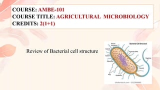
- 1. COURSE: AMBE-101 COURSE TITLE: AGRICULTURAL MICROBIOLOGY CREDITS: 2(1+1) Review of Bacterial cell structure
- 2. Cells are basically of two types Prokaryotic cells and Eukaryotic cells Prokaryotic cells are primitive type of cells • Lack true nucleus and other cell organelle (mitochondria, chloroplasts, endoplasmic reticulum, golgi complex, etc.) • The word prokaryotes is derived from Greek word (pro=before, karyo=nucleus) Eukaryotic cells have true nucleus and it also contains cell organelles • The word eukaryotes is derived from Greek word eu means true and karyo means nucleus
- 3. Prokaryotic cells: • Lacks a defined nucleus hence hereditary material not enclosed in an envelope has a relatively simple internal organization • As cell divide by fission and do not contain numerous chromosomes, no complex cell division machinery has evolved • consist of a single closed compartment that is surrounded by the plasma membrane- it is selectively permeable with in pockets (mesosomes) • Outside the membrane, a rough cell wall rich in carbohydrates and aminoacids may be present
- 4. • Eukaryotic cells, unlike prokaryotic cells, contain a defined membrane-bound nucleus and extensive internal membranes that enclose other compartments, the organelles • The region of the cell lying between the plasma membrane and the nucleus is the cytoplasm, comprising the cytosol (aqueous phase) and the organelles. • Eukaryotes comprise all members of the plant and animal kingdoms, including the fungi, which exist in both multicellular forms (molds) and unicellular forms (yeasts), and the protozoans (proto,primitive; zoan, animal), which are exclusively unicellular.
- 7. Prokaryotic cell Eukaryotic cell 1 A true nucleus is absent A true nucleus is present 2 Nuclear membrane is absent Nuclear membrane is present. 3 Nucleolus is absent Nucleolus is present. 4 Chromosome is single, circular. Chromosomes are many and linear 5 Chromosome present freely in the cytoplasm Chromosomes are enclosed inside the nuclear membrane 6 Histone proteins are absent in the organisation of chromosome Chromosomes are well organised with histone protein 7 In photosynthetic cells, chlorophyll pigments are present in cell membrane In photosynthetic cells, the chlorophyll pigments are present in the plastids of Chloroplast 8 The cell wall contains amino sugars & muramic acid When cell wall present, it does not contain amino sugars & muramic acid
- 8. Prokaryotic cell Eukaryotic cell 9 Ribosomes are 70s type Ribosomes are 80s type 10 Cell organells are absent. Cell organells are present 11 Enzymes necessary for respiration are present in plasmamembrane Enzymes necessary for respiration are present in mitochondria 12 Cell division by the process of amitosis i.e., mitosis and meiosis is absent Cell division by the process of mitosis and meiosis 13 mRNA will not have the 5' methyl cap and 3' poly A tail. mRNA having 5' methyl cap and 3‘poly A tail 14 Transcription and translation are combined process, both takes place in cytoplasm Transcription and translation are not combined process. Transcription takes in nucleus, while translation in cytoplasm 15 In genes non-coding regions are absent In genes non-coding region (i.e., introns) are present.
- 10. 0.5 to 1.0 μm in diameter, surface area/ volume ratio is exceedingly high favoring unusually high rate of growth and metabolism of bacteria. No circulatory mechanism is needed to distribute the nutrients that are taken in, due to this high surface to volume ratio.
- 11. The average diameter of spherical bacteria is 0.5-2.0 µm. For rod-shaped or filamentous bacteria, length is 1-10 µm and diameter is 0.25-1.0 µm. E. coli , a bacillus of about average size is 1.1 to 1.5 µm wide by 2.0 to 6.0 µm long. Spirochaetes occasionally reach 500 µm in length and the cyanobacterium Oscillatoria is about 7 µm in diameter. The bacterium, Epulosiscium fishelsoni , can be seen with the naked eye (600 µm long by 80 µm in diameter).
- 13. One group of bacteria, called the Mycoplasmas, have individuals with size much smaller than these dimensions. They measure about 0.25 µ and are the smallest cells known so far. They were formerly known as Pleuro Pneumonia Like Organisms (PPLO). Mycoplasma gallicepticum, with a size of approximately 200 to 300 nm are thought to be the world smallest bacteria. Thiomargarita namibiensis is world’s largest bacteria, a gram-negative Proteobacterium found in the ocean sediments off the coast of Namibia. Usually it is 0.1—0.3 mm (100—300 µm) across, but bigger cells have been observed up to 0.75 mm (750 µm).
- 15. Structure of bacteria has two aspects, arrangement and shape. Due to the presence of a rigid cell wall, bacteria maintain a definite shape, though they vary as shape, size and structure. When viewed under light microscope, most bacteria appear in variations of three major shapes: the rod (bacillus), the sphere (coccus) and the spiral type (vibrio). So far as the arrangement is concerned, it may Paired (diplo), Grape-like clusters (staphylo) or Chains (strepto).
- 16. •Cocci (or coccus for a single cell) are round cells, sometimes slightly flattened when they are adjacent to one another. •Bacilli (or bacillus for a single cell) are rod-shaped bacteria. •Spirilla (or spirillum for a single cell) are curved bacteria which can range from a gently curved shape to a corkscrew- like spiral. Many spirilla are rigid and capable of movement. A special group of spirilla known as spirochetes are long, slender, and flexible.
- 27. Glycocalyx, literally meaning "sugar coat" (glykys = sweet, kalyx = husk), is a network of polysaccharides that project from cellular surfaces of bacteria, which classifies it as a universal surface component of a bacterial cell, found just outside the bacterial cell wall. A distinct, gelatinous glycocalyx is called a capsule, whereas an irregular, diffuse layer is called a slime layer. This coat is extremely hydrated and stains with ruthenium red. Many bacterial cells secreted at the time of their active growth.
- 28. When the glycocalyx forms a well-defined persistent layer, it is called capsule. The capsule is a major virulence factor in the major disease-causing bacteria, such as Escherichia coli and Streptococcus pneumoniae.
- 29. The functions of the capsule depend on bacterial species (1) they may block attachment of bacteriophages (2) they may be antiphagocytic (3) they may provide protection against temporary drying by binding water molecules (4) they may promote attachment of bacteria to surfaces.
- 30. When the glycocalyx does not form a persistent layer, but is present more diffusely forming a loose mass around the bacterial cell, it is called slime layer. It is easily deformed, difficult to see, and can be very easily removed by washing the bacterial cells Gliding bacteria often produce slime, which aids in their mobility. It helps bacteria to attach them to the substratum and lubricates the surface for more efficient movement.
- 31. Flagella (singular, flagellum) are hairlike structures that provide a means of locomotion for those bacteria that have them. They can be found at either or both ends of a bacterium or all over its surface. The flagella beat in a propeller-like motion to help the bacterium move toward nutrients; away from toxic chemicals; or, in the case of the photosynthetic cyanobacteria; toward the light.
- 32. A flagellum is composed of 3 parts a). Basal body associated with cytoplasmic membrane and cell wall b). a short hook and c). A helical filament which is usually several times longer than the bacterial cell. The hook and filament are made up of protein whereas the composition of basal body is not known. The protein of the filament is known as flagellin.
- 33. Based on the arrangement of flagella, there are four types of bacteria: (i) Monotrichous: A single polar flagella. e.g. Pseudomonas aeruginosa. (ii) Lophotrichous: A cluster of polar flagella. e.g. Pseudomonas fluorescens. (iii) Amphitrichous: Either a single or cluster of flagella at both the cell poles. e.g. Aquaspirillum serpens. (iv) Peritrichous: Flagella arranged along the sides of the bacteria. e.g. Salmonella typhi.
- 35. Many species of bacteria have pili (singular, pilus), small hairlike projections emerging from the outside cell surface. These outgrowths assist the bacteria in attaching to other cells and surfaces, such as teeth, intestines, and rocks. Without pili, many disease-causing bacteria lose their ability to infect because they're unable to attach to host tissue. Specialized pili are used for conjugation, during which two bacteria exchange fragments of plasmid DNA.
- 36. Each bacterium is enclosed by a rigid cell wall composed of peptidoglycan, a protein-sugar (polysaccharide) molecule. The wall gives the cell its shape and surrounds the cytoplasmic membrane, protecting it from the environment. It also helps to anchor appendages like the pili and flagella, which originate in the cytoplasm membrane and protrude through the wall to the outside. The strength of the wall is responsible for keeping the cell from bursting when there are large differences in osmotic pressure between the cytoplasm and the environment.
- 37. Most of the bacteria retain their original cells even after subjected to very high pressure or severe physical conditions. It accounts for 10-40% of dry weight of the cell. Cell walls can be broken by sonic or ultrasonic treatment or by subjecting the cells to extremely high pressure and subsequent sudden release of pressure.
- 38. Cell wall composition of eubacteria is different from that of archaebacteria. Eubacteria cell wall is made up of peptidoglycan (murein and insoluble, porous cross linked polymer of enormous strength and rigidity. Peptidoglycan is basically a polymer of N-acetylglucosamine, N- acetylmuramic acid, L- alanine, D-alanine, D-glutamate and a diamino acid. The peptidoglycan is present only in prokaryotes. The cell walls of archaebacteria are generally made up of proteins, glycoproteins or polysaccharides.
- 39. Cell wall composition varies widely amongst bacteria and is one of the most important factors in bacterial species analysis and differentiation. For example, a relatively thick, meshlike structure that makes it possible to distinguish two basic types of bacteria. A technique devised by Danish physician Hans Christian Gram in 1884, uses a staining and washing technique to differentiate between the two forms. When exposed to a gram stain, gram-positive bacteria retain the purple color of the stain because the structure of their cell walls traps the dye. In gram-negative bacteria, the cell wall is thin and releases the dye readily when washed with an alcohol or acetone solution.
- 42. CYTOPLASMIC MEMBRANE PROTOPLAST AND SPHAEROPLAST MESOSOMES CYTOPLASM CHROMOSOME RIBOSOMES SPORES CONIDIOSPORES AND SPORANGIOSPORES CYSTS
- 43. This is about 7.5 ηm thick and is immediately beneath the cell wall. This is primarily composed of phospholipids (20-30%) and proteins (60-70%). This membrane contains various enzymes involved in respiratory metabolism and in the synthesis of capsular and cell wall components. It is the site of generation of proton motive force, which drives ATP synthesis, certain nutrient transport systems and flagellar motility. Damage to this membrane may result in the death of the cell.
- 44. A protoplast is that portion of a bacterial cell consisting of the cytoplasmic membrane and the cell material bound by it. This can be prepared from Gram positive bacteria by treating the cells with an enzyme such as lysozyme, which selectively dissolves the cell wall or by culturing the bacteria in the presence of an antibiotic such as penicillin.
- 45. Sphareoplast is a protoplast surrounded by the outer membrane of cell wall. In gram-negative bacteria only peptidoglycan layer can be removed but outer membrane is still intact surrounding the protoplast
- 47. • In many bacteria, especially Gram- positive bacteria, the cytoplasmic membrane appears to be infolded at more than one point. • Such infoldings are called mesosomes. • Mesosomes are thought to be involved in DNA replication, cell division and export of exocellular enzymes.
- 48. The cytoplasm, or protoplasm, of bacterial cells is where the functions for cell growth, metabolism, and replication are carried out. It is a gel-like matrix composed of water, enzymes, nutrients, wastes, and gases and contains cell structures such as ribosomes, a chromosome, and plasmids. The cell envelope encases the cytoplasm and all its components. Unlike the eukaryotic (true) cells, bacteria do not have a membrane enclosed nucleus. The chromosome, a single, continuous strand of DNA, is localized, but not contained, in a region of the cell called the nucleoid. All the other cellular components are scattered throughout the cytoplasm
- 49. Volutin granules (reserve source of phosphate), poly-β- hydroxybutyrate (PHB) and glycogen (both serving as source of carbon and energy) are some of the granules present in the cytoplasm of some bacteria. Gas vesicles are present in bacteria that grow in aquatic habitat
- 51. The nucleoid is a region of cytoplasm where the chromosomal DNA is located. It is not a membrane bound nucleus, but simply an area of the cytoplasm where the strands of DNA are found. Most bacteria have a single, circular chromosome that is responsible for replication, although a few species do have two or more. Smaller circular auxiliary DNA strands, called plasmids, are also found in the cytoplasm.
- 52. The very existence of plasmids in bacterial cytoplasm was revealed by Lederberg in 1952 while working on conjugation process in bacteria. A plasmid is a small, circular piece of DNA that is different than the chromosomal DNA, which is all the genetic material found in an organism’s chromosomes. It replicates independently of chromosomal DNA. Plasmids are mainly found in bacteria, but they can also be found in archaea and multicellular organisms. Plasmids usually carry at least one gene, and many of the genes that plasmids carry are beneficial to their host organisms.
- 53. Ribosomes are 70 S type consisting of 50 S and 30 S sub- units. Some ribosomes are free in the cytoplasm and some are attached to inner surface of the cytoplasmic membrane.
- 54. Spore is a metabolically dormant form, which under appropriate conditions can undergo germination and grow out to form a vegetative cell. Spores produced within the cell are called endospores and the spores produced external to cell are called exospores.
- 55. Endospores are thick walled, highly refractile bodies that are produced (one per cell) by Bacillus, Clostridium, Sporosarcina and few other genera. They are generally formed at the end of the active growth or during stationary phase. They are extremely resistant to desiccation, staining, disinfecting chemicals, radiation and heat.
- 56. Exospores are formed external to the vegetative cell by budding at one end of the cell in the methane oxidizing genus Methylosinus. They are desiccation and heat resistant.
- 58. CONIDIOSPORES AND SPORANGIOSPORES The bacteria, actinomycetes form branching hyphae. From the tips of these hyphae spores develop singly or in chains. If the spores are contained in an enclosing sac (sporangium), they are termed SPORANGIOSPORES, if not they are called CONIDIOSPORES. These spores can survive long periods of drying but they do not have high heat resistance.
- 60. • thick walled, desiccation resistant, dormant forms that develop by differentiation of vegetative cells. • EX: AZATOBACTER
- 63. • Draw a neat labelled diagram of bacterial cell and explain the internal and external structures of the bacterial cell. • Explain the types of flagella with diagrams and examples • Write the difference between 1. Eukaryotic cell and Prokaryotic cell 2. Gram positive and Gram negative cell walls 3. Endospore and Exospores 4. Spore and Cysts 5. Flagella and Pili 6. Protoplast and Sphaeroplast