Zeine et al. Poster 2007 Cancer Associated Fibroblasts in Neuroblastoma
•
2 gefällt mir•564 views

Cancer Associated Fibroblasts in Aggressive Neuroblastoma Tumors
Empfohlen

Empfohlen
Weitere ähnliche Inhalte
Was ist angesagt?
Was ist angesagt? (20)
Ähnlich wie Zeine et al. Poster 2007 Cancer Associated Fibroblasts in Neuroblastoma
Ähnlich wie Zeine et al. Poster 2007 Cancer Associated Fibroblasts in Neuroblastoma (20)
Mehr von Rana ZEINE, MD, PhD, MBA
Mehr von Rana ZEINE, MD, PhD, MBA (18)
Kürzlich hochgeladen
Kürzlich hochgeladen (20)
Zeine et al. Poster 2007 Cancer Associated Fibroblasts in Neuroblastoma
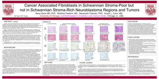
- 1. Cancer Associated Fibroblasts in Schwannian Stroma-Poor but not in Schwannian Stroma-Rich Neuroblastoma Regions and Tumors Rana Zeine MD, PhD, Radhika Peddinti, MD, Alexandre Chlenski, PhD, Susan L. Cohn, MD. University of Chicago, and Northwestern University Lurie Cancer Center, Chicago, IL, USA ABSTRACT updated Context: Cancer-Associated Fibroblasts (CAFs) promote tumor angiogenesis and are associated with aggressive clinical behavior in prostate, lung, colon, gastric and breast carcinomas. CAFs represent a population of stromal 'activated fibroblasts‘ / myofibroblasts that are routinely identified by alpha-Smooth Muscle Actin ( SMA) expression and distinguished from mature pericytes by lack of expression of heavy MW-Caldesmon (h-CD). Neuroblastomas are pediatric neoplasms that exhibit seven histologic subtypes classified as either Schwannian stroma (SS)-poor or SS-rich / dominant. Design: To investigate whether CAFs also play a role in mediating angiogenesis in Neuroblastoma, we histologically examined 82 tumors diagnosed at Children's Memorial Hospital in Chicago. Adjacent paraffin-embedded sections were stained with H&E, Masson-Trichrome; and immunohistochemistry was performed for SMA, h-CD and CD31. Microvascular Proliferation was defined as vessels with hypertrophic CD31 + endothelial cells and additional layers of vascular mural cells including SMA + cells. Results: Light microscopic examination revealed the presence of a population of stromal cells that were strongly positive for SMA and were negative for CD31 and h-CD consistent with CAFs. CAFs were abundant within extensive reactive stromal bands in SS-poor undifferentiated (n=6), poorly differentiated (n=14) and differentiating (n=33) Neuroblastomas. CAFs were associated with microvascular proliferation. By contrast, CAFs were less widely distributed in Ganglioneuroblastomas and were rare in SS-rich regions (intermixed n=13, nodular n=5). In SS-dominant Ganglioneuromas (n=11) CAFs were rare. Pericytes in normal vessels were SMA + /h-CD + . Conclusion(s): CAFs were identified in Neuroblastoma and were associated with SS-poor regions and microvascular proliferation. Our data indicate that CAFs promote angiogenesis in SS-poor Neuroblastomas. METHODS Patients were diagnosed between 1987 and 2005 at The Children’s Memorial Hospital, Northwestern University in Chicago, and this study was approved by the Institutional Review Board. Adjacent Formalin-Fixed-Paraffin-Embedded tissue sections were stained with H&E and with Masson-Trichrome special stain according to standard procedure. Immunohistochemistry was performed on additional adjacent sections for alpha-Smooth Muscle Actin (1A4), CD31 and for heavy Molecular Weight Caldesmon (h-CD) (mouse anti-human mAbs, DakoCytomation, Carpenteria, CA). Target-retrieval was done according to the manufacturer’s instructions. The EnVision+/HRP anti-mouse detection system was used to visualize binding with 3,3'-diaminobenzidine (DAB; DakoCytomation). Sections were counterstained with Gill's hematoxylin. Two investigators, one pathologist (RZ), and (RP), evaluated entire sections from one block for each tumor. The extent of tumor infiltration by SMA positive cells was scored as rare, few or many. DISCUSSION The behavior of Neuroblastoma tumors is influenced by the type of microenvironment that gets established. The more aggressive neuroblastomas exhibit varying degrees of Reactive Fibrovascular Stroma, whereas, benign tumors develop a matrix rich in Schwannian Stroma. We observe relative exclusion of Cancer-Associated Fibroblasts from regions rich in Schwannian Stroma. We also observe that the populations of infiltrating CAFs co-localize with angiogenesis and Microvascular Proliferation as described for this set of Neuroblastoma tumors (1,2). In Schwannian Stroma-Rich Neuroblastoma, there appears to be an inhibition of these tumor promoting processes that are associated with the development of Fibrovascular Stroma. It is possible, therefore, that strategies targeting Cancer-Associated Fibroblasts (6,8) may have a role in the treatment of patients with Schwannian Stroma-Poor Neuroblastoma. BACKGROUND We have recently reported on the clinical significance of Microvascular Proliferation in Neuroblastoma, and described its presence in Schwannian Stroma-Poor but not in Schwannian Stroma-Rich Neuroblastoma tumors and regions (1,2). This provided further support for the anti-angiogenic properties ascribed to Schwannian-derived factors (3). Indeed, Schwannian stroma predominates histopathologically in the benign Ganglioneuromas, and in the Intermixed Ganglioneuroblastomas which carry an excellent prognosis. In this study, we examine the presence and distribution of Reactive Fibrovascular Stroma, and describe tumor infiltration by Cancer-Associated Fibroblasts (CAFs) in the 7 diagnostic categories designated by the International Neuroblastoma Pathology Committee Classification. CAFs are ‘activated’ fibroblasts that have acquired SMA expression and developed myofibroblastic properties. Studies on human carcinomas including prostate (4), breast (5) (6) and colon (7) have revealed significant correlations between clinically aggressive tumor behavior and the extent of tumor infiltration by CAFs. Because of their clinicopathologic significance in the malignant progression of cancer, CAFs may represent a new target for emerging cancer therapies (8). Myofibroblasts in granulation tissue and cancer are primarily derived from local mesenchymal cells; and, in invasive carcinoma, can undergo further cytodifferentiation similar to smooth muscle metaplasia (9). Vascular smooth muscle cells (Pericytes) have also been demonstrated to have mesenchymal cell progenitors in cancer (10). Pericytes can be distinguished from CAFs by their differential expression of heavy Molecular Weight Caldesmon (h-CD) (11). CAFs are also characterized by their expression of a serine protease, Fibroblast Activation Protein. Some of the mechanisms mediating the role of CAFs in promoting tumor growth, angiogenesis, and metastasis involve stromal cell-derived factor 1 (SDF-1) through it’s cognate receptor CXCR4 (6). In order to evaluate tumor infiltration by CAFs, we examined tissue sections from human Neuroblastoma tumors for the expression of SMA, CD31 and h-CD. We observed that CAFs constituted a major component of the Reactive Fibrovascular Stroma which was more abundant in Schwannian Stroma-Poor regions and tumors. Furthermore, CAFs co-localized with Microvascular Proliferation (vessels with hypertrophic CD31+ endothelial cells and additional layers of vascular mural cells including aSMA+ cells) where present within aggressive Neuroblastoma tumors. CONCLUSIONS 1) SMA-expressing, ‘activated’, fibroblasts (Cancer-Associated Fibroblasts, CAFs) are widely distributed within bands of Reactive Fibrovascular Stroma in Schwannian Stroma-Poor Neuroblastoma regions, but confined to thin septae within Schwannian Stroma-Rich tumors. 2) Heavy molecular weight Caldesmon (h-CD) expression distinguishes tumor Pericytes from CAFs in Neuroblastoma. 3) CAFs are located in the vicinity of blood vessels and are spacially related to Microvascular Proliferation in Neuroblastoma, consistent with their presumed role in promoting tumor progression and angiogenesis. 5) The mechanism(s) for relative exclusion of CAFs from Schwannian Stroma-Rich regions remain to be elucidated. 6) Clinicopathologic correlation for Neuroblastoma with extent of infiltration by Cancer-Associated Fibroblasts is warranted. 7) Histopathologic evaluation for SMA-positive stromal cells in Neuroblastoma may guide patient stratification for clinical trials with novel treatments targeting Cancer-Associated Fibroblasts. REFERENCES 1. Zeine R. et al. Vascular Endothelial Proliferation in Schwannian Stroma-Poor but not in Schwannian Stroma-Rich Regions of Neuroblastoma. Brain Pathol. 2006, 16(s1):S154 2. Peddinti R., Zeine, R. et al. Prominent Microvascular Proliferation in Clinically Aggressive Neuroblastoma Tumors. Clin. Cancer Res. 2007, 13(12):3499 3. Chlenski A. et al. SPARC is a key Schwannian-derived inhibitor controlling neuroblastoma tumor angiogenesis. Cancer Res. 2002, 62:7357 4. Tuxhorn JA. et al. Reactive Stroma in Human Prostate Cancer: Induction of Myofibroblast Phenotype and Extracellular Matrix Remodeling. Clin. Cancer Res. 2002, 8:2912 5. Yazhou C. et al. Clinicopathological Significance of Stromal Myofibroblasts in Invasive Ductal Carcinoma of the Breast. Tumor Biol. 2004, 25:290 6. Orimo A. et al. Stromal Fibroblasts Present in Invasive Human Breast Carcinoma Promote Tumor Growth and Angiogenesis through Elevated SDF-1/CXCL12 Secretion. Cell 2005,121:335 7. Henry LR. et al. Clinical Implications of Fibroblast Activation Protein in Patients with Colon Cancer. Clin. Cancer Res. 2007, 13:1736 8. Kalluri R. and Zeisberg M. Fibroblasts in Cancer. Nature Rev. Cancer 2006, 6:392 9. Sch ürch W. et al. Myofibroblast in Mills SE. (Ed.) Histology for Pathologists LWW 2007, ch.6 10. De Palma M. et al. Tie2 Identifies a Hematopoietic Lineage of Proangiogenic Monocytes Required for Tumor Vessel Formation and a Mesenchymal population of Pericyte Progenitors . Cancer Cell 2005, 8:211 11. Nakayama H. et al. Differential Expression of High Molecular Weight Caldesmon in Colorectal Pericryptal Fibroblasts and Tumor Stroma. J. Clin. Pathol. 1999, 52:785 FIGURE LEGEND H&E (A-F), Masson-Trichrome special stain (G-L), and Immunohistochemistry for SMA (M-U) and heavy-MW Caldesmon (V-Z) of representative sections from Ganglioneuroma (A,G,M,V); Ganglioneuroblastoma Intermixed (B,H,N,W) and Nodular (C,I,O,X); Differentiating (D,J,P,Q,Y), Poorly Differentiated (E,K,R,S,Z) and Undifferentiated (F,L,T,U) Neuroblastoma Tumors. Magnification x200 P&Y x400. SMA immunostain revealed very few positive stromal cells within Schwannian Stroma-Dominant Ganglioneuroma (M) and Schwannian Stroma-Rich Ganglioneuroblastoma (N). By contrast, SMA positive CAFs (Cancer-Associated Fibroblasts) were widely distributed in the Schwannian Stroma-Poor region of a nodular Ganglioneuroblastoma (O) and in the Differentiating (P,Q), Poorly Differentiated (R,S) and Undifferentiated (T,U) Neuroblastoma Tumors. Abundance of Fibrovascular Stroma in Schwannian Stroma-Poor Neuroblastomas and regions could be appreciated on H&E (C-F) and Masson-Trichrome special stain (I-L). Images of CD31 are provided in Peddinti, Zeine et al. ’07 (2). Heavy Molecular Weight Caldesmon (h-CD) positivity was specific to blood vessels, distinguishing tumor pericytes from CAFs (V-Z vs. M-U). GANGLIONEUROMA INTERMIXED NODULAR NEUROBLASTOMA DIFFERENTIATING J D E B H H&E x200 h-CD x200 C F I L K O N NEUROBLASTOMA POORLY DIFFERENTIATED NEUROBLASTOMA UNDIFFERENTIATED Q S U RESULTS MASSON TRICHROME x200 SMA x200 W Y Z A G M V Schwannian Stroma-Dominant Focal Reactive Stroma Few Cancer-Associated Fibroblasts plus Pericytes Only Pericytes h-Caldesmon positive (same field as A&M) Only Pericytes h-CD positive (same field as B&N) Few Cancer-Associated Fibroblasts in Vicinity of Neuroblastic Cells Thin Reactive Stromal Septae Schwannian Stroma-Rich, Intermixed Neuroblastic Cells Fibrovascular Tumor Stroma in Schwannian Stroma-Poor Region Abundant Fibrovascular Stroma in Schwannian Stroma-Poor Region Many Cancer-Associated Fibroblasts plus Pericytes Abundant Fibrovascular Stroma Many Cancer-Associated Fibroblasts within Reactive Fibrovascular Stroma GANGLIONEUROBLASTOMA Many Cancer-Associated Fibroblasts with Pericytes and Microvascular Proliferation Abundant Reactive Stroma with Microvascular Proliferation Microvascular Proliferation within Schwannian Stroma-Poor Tumor Reactive Stroma within Schwannian Stroma-Poor Tumor R P T Fibrovascular Stroma within Schwannian Stroma-Poor Tumor Abundant Fibrovascular Stroma Many SMA positive cells within networks of Fibrovascular Stroma X Only Pericytes h-CD positive (same field as C, I &O) CAFs negative for h-CD (same field as P) Only Pericytes positive for h-CD Other stroma negative for h-CD x400 x400 CAP Sept. 2007 Chicago