2. Contents
Inroduction
Embryology
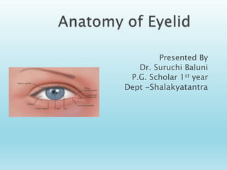
Anatomy
Layers of Eyelid
Muscle of Eyelid
Gland of Eyelid
Function of Eyelid
Nerve supply
Vascular supply
Venous drainage
Lymphatic drainage
3. The eyelids are the mobile tissue in front of
the eyeballs. (ocular appendages).
Eyelids are thin curtains of skin ,muscle,
fibrous tissue and mucous membrane.
Two in number: Upper and lower eyelid
4. Functions:
Act as shutters protecting eyes from injuries and
excessive light.
Spread the tear film over the cornea and
conjunctiva.
Contribute to the facial feature and information
regarding the state of wakefulness and attention.
Its is mobile, multilamellar structure that covers
eyeball anteriorly.
5. It is formed by reduplication of surface ectoderm
above and below the cornea during 2nd month of
gestation.
The folds enlarge and their margin meet and fuse
with each other by 3rd month.
A closed space, conjunctival sac is formed.
The folds thus formed contain some mesoderm
which would form the muscles of the lid and the
tarsal plate.
Lid separate after seventh month of intra uterine
life
7. Tarsal glands are formed by ingrowth of a
regular row of solid columns of ectodermal
cells from the lid margins.
Cilliary glands are outgrowth from the ciliary
follicles.
Cillia develop as epithelial buds from lid
margins.
8.
9. Extent :
Upper eyelid extends from
the eyebrow downward to end
in a free margin which forms
the superior boundary of
palpebral fissure.
Lower eyelid merge into the
skin of the cheek.
Parts
Each eyelid is divided by an
horizontal furrow into an
orbital and tarsal plate.
Additional folds in lower lid are
Nasojugal fold medially and the
malar fold laterally.
These folds limit the spread of
blood downward from eyelids
to cheek.
10. Upper eyelid covers 1/6th part of the cornea.
Lower lid just touchs the limbus.
Palpebral aperture
It is the elliptical space between the
upper and the lower lid. When the
eyes are open,it measures about
vertically-10-11mm.
horizontally 28-30 mm.
At Birth
horizontal 18-21mm.
vertical 8 mm.
11. Eyelids meet at medial and lateral canthi.
Medial canthus: it is rounded and is separated from the globe
by tear lake (lacus lacrimalis). In this area, there is caruncle
and plica semilunaris.
Lateral Canthi
It is about 5-7 mm from the
lateral orbital margin.
It forms an acute angle of
about 60 degree with eyes
wide open and 30-40 degree
with eyes open in normal way
12. caruncula lacrimalis:
its is a small
pink,globular nodule at
the inner corner (medial
canthus) of the eye
.consist of skin,hair
follicle,sweat gland and
sebaceous gland.
A semilunar fold called plica
semilunaris lies on lateral
side of caruncle represents
the third eye lid of other
vertebrae.
13. Each lid margin is divided into two parts by the
lacrimal papilla(a small elevation present on the
medial side, which contains a hole- the lacrimal
punctum in its centre).
The medial portion of the eyelid margin,
extending from the punctum medially to the
medial canthal angle termed as lacrimal portion
is rounded and devoid of lashes or glands.
The lateral, ciliary portion of the eyelid margin
consists of a rounded anterior border, a sharp
posterior border (placed against the globe) and
an intermarginal strip between the two borders.
14. Grey line which marks the junction of skin
and conjunctiva,divides the intermarginal
strip into an anterior strip bearing lashes
and a posterior strip which contains
opening of meibomian glands arranged in a
row and a lipid strip.
15. Arranged in 2-3 row.
Upper lid 100-150 directed forward,upward and
backward
Lower lid 50-75 directed forward downward and
backward.
Cilia
Cillia vary in size ranging from 20-120mm in diameter
and from 6-12mm in length
Taper throughout their length to end in fine sharp point.
Each cilium has a life span of some 3- 4 months. At the
termination of this period the old cilium drops away the
follicle test for several week and then a new cilium grows
out.
Each follicle is surrounded by a dense plexus of vessels
and nerves, the latter provide the tactile sensibility to
each cilium.
16. Trichiasis:Acquired misdirection of
eyelashes.
Madarosis: Decrease in number of
eyelashes.
Lash Poliosis: Premature graying of the
lashes.
Trichomegaly: Excessive eyelash
growth.
CONGENITAL /DEVELOPMENTAL ANOMALIES
Coloboma of lid: Notch in the edge of eyelid
Cryptophthalmos: partial or complete loss of
brows, palpebral fissure, lashes,conjuctiva
and absence of eyelid.
18. Euryblepharon: vertical shortening
and horizontal lengthening of
eyelids.
Epiblepharon: lower lid Pretarsal
muscle and skin ride above the
lower lid margin to form a
horizontal fold of tissue.
Ankyloblepharon: partial or
complete fusion of eyelids by
webs of skin.
Symblepharon: adhesion of lid
to the gobe.
19. Distichiasis : extra
rows of
eyelashes.
Blepherophimosis:
condition in
which palpebral
fissure appear to
be smaller
20. Skin
The skin covering the eyelid is elastic, having a
fine texture, is thinnest in the body and folds
easily thereby contributing to the ease and
speed of mobility of the upper eyelid.
Nasal part of the skin is smooth,shining and
greasy in comparison to temporal part.
Fine hair are seen on the temporal part of skin.
22. Beneath the skin is a layer of loose aerolar connective
tissue, containing no fat. It is thus readily distended by
oedema or blood.
This layer is non- existent near the ciliary margin, at the
lid folds and at medial and lateral angles where the skin
is attached to the underlying ligaments.
23. Layer of striated muscle
• This layer consists of orbicularis muscle which forms a thin oval sheet
across the eyelids. It comprises three portions: the orbital, palpebral and
lacrimal.
• The Orbital part forms the most peripheral fibres of the
orbicularis which arise from the anterior part of the medial
palpebral ligament and the adjacent bones( upper orbital margin,
the maxillary process of frontal bone, frontal process of maxilla
and the lower orbital margin medial to the infra-orbital foramen.
24. • The Palpebral part of orbicularis are preseptal and pretarsal
portions. The fibres of pretarsal portions helps in drainage of tear
by lacrimal sac and are called as pars lacrimalis.(Horners muscle)
• It is supplied by zygomatic branch of facial nerve. Therefore in
paralysis of facial nerve there occurs lagophthalmos.
25. Function of orbicularis oculi muscle
• Closure of eyelids
• Orbital portions
Forced closure of eyelids
Reflex blinking
• Palpebral portion
Helps in gentle closure during blinking and sleep.
26. Also known as oculosympathetic paresis
Characterised by classical triad
Miosis (constricted pupil)
Partial ptosis
Apparent anhidrosis (decreased sweating)
27. • Small bundle of striated muscle fibers
• At the eyelid margin
• Extension of pretarsal portion of
orbicularis oculi fibers
• Function:
• Keep the lids in close apposition to the globe
28. • Levator palpebrae Superioris( major eye
lid retractor)
• Origin- Arises from the apex of the orbit,above
annulus of zinn.
• It is a flat muscle that broadens as it passes forwards.
29. Other sites of insertion
Some fibres are attached to:-
• The skin of upper lid.
• The superior conjunctival fornix.
• The upper edge of the superior tarsus(
superior tarsal muscle)
Primary insertion- Primary
point of insertion is into superior
surface of the tarsus.
30. Superior Tarsal Muscle
• A thin sheet of smooth
muscle lies beneath the main
tendon of levator palpebrae
superiors.
• This group of smooth muscle
fibres help to maintain eyelid
elevation.
• Loss of function of the
superior tarsal muscle results
in drooping of upper eyelid.
Course & attachment
oPasses forward below the
roof of the orbit, above the
superior rectus
oAt septum orbitale, it fans out
into white tendon called
aponeurosis of LPS and forms
medial and lateral horns
31. Superior division of the oculomotor nerve
supplies the muscle.
Loss of oculomotor nerve function result in
complete ptosis or drooping of the superior
eyelid
Whereas loss of sympathetic innervation to
the superior tarsal muscle result in partial
ptosis.
32. Whitnall ligament- it is located at
transition zone- act as a fulcrum for
levator transferring its vector from
ant-post to sup-inf direction.
Its analogue in lower lid is Lockwood
Ligament
CAPSULOPALPEBRAL
FASCIA
Fibrous sheet in the lower eyelid
,that arises from the lockwoods
ligament
33. This layer splits the eyelid into two- The
anterior lamina and posterior lamina- which
are easily approachable through the grey line.
It’s a layer of loose connective tissue. The
nerve and vessels of the eyelids lie in this
layers and so to anaesthetise the lid, injection
is made in this plane.
34. It is the framework of the lid
which consist of central thick part
the tarsal plate and Peripheral thin
part the septum orbitale.
The fibrous layer also include the
medial and palpebral ligaments.
Tarsal plate are firm plates that
form the skeleton of the eyelids
giving them shape and firmness.
29 mm long and 1mm thick
Anterior surface -convex
Posterior surface-concave
-lined by conjunctiva which is firmly
adherent to theTarsal plates.
35. Septum orbitale is a thin
floating membrane of
connective tissue which take
parts in all movements of the
eyelid.
Applied aspect
With age orbial septum
weakens orbital fat herniats
the condition manifest as
DERMATOCHALASIS
36. Layer of non striated muscle fibres
• Sympathetics accessory
retractor of upper eyelid.
• Modulates the position of the
upper and lower eyelids when
the eye is open
• Origin –under surface of the
levator muscle just anterior to
whitnall’ligament.
• Insertion –anterior edge of the
superior tarsal border
Mullers muscle
37. o Transparent vascularized membrane
covered by a non keratinized epithelium that
lines the posterior surface of the eyelids
(palpebral conjunctiva)and the anterior
surface of the globe (bulbar conjunctiva)
o Firmly adherent to the tarsus
38. • Tarsal / Meibomian Glands.
• Gland of Zeis.
• Gland of Moll.
• Accessory lacrimal
gland.
39. Modified sweat gland
Present on the posterior part of
stroma of tarsal plate
30 -40 no. in upper eyelid & 20-
30 no. in lower eyelid
Oily secretion
40. Gland of Zeis
Modified sebaceous glands Attached to
eyelash follicles
(usually two glands with each cilium)
Sebum secretion
Gland of Moll
Modified sweat gland Lies
between cilia
Numerous in lower lid than upper lid
41. A/k as Ciaccio’s glands or wolfring’s glands
Found in lacrimal caruncle of eyelids
Located in upper border of superior tarsus
and lower border of inferior tarsus
Functions:
Production of tear which are secreted onto
the surface of conjunctiva
44. Sensory Nerve Supply:
Derived from branches of trigeminal nerve.
Upper eyelid - supraorbital, supratrochlear &
lacrimal nerves (ophthalmic division)
extreme medial portion of both upper & lower
eyelid - infratrochlear nerve
lateral portion of upper eyelid -
zygomaticotemporal branch of the maxillary nerve
lower eyelid - infraorbital nerve (maxillary division)
lateral portion of lower eyelid - zygomaticofacial
branch of the maxillary nerve
45.
46. Aeteries of the lid (medial and lateral
palpebral)form marginal arterial arcade, 2mm
away from lid margin in upper eyelid and
4mm away from lower lid margin.
47. Another arcade ( superior
arterial arcade ) is formed in
upper eyelid .
Branches go forward and
backward from these arches to
supply various structures.
48. These are arranged in two plexus-
1- A post- tarsal which drain into ophthalmic
vein
2- A pretarsal opening into subcutaneous
vein.
49.
50. Arranged in two sets pretarsal and post tarsal
Those from lateral half of the lids drain into
pre auricular lymphnodes and those from
medial half of the eyelids drain into
submandibular lymph nodes.
51. External Hordeolum (Common Stye)
Localized suppurative inflammation of gland of
Zeis and glands of Moll’s at lid margin at ciliary
follicle.
52. Internal Hordeolum( Meibomian stye)
Internum Hordeolum is a suppurative inflammation of
meibomian gland associated with the blockage of the
duct.
53. Chalazion
Chronic granulomatous inflammation of meibomian
gland or sometimes Zeis glands caused by retained
sebaceous secretions
Ocurrs secondary to obstruction of the gland duct.
More common in upper eyelid appearing as hard,
immobile, painless, roundish lump.
54. Blepharitis
Blepharitis is chronic inflammation of lid margin
occurring as true inflammation.
Bilateral and often misdiagnosed as conjunctivitis