EAR AND AUDITORY PATHWAY ANATOMY
•Als PPT, PDF herunterladen•
49 gefällt mir•13,109 views

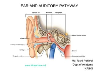
The document provides information on the anatomy of the ear and auditory pathway. It discusses the three parts of the ear - external ear, middle ear, and inner ear. The external ear includes the pinna and external auditory meatus. The middle ear contains the tympanic membrane, three ossicles (malleus, incus, stapes), and two small muscles (tensor tympani and stapedius). The inner ear is made up of the cochlea and vestibular system. The auditory pathway transmits signals from the inner ear to the brainstem and auditory cortex.

Empfohlen
Weitere ähnliche Inhalte
Was ist angesagt?
Was ist angesagt? (20)
Andere mochten auch
Andere mochten auch (20)
Ähnlich wie EAR AND AUDITORY PATHWAY ANATOMY
Ähnlich wie EAR AND AUDITORY PATHWAY ANATOMY (20)
Mehr von Nepalese army institute of health sciences
Mehr von Nepalese army institute of health sciences (20)
Kürzlich hochgeladen
Kürzlich hochgeladen (20)
EAR AND AUDITORY PATHWAY ANATOMY
- 1. EAR AND AUDITORY PATHWAY Maj Rishi Pokhrel Dept of Anatomy NAIHS www.slideshare.net
- 2. • One structure of human body 0.1 mm thick and derived from all 3 germ layers? 2
- 3. 3Cochlea Sec tympanic membrane Promontory Middle ear Stapes Semicircular canal Vestibule of inner ear THREE PARTS OF EAR Ext auditory meatus Tympanic membrane External ear
- 4. EXTERNAL EAR 4 • Pinna • External auditory meatus PINNA • Concha • Helix • Antihelix • Scaphoid fossa & triangular fossa • Tragus • Antitragus • Intertragic notch • Lobule
- 6. EXTERNAL AUDITORY MEATUS Extends from bottom of concha to TM (Length: 25 mm) Parts – Cartilaginous: lateral 1/3rd – Bony: medial 2/3rd Bends – B/T lateral 1/3rd & medial 2/3rd – 5 mm from tympanic membrane Direction – Backwards & medially Epithelium (skin) – Adherent to bone & cartilage – Ceruminous glands – Secretions prevent entry of bacteria 6
- 7. 7 Nerve supply • ATN (V3) • Great auricular (C2,3) •Lesser occipital (C2) •Facial •Vagus •Motor- Facial Blood supply •Post auricular •Superficial temporal •Ant tympanic •Deep auricular
- 8. TYMPANIC MEMBRANE Size Diameter – 1cm Thickness – 0.1 mm Position – Lie obliquely 55 deg to EAM – Faces downward, forward & laterally Umbo – Concavity due to attachment of handle of malleus 8
- 9. TYMPANIC MEMBRANE Subdivisions – Pars flaccida (Shrapnel’s membrane) • between folds • lax area – Pars tensa • Rest of the membrane • tense due to – attachment of handle of malleus – radiating fibres of intermediate layer 9 PF Pars Tensa AMF PMF Line of attach of handle of malleus on medial surface
- 10. TYMPANIC MEMBRANE 10 Surfaces Lateral surface • Concave, directed down, forward & laterally Medial surface • Convex, maximum at umbo • Handle of malleus attached here • Chorda tympani is medial to handle of malleus
- 11. TYMPANIC MEMBRANE Structure From lateral to medial – Outer cuticular layer: stratified squamous nonkeratinized epithelium – Intermediate fibrous layer: Outer radiating, inner circular fibres – Inner mucous layer: columnar epithelium; patchy ciliated Handle of malleus & chorda tympani lie b/t mucous & intermediate fibrous layer 11 Outer cuticular layer Middle fibrous layer Inner mucous layer Deep circular fibres Superficial Radiating fibres Pars flaccida
- 12. Tegmen tympani Tensor tympani Petrotympanic fissure Anterior canaliculus Chorda tympani Tympanic membrane Handle of malleus Processus cochleariformis Aditus LATERAL WALL OF LT MIDDLE EAR WITH CONTENTS
- 13. TYMPANIC MEMBRANE Nerve supply – Lateral surface • ATN • Vagus (auricular br) – Medial surface • IX CN (tympanic br) • Chorda tympani Blood supply – Deep auricular – Anterior tympanic – Posterior auricular: stylomastoid br 13
- 15. CLINICAL ANATOMY • ASOM – Pus discharged laterally • Myringotomy • Incision at posteroinferior quadrant –Prevent damage to chorda tympani –Rich blood supply: healing faster 15
- 16. MIDDLE EAR 16 Canal for T T Auditory tube Aditus to antrum Mastoid antrum Mastoid cells Mastoid process A P
- 17. 17 Left ear: TM removed Roof Floor Medial wall Ant wall Post wall
- 18. 18
- 19. ROOF 19 Petrous temporal Formation – Tegmen tympani: thin bony plate of petrous temporal • Separates tympanic cavity from MCF • Pierced by lesser & greater petrosal nerves Applied – If unossified: spread of infection to meninges
- 20. FLOOR Formation – Floor of jugular fossa (posteriorly) – Posterior wall of ascending part of carotid canal (anteriorly) - Tympanic Canaliculus for tympanic br of IX CN Applied – Spread of infection to IJV: thrombosis 20 Epitympanic recess Aditus Auditory tube ICA Sup bulb of IJV VII CN
- 21. ANTERIOR WALL • Shortened by approximation of roof & floor • Anteriorly shows posterior wall of carotid canal • Canal for • Tensor tympani (above) • Auditory tube (below) • Processus trochleariformis • Bony shelf extends back on medial wall & turns laterally; pulley for tensor tympani 21
- 22. 22
- 23. POSTERIOR WALL • Wider above than below • Aditus to mastoid antrum • Facial canal • Pyramid: tendon emerges from apex • Posterior Canaliculus of chorda tympani 23
- 24. MEDIAL WALL Promontory: due to basal turn of cochlea – Tympanic plexus (IX CN) lies over promontory Oval window (fenestra vestibuli ) – behind & above promontory – closed by base of stapes & annular ligament Round window (fenestra cochleae ) – below & behind promontory – closed by secondary TM Facial canal: above & behind promontory – oblique part, run back & down above oval window Prominence of lateral semicircular canal – above facial canal 24
- 25. 25 Left ear: TM removed Medial wall
- 26. 26 MEDIAL WALL
- 27. LATERAL WALL • Tympanic Membrane • Epitympanic recess – Part above tympanic cavity – Upper half of malleus – Greater part of incus 27
- 28. MIDDLE EAR: COMMUNICATIONS • Anterior wall : auditory tube • Posterior wall: mastoid antrum • Medial wall : inner ear • Lateral wall shows : tympanic membrane • Roof and floor: none 28
- 29. EAR OSICLES
- 30. MALLEUS Head In epitympanic recess Post surface: facet for incus- Incudomalleal joint (saddle) Neck Against pars flaccida Chorda tympani cross medial to neck • Handle Project down & back till umbo Processes: lateral pr: Upper end, handle & lat pr attached to fibrous layer of TM, malleolar folds Anterior pr: attachment of ant lig of malleus 30
- 31. INCUS Body • In epitympanic recess • Articulates anteriorly with head of malleus Short process • Lig attached to fossa incudis (post wall of TC) Long process • Hook medially, articulate with stapes • Incudostapedial joint: ball & socket 31
- 32. STAPES • Head – Articulates with lenticular nodule of incus • Neck – Stapedius attached to back of neck • Ant & post limbs • Foot plate – anchored to fenestra vestibuli by annular lig – syndesmosis 32
- 33. TENSOR TYMPANI Origin • Bony & cart part of AT, becomes tendon, hooks around processus trochleariformis Insertion • Handle of malleus: upper part Nerve supply • Nerve to medial pterygoid Actions • Pull handle of malleus, TM concave, makes tense • Increase tight adhesion of footplate to fenestra vestibuli – dampens sound 33
- 34. STAPEDIUS Origin • Interior of hollow pyramid Insertion • Back of neck of stapes Nerve supply • Facial nerve Actions • Retract neck of stapes from fenestra vestibuli • Paralysis: hyperacusis 34
- 35. 35
- 36. 36 CLINICAL ANATOMY Normal TM Wax
- 38. MYRINGOTOMY MYRINGOTOMY & SYRINGING EAR SYRINGING
- 39. 39 OTITIS MEDIA Serous otitis mediaASOM
- 41. TYMPANOSCLEROSIS
- 42. Internal ear 42
- 43. 43
- 44. 44
- 47. 47
- 49. 49
- 50. 50
- 51. 51
- 52. 52
- 53. 53
- 55. ? 55