2. 622 WOESE ET AL.
INTRODUCTION
Translation physically links the genotype and
the phenotype and thereby defines them. The
process is a necessary precondition to the evolu-
tion of all macromolecular structure. The size of
enzymes, their complexity, and their specificity
reflect an underlying accuracy in the translation
process. Is it not true, therefore, that translation
embodies the essence of the cell?
Translation occurs in the framework of the
ribosome, an enormous and complex molecular
aggregate that comprises two ribonucleoprotein
subunits (12). The smaller of these (in bacteria)
contains about 20 separate protein components,
positioned around a large ribonucleic acid
(RNA) some 1,500 nucleotides in length. The
larger subunit comprises an RNA of about twice
that size (2,900 nucleotides), in addition to a
much smaller 5S RNA (120 nucleotides) and
about 30 distinct proteins (26, 40).
For almost three decades, biologists have
sought to understand the molecular mechanics
of translation, but with little beyond descriptive
success. In approaching translation, we have
tended to focus on the protein components as
the elements that define ribosome function-the
ribosomal RNAs (rRNAs) were seen as basically
structural. With the advent of nucleic acid se-
quencing technology, however, has come an
interest in the possible functional roles for
rRNAs. If we can transform their linear molecu-
lar sequence into a detailed and dynamic three-
dimensional form, we will almost certainly un-
derstand the all-important translation process.
The earliest speculations concerning rRNA
secondary structure were based upon a partial
(and, it turns out, incorrect) sequence from one
organism (20) and therefore need not be consid-
ered here. Studies of transfer RNA (tRNA) and
of 5S RNA (25) have forcefully demonstrated
that the only reliable way to determine second-
ary structure presently available (outside of X-
ray crystallography) is through comparative
analysis of primary structure. A comparative
approach to the 16S rRNA sequence, based
upon the Escherichia coli sequence (9, 11), the
nearly complete 16S rRNA sequence from Bac il-
lus brevis, and T1 ribonuclease (RNAse) oligo-
nucleotide catalogs from over 150 other bacte-
ria, gave us a first look at the true secondary
structure of the molecule (51, 98). The subse-
quent publication of 16S RNA sequences from
Zea mays chloroplasts (69) and mammalian mi-
tochondria (1, 21) permitted some refinement of
the original structure and provided further com-
parative proof for a number of helical elements
(54).
There now exist in the literature a number of
proposed secondary structures for 16S (and 23S)
rRNA (6-8, 30, 54, 79, 98, 105). All agree to a
first approximation (for all have used to some
extent a comparative approach). However,
there are differences in detail among them. Since
it is virtually impossible for interested biologists
to assess the relative merits of the various
models-indeed, it even requires considerable
effort to intercompare them-and since new
sequences add complexity as well as information
to the picture, it is of considerable value to
review the topic of the constraints in rRNA
sequence. The purpose of the present review are
to bring up-to-date and discuss in detail the
status of 16S rRNA secondary structure and to
present an overview on this rapidly developing
field.
The first part of the review concerns the
evidence for the individual helical elements. In
each case, extensive comparative evidence sup-
porting the double-helical stalk will be given,
and the structure will be further described in
terms of the susceptibility of various residues
therein to chemical modification and so on.
Also, the entire locale will, whenever possible,
be characterized in phylogenetic terms (conser-
vation of sequence, patterns of variability, etc.).
The data base used herein contains,the com-
plete sequences for the 16S-like rRNAs frqm E.
co/i (9), Z. mays (69) and tobacco chloroplasts
(86), and various mitochondria (1, 21, 39, 42, 74,
89: J. J. Seilhammer, G. M Olsen, and D. J.
Cummings, unpublished data); the unpublished
partial sequences from B. brevis (C. R. Woese
and H. F. Noller, unpublished data) and Bacillus
stearothermophilus (R. Gupta et al., unpub-
lished data); the 16S rRNA sequence from the
archaebacterium Halobacteriiin volcanii (33);
the 18S rRNA sequences from Sacchlaronyces
cerevisiae (64); Xenopiis laevis (65), and Dic-
tyostelium discoideum (R. McCarroll, G. J. Ol-
sen, Y. D. Stahl, C. R. Woese, and M. L. Sogin,
Biochemistry, in press) and the T, RNase cata-
logs for 16S rRNAs of over 200 organisms
(mostly eubacteria) (24 and references cited
therein; C. Woese et al., unpublished data).
Also, the susceptibility of various residues to
chemical modification has been measured by T1
and pancreatic RNase cataloging assays, using
bisulfite modification of C's (E. coli and B.
brevis 16S rRNAs), glyoxal substitution of G's
(E. coli 16S rRNA), m-chloroperbenzoic acid
modification of A's (E. coli 16S rRNA) (C. R.
Woese and L. J. Magrum, unpublished data),
and kethoxal substitution of G's (in E. coli active
[50] and inactive [36] 30S subunits and certain
ribonucleoprotein fragments), all reagents that
are known to respect secondary structure. Also
used to some extent are the relative sensitivity
of residues to enzymatic cleavage by T, and
pancreatic nucleases (which would detect un-
MICROBIOL. REV.
3. .- 7. -. :" _- .-, ,
-, .- -:: .:
p -.. "..
.7 7
2 C
2 2-
"I
-i
G I
A -1
- G :-IC 3 :;a
j
- .3 c
3 -
G - ;--4.
A
66 3
A
An 05c
0 C-4 C
C
G
A G
A A- G 391A i G j
OX
c c S 3 A G A C L, G A,.; U G U L,i G":3c G
G u
G -'6C G
A
-A I C A A
5ac- C A GG
a c 8 4
G c
A G A GL G G
A G G A A
u C-aso G
a 50'a 5?0 A
48- 66Q
52-0 -A
A A J
GA G G A G
A GGA-9C,A A A
A A.,9.0 A
94--
G-54C A L" 920 93C AG
I
G A A 'J GA C G GG G G r r CG A A A
A AG G G
L A 4fA A C
G 2O
A C Z G G 3
j A
.,3 % L,
G A C
G 'b A G'30 c
,3 ,
% ,
A AA
"I
A A
4 Ol A
A A G
G
3z.::
L -c
G S A -3 G
"C- 49C_1.3 A - I
CiO G A
C G "'DO -A
A
A G AAZCG'-AGG
42Q L, A .4 SC
32L
28-
4.C
-4.
44 7: "E.
4.;c e
p
A ,
.- : -; .,
1, 2 .. -- -
G GG C
r,
G- 3CC
A G
!3c
, -
"
I
o
.-I , %-
FICi. 1. Secondary Sti-LICIUI-e Of tVPICLd 16S-like i-RN.A. fi-on-i L (-()//. Numbering, is according to that III
reference 9. Canonical pairs Lil-e C(nnectcd bk- lines. Cl U pairs by dots. and SLIspected A-G pairs by opeii
circles. Helices considered to be proven by comparative criteria are shLided.
VOL. 47, 1983 16S-LIKE RIBOSOMAL RIBONUCLEIC ACID STRUCTURE 625
4. 16S-LIKE RIBOSOMAL RIBONUCLEIC ACID STRUCTURE 627
structured residues) or by cobra venom nuclease
(which attacks residues in double-helical confor-
mation) acting on the 30S subunit (90, 91).
PART 1. DETAILED CONSIDERATION OF
THE VARIOUS HELICAL ELEMENTS
In discussing 16S rRNA structure, we will
consider a double-helical element as definitely
existing only when it is supported by sufficient
comparative data. Specifically, a putative helix
is considered to exist when (i) it can be formed in
at least two different 16S rRNAs (involving
homologous segments in the molecule in all
cases), and (ii) a canonical base pairing covari-
ance can be demonstrated (i.e., a Watson-Crick
pairing in one case being replaced by a different
Watson-Crick pairing in some other case) for at
least two pairs in the helix. Although it is not a
necessary condition, bases in helical array
should be relatively unreactive to chemical re-
agents that respect secondary structure and to
nuclease attack, or sensitive to cobra venom
endonuclease.
Figure 1 is an overview of eubacterial 16S
rRNA secondary structure as it is now known,
to which the reader can refer in the detailed
discussion that follows. The molecule readily
structures into several major domains, each
comprising a number of helical elements and
subdomains. The 5' domain is defined by the
helix linking positions 30 and 550, the central
domain by that linking 565 and 885, the 3' major
domain by that linking 930 to 1390, whereas the
3' minor domain comprises the sequence beyond
the 1390 region. These domains and their subdo-
mains are also to some extent defined in terms of
certain ofthe ribosomal proteins, which stabilize
various parts of the structure and so protect
them from nuclease attack. For example, pro-
tein S4 in this way protects most of the 16S
rRNA domain enclosed by the helix linking
positions 30 and 550 (18, 19, 68, 102, 103) (Fig.
1). That this truly reflects structural organization
of the RNA is evident from the fact that under
carefully controlled conditions, the same do-
main is protected in the absence of protein (102,
103).
In the detailed discussion of the various heli-
cal regions, the numbering of residues will be
that of the E. coli 16S rRNA sequence (i.e., that
used for the rrnB operon from strain K) (8, 9).
In the analysis that follows, we will be using
the following terms and symbols. (i) An apex
loop is the (short) stretch of sequences that
connects one chain of a double helix with the
other, e.g., the anticodon loop in tRNA. (ii) A
bulge loop comprises one or more (nonpaired)
residues that protrude from one of the chains
of an otherwise simple double-stranded helix.
(iii) An interior loop can be considered adjacent
bulge loops in opposite strands ofa double helix.
(iv) An inner helix is one whose sequence lies
entirely within the loop defined by an outer
helix. (v) An irregular helix is one that, in
addition to canonical pairs and (a few) G * U
pairs, contains single residue bulge loops, non-
canonical "pairs," or excessive numbers of
G * U pairs. (In the text, canonical pairs and
G * U pairs are denoted by a dot [ ], whereas
noncanonical pairs are denoted by a hyphen
[e.g., A-G].) (vi) A catalog is the set of oligonu-
cleotides produced by complete digestion of a
(16S ribosomal) RNA with RNase T1. (vii) The
three primary lines of descent (24, 97) are re-
ferred to as eubacteria (e.g., E. coli, Bacillus
species, etc.), archaebacteria (e.g., methano-
gens, extreme halophiles), and eucaryotes. (viii)
Post-transcriptionally modified nucleotides are
designated by a superscript asterisk, e.g., A,
when not otherwise identified. (ix) Residues
TABLE 1. Helix 9-13/21-25a
Orpnism/
organeLle Sequence
10 20
E. coli ...... G A
ARzA G U Ul U G A U C A U IG G C U |A .
H. volcanii pA U IU C C G G Ul U G A U C C U IG C C G G A.
X. laevis pU A C U G G Ul U G A U C C U G C CAAU ...
Human mitochondria pA A U LA, G G U U G G U CCCUU[AGACCCU ...
aSecondary structural element(s) for region are boxed. (Breaks in boxes indicate either non-canonical pairs or
a bulged base; see, for example, Table 3.) Symbols are as follows: superscript *, nucleotide is modified;
superscript a, nucleotide is relatively resistant to chemical modification in free 16S rRNA; superscript .,
nucleotide is relatively sensitive to chemical modification in free 16S rRNA (or in the case of a few G residues in
the 30S subunit). Numbers indicate the position in the sequence. Chemical modification data involving C's
(bisulfite), A's (m-chloroperbenzoic acid), and G's (glyoxal) are from the unpublished study of C. Woese and
L. Magrum. References 10, 13, 35, 36, and 50 cover the interaction ofG's in the 30S subunit with kethoxal. (The
information in this footnote applies to Tables 1 through 38.)
VOL. 47, 1983
5. 628 WOESE ET AL. MICROBIOL. REV.
o) :<joj
': 0 < 0 1O
U U U k-i
3Li33
co
*
U C. U CUi
D
4) 4) n4i00r
4oOa
000N8
.I
-0
C44
a-
0
C.-
0
sensitive (resistant) to chemical modification are
indicated by a closed (open) circle superscript or
subscript. (x) The four normal nucleotides have
their usual designations, whereas R, Y, and N
refer respectively to purine, pyrimidine, and
unspecified nucleotides.
Helix 9-13/21-25 (Table 1)
The first helix in the molecule (helix 9-13/21-
25; Table 1) starts two to three residues from the
5' end in archaebacteria and eucaryotes. It has
the familiar form ofthe anticodon and TTC arms
in tRNA, i.e., a stalk of five to six pairs enclos-
ing a loop of seven residues. Sequence in the
structure is nearly constant for the three known
eubacterial examples, and the occurrence of the
T1 oligonucleotides AUCcUG21 and CUCAG27
in almost all eubacterial catalogs strengthens
this claim. In eucaryotes and archaebacteria, the
sequence in the helix is different, but in each
kingdom it appears to be nearly constant, as
judged from complete sequences and catalog
information; i.e., ... CYG11 covers all archae-
bacteria and eucaryotes so far characterized, as
does GAUCCUG21 (except for one archaebac-
terial example). Although the eubacterial exam-
ples contain five pairs, the archaebacterial ex-
amples and one eucaryotic example appear to
extend to a sixth pair (distal to the loop).
Helix 17-20/915-918 (Table 2)
One of the strands of helix 17-20/915-918
(Table 2) exists within the loop of the previous
helix, a situation somewhat analogous to the
codon-anticodon interaction within the antico-
don loop. This is the only documented example
of such a structure in the rRNAs thus far.
Whether this and the previous helix coexist in
the ribosome is not known. Both sides of the
helix are covered by T, oligonucleotides, and its
four canonical pairs can be formed in 99% of
catalogs. Variation in sequence in the helix
occurs but rarely, and even so is highly con-
strained. (Three of the four eubacterial oligonu-
cleotide examples shown in Table 2 are of multi-
ple phylogenetic occurrence.) In human
mitochondria, the helix may extend to 6 pairs,
i.e., 15-20/915-920, and in Aspergillus mito-
chondria to 10. These examples suggest that this
and the previous helix do not simultaneously
exist in 16S rRNA.
In B. brevis, CC19 is refractory to bisulfite.
Helix 27-37/547-556 (Table 3)
Helix 27-37/547-556 (Table 3) is the first ex-
ample ofan irregular helix. The structure occurs
in all three primary kingdoms, It is somewhat
variable in sequence. Examples of bulged resi-
dues (e.g., G31 in eubacteria) are common, as
0
40
r-
0c
-4
...
m.
u
u
0
c)
u
c:
U
0
0
0
0
0
0
C:-D
0
co
0
r..0
Cqo
X oo
le
o u
u u u
m:u
C) C) c
* *
.
u .
E*.
WI,
0%
.I
U 3U
000C
-u0:C
4 O~
ri aa0
n c
0
.Q
0
0c4
u)
A
0
1.
6. 16S-LIKE RIBOSOMAL RIBONUCLEIC ACID STRUCTURE 629
TABLE 3. Helix 27-37/547-556
Organism/
organelle
Sequence
30 550
0 0 * 0
E. coli A AI U G IA A C GC U G ......... A G Cl G
Paramecium U AU U [A A C G C U A ......... IU A G C G U uA A U C
mitochondria
Yeast A IG A U U A Al G G C Ul A ......... G IA G C G U U A A U CIA
mitochondria
Human
mitochondria U U C U A -U U AG C C.. AA G U C A A U A G AAGl
Chloroplasts A IG G A Ul G [A A C G C G ......... A IA G C G U U A U c CIG
H. volcanii A IG G U C A U U G C A ......... A A G U G A U G A CCG
* *
X.Ilaevis LUA G CA-UA U GC U|U.......... U|A GC GU A U A u u
are A-C "pairs" (e.g., mitochondria and some
eucaryotes) and modified residues (in eucary-
otes). The archaebacterial version of the helix is
not irregular.
The structure is one of several protected from
nuclease attack by ribosomal protein S4 (18, 19,
68, 103), and electron microscopic evidence
suggests that this may be a major binding site for
this protein (14). Chemical reactivity in and
around the helix is consistent with this structure.
Residues 557 and 558 are reactive with glyoxal,
as is bulged G31 with kethoxal (50). However,
residues 553 and 554 are relatively unreactive, as
would be predicted. C556, in the terminal base
pair of the helix, reacts with bisulfite, which is
consistent with the mechanism ofbisulfite attack
(3).
Eucaryotes modify A33 (2' 0-methyl [2'
OMe]); X. laevis modifies G548 (2'OMe) (65).
U553 (2'OMe) is modified in the Lemna catalog
(unpublished data), whereas S. cerevisiae modi-
fies A557. This density of modification is remark-
able, as is its variability (among eucaryotes) and,
indeed, its occurrence within a helix in the first
place.
The eucaryotic version of the structure ap-
pears to be one pair longer than the bacterial
versions, i.e., the pair at positions 26 and 557.
However, an A-G "pair" at these positions is
possible in E. coli and other organisms. (See
discussion of A-G pairs below.)
The unpaired sequence beyond the helix on
the 3' side is quite constant. The generalized
oligonucleotide GAuUUAYUG566 covers 96%
of the eubacterial catalogs.
Helix 39-47/394-403 (Table 4)
The eubacterial and archaebacterial structures
of helix 39-47/394-403 (Table 4) are the same,
with the exception of a bulged A397 residue
present only in the eubacterial (and mitochondri-
al) examples. (Note that the archaebacterial
version could also be made to conform strictly to
its eubacterial counterpart, i.e., with a bulged
A397, without any loss of pairing.) Although the
5' strand in eucaryotes seems analogous to the
procaryotic examples, a complementary (3')
strand has not been located with certainty. The
possibility shown in Table 4 is from a nonhomol-
ogous section of the sequence (covering position
520 in yeast 18S numbers). Therefore, the eu-
caryotic version of this structure cannot be
TABLE 4. Helix 39-47/394-403
Organism/organlele Sequence
40 400
E.coli G C G G C A G GCIC..........S C :CAFu G C C G CIG
Chloroplasts G C GG C A U G CGU......... |G C ; A rU G c c G G
Human mitochondria C U U A G U A A A U ......... G AU A C U A AC
Yeast mitochondria A AUAU A A G G A- -A ......... IG U 1 A U U A U Ul A
H. volcanii A U G G G G U C Cl G ......... G JG7G A C C C C A A G
X. laevis ru G U C U C AAGA. GAAuGA U AcA
VOL. 47, 1983
7. TABLE 5. Helix 52-58/354-359
Organism/organelie Sequence
50 60 360
E. coli C C U A A GCA CIA G CIA A G ...... ALG C A G U G G
B. brevis C C U A A[U AC A UGC A A ...... A G G
Human A U U -AC A C A[|U GC A A G ...... A G C A G A U
mitochondria
X. laevis A U U A AIG C C A LU G CI A C G ...... A LG C A G G G C
H. volcanii A U U U AG14; c Cl A IU G C U A G ...... A CA G G G C
considered proven. A possible (unproven) alter-
native is the pairing of (U)UGUC41 with GA-
CR(A)4w, producing a shortened version of the
helix seen in procaryotes. Sequence is nearly
constant among the eubacterial examples. Cata-
logs show it to be at least somewhat variable in
the archaebacteria.
C's at position 47 and 48 are unreactive with
bisulfite.
Helix 52-58/354-359 (Table 5)
Because of its near constancy of sequence,
helix 52-58/354-359 (Table 5) is difficult to dem-
onstrate by comparative evidence. The three
pairs beyond bulged A55 are not considered
proven for this reason. Indeed the entire se-
quence from position 48 to 68 is rather con-
served. For example, only one base replacement
separates the E. coli and chloroplast versions,
and 92% of the eubacterial catalogs are covered
by the general sequence GCYUAAYACAUG57.
Even the eucaryotic and archaebacterial ver-
sions are remarkably like their E. coli counter-
part. The general formula AYUNAccCCAUG57
covers all sequences and catalogs for both king-
doms. The mitochondrial examples show con-
siderable sequence variety in the region, and the
distal three pairs in the helix are not formed in
several such examples.
C54 iS unreactive with bisulfite. C52 is reactive,
as might be expected, since it is a terminal pair.
Helix 73-82/87-97 (Table 6)
Structure in region 60 to 110 is not completely
clear and is to some extent idiosyncratic. E. coli
and Proteus vulgaris have a helix, 73-82/87-97
(Table 6), which includes a bulged G94, which
appears proven except for the three pairs out-
side the bulge. Also, residues 71 to 81 are known
to be unreactive with modifying reagents. G86
and G94 (in the apex loop and bulge loop,
respectively) are reactive with kethoxal in active
30S subunits, however (50). Cobra venom
RNase cuts after positions 72, 74, 78, 89, and 95
(90). Chloroplasts delete most of this structure,
and the archaebacteria and eucaryotes appear to
as well. However, archaebacteria may have a
somewhat different, seven-pair helix in the area
(65-71/98-104), which may have a counterpart
in D. discoideum, but we consider these as yet
unproven.
Helix 113-115/312-314
The small helix 113-115/312-314 occurs in two
versions: GUG11S/CAC314 in eubacteria and all
of the mitochondria, and CUC115/GAG314 in
archaebacteria and eucaryotes. The sequences
are in entirely analogous positions in all cases,
i.e., they are defined by surrounding homolo-
gous sequence or secondary structure or both.
Conservation of sequence in the helix is further
implied by the eubacterial catalogs, 92% of
which contain an oligonucleotide of the form
...YCACAYUG318. In yeast mitochondria, the
pairing can be extended, i.e., AACGUG115/
CACGUU317, creating two adjacent A - U pairs
that ostensibly conflict with another helix (see
Table 12); however, see discussion of coaxial
helices in part 2 below.
TABLE 6. Helix 73-82/87-97
SequenceOrganism/organelle
80
0 0 0 0 0 0 c . O 0
E. coliB A A AR AC CUG UGUE.coliB A A g~~~~~~~~~~~~~~~~~~~~~~~~~IA G G A A 0 C A-G. C U U G CUG C U U q GaU
E. coliK-12 A A I A G A A G AAA C U U G U GC U U G
Proteus vulgaris A AIC AG G A G A A A GIC U U U U C U YlG CU
630 WOESE ET AL. MICROBIOL. REV.
8. 16S-LIKE RIBOSOMAL RIBONUCLEIC ACID STRUCTURE 631
Helices 122-128/233-239 and 136-142/221-227
(Table 7)
Helices 122-128/233-239 and 136-142/221-227
(Table 7) occur in all three kingdoms. However,
in all eucaryotes except for Dictyostelium, the
structures are irregular, especially the inner he-
lix. (A slight irregularity is noted also in the two
chloroplast examples of the outer helix, i.e., the
pairing A126-C235.)
The interior loop defined by the two helices
appears to be structured, as Table 7 shows; a
canonical pairing covariance exists for positions
131 versus 231. Note also the (A,G)129 versus
(G,A)232 covariance, suggesting additional struc-
ture. Sequence in the interior loop seems con-
strained, as does that preceding the helices (i.e.,
positions 116 to 121). However, within the heli-
ces themselves, some positions are readily vari-
able. (Note that the catalog examples and one
sequence are all taken from within the same
[phylogenetically defined] genus.)
Many residues within each of the helices have
been shown to be resistant to chemical modifica-
tion, whereas the flanking sequences, at posi-
tions 119 or 120, 129 to 131, 132, and 134, are
reactive with the modifying reagents.
In the mitochondria, only the fungal and pro-
tist examples clearly demonstrate both helices.
For future reference, note that the archaebacte-
rial sequence inserts an A residue after position
121 and a G after position 239.
Helices 144-147/175-178 and 153-158/163-168
(Table 8)
Helix 144-147/175-178 (Table 8) is not con-
vincingly present in E. coli or most eucaryotes;
however, it does form in B. brevis, chloroplasts,
Aspergillus mitochondria, and the archaebacter-
ium H. volcanii. Its extension by two pairs plus
a G-A juxtaposition, i.e., GAU15s/AUA174, is
possible in all but chloroplasts, but remains
unproven.
The second helix, 153-158/163-168 (Table 8),
is quite variable in sequence except for the
terminal C * G and G * C pairs; in the genus
Bacillus alone at least six versions exist. Varia-
tion may be under (complex) constraint, howev-
er, for position 157 versus 164 is rarely a canoni-
cal pairing (it is U * G, G * U, or, in
D. discoideum, A -
C). Moreover, all eucaryotic
sequences exhibit a G -
G166 "pair." Sequence
within the apex loop seems constant between
eubacteria and archaebacteria, whereas most
eucaryotes vary it somewhat and insert a residue
therein.
All mitochondrial examples have an abbrevi-
ated form of this composit helix (and the mam-
malian versions delete it completely).
The resistance ofresidues 175 to 178 to chemi-
-
-)
c~ c c c c
'E ; E
~ ~ ~ :)n>. > E
~~~~~ :> c c
~ ~ >Ia
C CC CC C C C C
ee:W00:ICIF:
0000oC: W# 0
C
H
n > >
CC:
a0
:E
> c
C
a
c
VOL. 47, 1983
c;
3
Dn
0
9
3
A.
3
(A
C
co
n
co
'a
0
2'
0
0
0
0~
00
le
I
o
0
0
0
o0
,0
, 0(0, o
) o
D o
r
(D
P-A
t,
oL
v0
w
w
t!wri
9. 632 WOESE ET AL.
.< 0 < CD <
OR:
c;
D53
O UIQ)3:kl
0
-
U C C U * U
o I0kk 0
-
l li u 0L
II I s
<:<< 3 <:
000 3
1 H 0
3 U 3 3:)
"O
C: O< 3:
:: < u <:
0<0
0
0
0
Ow
0 00
U U
3 3 3
3 3 D
z C Z
0 z0
l 0 l
u u
0: 0 u0
U. U <
U U U
< 3: U
's1 U. 1
C)~~~~~~~~~~C
C' 0 -
cal modification is consistent with the first of
these two helices. Residues in helix 153-158/
163-168 are also protected against chemical
modification, whereas those immediately flank-
ing the helix, i.e., A's at positions 151 and 152,
and C169, are readily modified. The A residues in
the apex loop are only moderately reactive (with
m-chloroperbenzoic acid). Cobra venom nucle-
ase cleaves after residues 155 and 156 of the
inner helix (90).
The 180-220 Region (Table 9)
Structure in the 180 to 220 region (Table 9) is
somewhat variable, as is the number ofresidues.
The eucaryotic versions are the largest, all being
over 100 residues in length. The version in B.
brevis is also slightly larger (50 residues) than its
E. coli counterpart. The two helices shown in
Table 9 both have a reasonable amount of com-
parative data to support them. However, the
first is not formed convincingly in E. coli (al-
though the three-pair version indicated is possi-
ble), and the second is not formed convincingly
in the archaebacterium H. volcanii (although,
again, a three-pair version seems possible). B.
brevis and all eucaryotes will form both, and the
latter may have additional structure in the region
not included in Table 9. The second helix is well
defined by the sequence or structures (or both)
surrounding it. In most cases, the helix is imme-
diately preceded by a stretch ofthree A residues
(or three purines in eucaryotes), and in all cases,
the helix ends with position 219 (which can be
defined not by this helix but by the following one
[see Table 7] which begins at, and so defines,
residue 221).
A number of positions in this region (184, 191,
193, 194, 214, 215, 217, 220) are known to be
protected against chemical modification, where-
as others (181, 182, 183, 196, 198, 204, 206, 207,
210) are reactive. The region is striking for its
high ratio of purines to pyrimidines. E. coli
contains a stretch of 11 contiguous purines, B.
brevis 12, and chloroplasts 11 in approximately
the same area (positions 190 to 205 in E. coli).
Helix 240-259/267-286 (Table 10)
Helix 240-259/267-286 (Table 10) has three
sections separated by bulge loops. The two
procaryotic versions resemble one another more
than they do the eucaryotic version. The inner-
most helix (252-259/267-274) is highly con-
strained in sequence, as is the apex loop. The
generalized oligonucleotide CYYACCAuG275
covers at least 95% of eubacterial and the eu-
caryotic examples. Most archaebacterial exam-
ples are also covered by the same general se-
quence if a G273 possibility is included. The
oligonucleotide AUCCCUAG289, which would
measure variability in the outermost helix, is not
00
-4
00
4-
c)
00
-
t-
r-
c)
r-
4
00
m)
4)
C.)
4.)CIO)
V
0
0
.4
eo
MICROBIOL. REV.
10. 16S-LIKE RIBOSOMAL RIBONUCLEIC ACID STRUCTURE 633
0--
>
a
3> C C C C
C Q Is
>' :> Q
Q >
>QC
a C C:
>
:> >
:E > no
> )01>Q >
>
0
>Q nC
> :> > >
a . a 0
C C C) c
aQ nQ a
> C): C)
C
000C >aa
> 0
I aI II
VOL. 47, 1983
o0
a Q
I to
C)
.0
C
0
CD
C
a
3
w
14C
0
'CI
CD
0t, 0
S : 0.>U>
000
>-~ >. >zC: I I >
0 0 0
c> > >
C C(No
0 0 > 0.
Ih )
C) C) C) )oO'00
C
C')
~00> > 3> >
CC)
CC0 C)C
0
x
00
A
0
30
0-
r
t7l
00
0
co
000
I.-
a
0
0
D
DI
D
9
9
p
p
p
3
11. 634 WOESE ET AL.
conserved over any substantial phylogenetic dis-
tances, nor can relatives be recognized-imply-
ing a variable sequence.
The tested residues in the helices are not
reactive to the modifying reagents, whereas four
of the five tested residues in the apex loop are
reactive, the exception is C264. Cobra venom
nuclease cuts after residue 272 in the innermost
helix (90).
The outermost, but not the innermost, helix is
retained in the RNA fragment protected from
nuclease digestion by ribosomal protein S4 (18,
19, 68, 103). The latter helix (252-259/267-274) is
protected, however, by the addition of protein
S20 (18, 19, 102-104) and so appears to be (part
of) the binding site for this protein; consistent
with this is the observed photochemical cross-
linking of S20 to this region of 16S RNA (18).
Helix 289-292/308-311 (Table 11)
The four-pair helix 289-292/308-311 shown
boxed in Table 11 has ample comparative sup-
port. The region, however, has further, seeming-
ly complex structure. All examples will form the
helix of four pairs indicated by overlining (if an
A-G pair is admitted in Paramecium mitochon-
dria). The seven examples shown all have differ-
ent sequences in this helix. However, another
helix, which is an uninterrupted extension of the
boxed helix (shown by underlining), also seems
possible in four of the examples (E. coli, chloro-
plasts, H. volcanii, and Paramecium mitochon-
dria). Note also that some of the bases that
would be (exclusively) in this latter helix are
relatively unreactive with the modifying re-
agents. Since this and the previous helix are
mutually exclusive, we do not consider the
comparative evidence sufficiently compelling to
consider it (the smaller helix) proven. It is
possible, of course, that these two helices each
exist at different stages in the translation cycle,
i.e., together they constitute a switch.
Sequence in the apex loop tends to be con-
served (see E. coli versus H. volcanii), and the
bases therein are strongly reactive with modify-
ing reagents (unpublished experiments).
Helices 316-322/331-337 and 339-342/347-350
(Table 12)
Although its apex loop seems almost constant
in sequence among the three kingdoms, there is
some variation in sequence in helix 316-322/
331-337 proper, enough to demonstrate its exis-
tence by sequence and oligonucleotide compari-
sons (Table 12). The eubacterial and eucaryotic
versions of the structure each contain an A-G
pair (at different positions), whereas their ar-
chaebacterial counterpart seems to add an
eighth pair, U323 * A330. Residues flanking the
helix and in the apex loop (seven) that can be
tested are quite sensitive to chemical modifica-
tion. The three or four tested C residues in the
stalk were resistant. A cobra venom nuclease
cut at position 337 is prevented upon 30S-50S
subunit association (90).
Helix 339-342/347-350 and its apex loop are
invariant in sequence among eubacteria and
among archaebacteria, although the sequence in
the helix proper is not the same in the two
kingdoms. Their eucaryotic counterpart would
contain an abnormal pairing A3Q-A349, except
for D. discoideum (not shown), which has four
canonical pairs. The C residues in the helix (E.
colt) are surprisingly sensitive to bisulfite modi-
fication in the free RNA, and the area is sensi-
tive to nuclease attack. Yet cobra venom cuts
have been reported for the region, which sug-
gests secondary structure (90).
The residues flanking and between the two
helices tend to be highly conserved within a
TABLE 11. Helix 289-292/308-311
Organism/organelle Sequence
300
_______0 . 0 0 0 0 0 0 0
E. coli U Al G C UG|- G U C U G A G A G G A U G A C |C A G Cl
Chloroplasts U AG C U G- G U C C G A G A G G A U G A UJC A G
H. volcanii U A C G G G -U U G U G A G A G C A A G A GC C C
Paramecium U Al G C U G- A U U U G U G A G A A G A A U IC A G Cl
mitochondria
Aspergillus U A|G U C G| U G A C U G A G A G G U C G A U C G A C|
mitochondria
Yeast U A A U C GA U A A U G A A A G U U A G A AIC G A U|
mitochondria
X. Iaevis A AC G G GI-G A A U C A G G G U U C G A U luC C G
MICROBIOL. REV.
12. 16S-LIKE RIBOSOMAL RIBONUCLEIC ACID STRUCTURE 635
IC' >'IQ
C C
C^
IC'
IQ
C> C)
1O1EC)3 IQ
> 3
c)c)c)c)c)
C) C)C) C)CQ
Si 0
C: C m n
> > 3>> >
C) C) n
3> > >b > >
Q~5' C')Q .o
C)
Iqr5C) > C)C)
> n n no n
C) Q C)> >C
> 3> I> > 3>
> :E > C: (I:> (n C
n ftjL ].al
> no3
C') C' (m).
KOI)
>.>
0
C,
C)
co
0
C,
-3
tz
t-I
m
1--A
!IJ
x
2.
C)
(10
Qh
w
1.-&
aN
(.j
w
w
17,w
w
--j
p
1=
w
w
110
t.jPh.
ui
.1h.
;j
w
tA
C)
VOL. 47, 1983
13. Table 13. Helices 368-371/390-393 and 375-379/384-388
Sequence
370 3900 0 S *
GAAUAIUUGC CAAIUGGG CIGCAAR;C CU AJC G1C C A0 0 *
GA AUU[UUC C 4C AAIUGGAC1G AAAGUCAUAUGG AGIC AA
UAACCLUUUA C4CAAIUAAA C]GAAA|G U U U A,UAA G C U A
G A A A|C C U U U)OC A CIU G C A CIG C A A[GU AIU A A G G GIG A
GCAAIAUUACCICACUCCCGA C G -C GGGGGGAf GGU A G U1G A
Catalog
Various eu-
bacteria
G A A U A U U G G A C A A U G
Certainpur- GAAUCUUAGACAAUG
ple bacteria
given kingdom, but vary to some extent among
kingdoms.
Helices 368-371/390-393 and 375-379/384-388
(Table 13)
The composite structure of helices 368-371/
390-393 and 375-379/384-388 (Table 13) is per-
haps best considered a single irregular helix
containing an interior loop. The bulged residues
are largely conserved in sequence among the
three kingdoms, whereas sequence in the cap-
ping loop is conserved between archaebacteria
and eubacteria. Both helices vary somewhat in
sequence, but variation seems constrained. Cat-
alog data show that when G occurs at position
370, C always occurs at position 391 (in the
oligonucleotide AUCCAG). In the same way,
A369 covaries with U392. Bulged C372 and the A's
at 373 and 374 are reactive in chemical modifica-
tion tests.
Although the pair between positions 367 and
394 will potentially form in all cases, G394 in
GAUC C AG
GAUC UAG
eubacteria is taken to pair with C47 instead
(Table 4). Archaebacteria and eucaryotes can
form a sixth pair between positions 366 and 395,
again overlapping the helix of Table 4 in the
archaebacterial case. (See discussion of coaxial
helices in part 2.)
The flanking sequence covering position 365
seems highly conserved among eubacteria; the
T1 oligonucleotide covering the region contains
the general sequence GAAUyUU368 in 97% of
cases. The fungal, protist, and murine mitochon-
dria, but not those of humans and cows, main-
tain the se*quence as well. Its eucaryotic equiva-
lent, GCAAAUU368, seems universal in that
kingdom, although the modification is not al-
ways present.
Helices 406-409/433-436 and 416-419/424-427
(Table 14)
Although both eubacteria and archaebacteria
can form helices in the region 406-409/433-436
and 416-419/424-427 (Table 14), the two ver-
TABLE 14. Helices 406-409/433-436 and 416-419/424-427
Organism/ Sequence
organelle
410 430
E. coli GUGUAUGAAGAAGG GC G U UG U A A AGUAC
B. brevis G U|G A A C|G A U G A AIGG U UUC GG A UUIG U A A A|G U U Cl
Chioroplasts GU|GGAGGUGGAAGGCCUACG GUCGU C A AC U UC|
H. volcanii G UIG C G A G G GCA U A U A - - - - - - - - - -|G U C C U CG
Organism/
organelle
Sequence
E. coli
B. brevis
Human mito-
chondria
H. volcanji
X. Iaevis
636 WOESE ET AL. MICROBIOL. REV.
14. 16S-LIKE RIBOSOMAL RIBONUCLEIC ACID STRUCTURE 637
sions are not much alike, the former having the
rather large interior loop, and the latter having a N
smaller structure, a single helix with no bulged t oO
residues. Comparative evidence for the eubac- *.
terial version is weak enough that only the outer
ofthe two helices can be considered phylogenet-
ically proven. Whether eucaryotes possess a S
true counterpart of these structures is uncertain
(see discussion re Table 15).
C C_ a
QC
Helices 437-446/488-497 and 455-462/470-477 >
(Table 15) a a c c ot
Again, the various kingdoms tend to structure C a c co
the areas 437-446/488-497 and 455-462/470-477 > C C r)
somewhat differently. Even within the eubac- Q >
teria there is some variation. For example, the > C
chloroplast version deletes the inner helix en- C a
tirely. However, note the sequence similarity in >
the outer helix between H. volcanii and chloro- 9 >
0 Q
plasts. Although the archaebacterial and eubac- c > >> >
terial versions of this structure seem to be truly > > > >
homologous, as judged by sequence homology ( 0> tQ r
and position in the molecule (the position of 0C
AAG500 can be defined by the succeeding helix
[Table 16]), their ostensible eucaryotic counter- Cc):
part does not occupy a strictly homologous C
portion ofthe sequence, and may be idiosyncrat- >C
ic rather than homologous. (The eucaryotic helix >C
in Table 15 precedes the 3' strand of that of
Table 4. This is the reverse of the order in 0 c
procaryotes.) The eucaryotic structure is sup- >
ported by some comparative evidence. > >
Cobra venom cuts have been reported for CC C
positions 461, 476, and 477 (90). A psoralen Q .
cross-link is reported between U458 and U473 (83,
88). I 1 c
It is remarkable that human mitochondria J
specifically and precisely delete this whole re- c
gion and the preceding one, i.e., 406 to 477 a no
(replacing them by a stretch offour C residues),
whereas fungal mitochondria precisely replace 0
C
both by a large idiosyncratic structure of high :> c c
A+U content. C C c
0 0 00
Helix 500-517/534-545 (Table 16) > > >
Helix 500-517/534-545 (Table 16) is a com- 0 0 0-
pound helix containing a bulge loop. The eubac- [M
teria (as evidenced by both sequences and oligo- c Q) C
nucleotide covariance) seem always to bulge six
> > >
_
bases starting with residue 505. Archaebacteria CI c C C 0
and eucaryotes, on the other hand, seem to n n c
bulge seven bases starting with residue 506. The c Q QQ
bulge loop in all cases [including catalogs] con- >
tains adjacent A residues-in E. coli, AA510. C
Given this invariance, one wonders whether the > > >
placement of these A's in the loop is also invari- c a >
ant, i.e., all examples of the bulge loop begin at a > > > >
the same residue, 505, and contain six residues 0O > > > >
only. To accomplish this, the archaebacterial, 0 0 0 0
eucaryotic, and some mitochondrial versions
VOL. 47, 1983
15. 638 WOESE ET AL. MICROBIOL. REV.
<0 <
i0
< <:
<
*<
V 0
ou u
7: 7
ou o
o
0 C
00
OQ U
00 V
00u
a
U U
:
< <
o o
00
U U
Z)
00
o U
U U
0 0
U <
U U
00
U U
V V
0 0
o <
< <
U U
0
0 D 0
c <:
< <: <:
<: <: 6
*0 0 *0
U U U
* O * C* V
U U U
0 0 0
0
::
C 0 *0
u < U
<
<: < <1
U U U
000
lU
00
0
0
u
u
U
C)
0
U
u
U,
0
U
0
U
0
0
0
C)
19
:
:D
t a
.
a
u
r
cx
U
.0
a
16. 16S-LIKE RIBOSOMAL RIBONUCLEIC ACID STRUCTURE 639
need to bulge an additional residue in the inner
helix at position 510; Table 16. The mitochondri-
al examples all seem abnormal and idiosyncratic
with respect to the bulge loop.
The entire 500 to 545 structure seems ex-
tremely important to ribosome function. It is
present in all organisms and organelles, and
sequence is highly conserved, being very nearly
universal in the apex loop.
GCCm7GCG529 (m7G is 7-methylguanine) is
found in all but one of the eubacterial catalogs.
The generalized sequence GCUAACUcYG51s,
which covers the bulge loop and part of the
upper stalk, is found in 91% of the eubacterial
catalogs, whereas GUAAUACR537, which
crosses the apex loop and stalk, is found in 93%
of them. In all cases, a pairing relationship holds
between the latter two oligonucleotides, e.g.,
CUAA KUAUGl and UAAUA IFAUAG (mi-
nor variants of both oligonucleotides found in
mycoplasmas; Table 16).
The chemical reactivity of various residues in
this region is consistent with the structures
given. C507, C525, C526, and C528 are highly
reactive with bisulfite, as is G529 with glyoxal.
Note that G530 is reactive with kethoxal in active
30S subunits and 70S ribosomes (13, 50), but
becomes shielded in polysomes (10), underscor-
ing the probable functional importance of this
region. Residues 501, 503, 504, 511, 513, 514,
536, and 537, all in helical conformation, are not
reactive with the modifying reagents.
The patterns of post-transcriptional modifica-
tion in this region are notable. m7G527 is found in
all eubacteria and in two of the archaebacterial
examples. Moreover, it is the first modification
introduced into eubacterial 16S rRNA (73).
2'OMeU531 is found in a few eubacteria and in
all eucaryotic examples. G506 is modified in X.
laevis 18S rRNA (at least). One eubacterial
example modifies G515 (Table 16).
A potential helix can form between positions
564 to 570 and 880 to 886. Sequence in the area is
too conserved to provide convincing phyloge-
netic support for the structure. Moreover, mito-
chondrial, archaebacterial, and eucaryotic ex-
amples contain at least one mispair. Although
the existence of this helix is therefore doubtful,
it is mentioned here for the following reason.
When prepared under the proper conditions, the
16S rRNA fragment protected from nuclease
attack by protein S4 terminates at position 575
rather than at position 557 (19). In that case, the
protected fragment also includes residues 819 to
858 and 870 to 898 (see Table 21). This indirect
evidence is consistent with, if not suggestive of,
the proposed helix. In addition, electron micro-
graphs show that protein S4 sequesters the ends
of a loop corresponding in approximate size and
position to residues 570 through 880 (14).
Helices 576-580/761-765 and 584-587/754-757
(Table 17)
Helices 576-580/761-765 and 584-587/754-757
(Table 17) are seen in eubacteria and archaebac-
teria. Their counterpart in eucaryotes is uncer-
tain. Table 17 shows two possibilities, the first of
which is homologous with the procaryotic exam-
ples. The second, however, appears to be a
better match, forming seven to eight contiguous
canonical pairs. The second (647-653/752-759)
is in effect an extension of the outer helix shown
in Table 19. Sequence constancy in the eucary-
otes prevents there being any comparative evi-
dence to bear on the situation. This is the third
example of eucaryotes having an ostensibly
analogous helix in which one of the strands is
from a nonhomologous area in the sequence (the
TABLE 17. Helices 576-580/761-765 and 584-587/754-757
Organism/organelle Sequence
580 760
* 0* 0 0 0 0 0 0
E. coli AAAGICGCAC|GCAGGCG|...
GA|CGC7UCAGIGGUGC GlAAA
Chloroplasts AAAGCGUCUIGUA|GGU Gl ... GAICACU|GAGAGACG|AAA
H. volcanii AAAGCG7UCCGUA|GCCG ... GACGGUGAGGG ACGAAA
580 760
AAAAAGCUCGUAGUUG ... RUUAAUCAAGAACGAAA
Eucaryotes 650 760
AUGAUUAAuAGG...RUUAAUCAAGAACGAAA
VOL. 47, 1983
17. 640 WOESE ET AL.
other two being the helices shown in Tables 4
and 15).
Helix 588-617/623-651 (Table 18)
The compound structure 588-617/623-651
(Table 18) can be considered a single irregular
helix with somewhat similar interior loops in the
various cases. The outer part (588-606/633-651)
seems to be the binding site for ribosomal pro-
tein S8 (67, 104). E. coli protein S8 will bind to
the archaebacterial site in spite of the fact that
the two sites are quite different in sequence and
appear somewhat dissimilar in secondary struc-
ture (84).
The inner helix 612-617/623-628, which is not
protected from nuclease attack by protein S8,
appears to be regular in structure. Both its apex
loop and (innermost) terminal base pair are
highly conserved in sequence. A generalized
oligonucleotide sequence GCUYAACN624 fits
90% of the eubacterial catalogs and seems also
to account for almost all archaebacterial exam-
ples as well. This helix provides a particularly
good example ofresidues in stalks being protect-
ed against chemical modification (eight tested),
whereas residues in accompanying loops and
flanking sequences are not (at least six).
Eucaryotes have an idiosyncratic structure in
this region; the 170 extra residues they contain
account for much of the increased size of the
eucaryotic 18S rRNA.
Helices 655-672/734-751 and 677-684/706-713
(Table 19)
The outer of the two helices 655-672/734-751
and 677-684/706-713 (Table 19) is, like the pro-
tein S8 binding site, an irregular helix ofvariable
sequence. The position of the bulged residue
seems to be phylogenetically variable. This helix
too binds a ribosomal protein, S15 (48, 104). The
helix is found in all three kingdoms. It is notable
for the number of A-G pairings that seem to
occur in or around its interior loop. The archae-
bacterial example exhibits four of these, the
eucaryotic examples two or three. These can
replace bona fide pairs in the E. coli version,
which has two A-G juxtapositions itself.
As was the case for the structure in Table 18
(defined by ribosomal protein S8), the inner
helix is not protected from nuclease attack by
the ribosomal protein (48, 104) and is of rather
conserved sequence. This is particularly true of
the loop sequence between positions 690 and
700. The generalized sequence GAAAUG698 can
be located in 82% of eubacterial catalogs.
GAAAUC698 covers most archaebacterial exam-
ples, whereas eucaryotes are described by
GAAAUUCU700.
The apex loop defined by 677-684/706-713
contains further structure. The canonical covari-
ance between positions 690 (A,G,C,U) and 697
(U,C,G,A) suggests the existence of the small
helix of three pairs shown in Table 19.
Structure in the large asymmetric interior
loop, i.e., positions 714 to 733, is uncertain. Its
sequence is somewhat conserved, and some
residues are mildly protected from chemical re-
agents. A convincing canonical covariance
involves positions 673 versus 717, and in archae-
bacteria and eucaryotes (but not eubacteria),
helices of three and five pairs, respectively, can
cover this region (Table 19). (The larger helix in
the eucaryotic example would compensate for a
shortening of the underlying helix, in this case,
i.e., 656-670/736-750.)
The possible functional importance of the
apex loop is underscored by its being highly
susceptible to chemical modification and by its
containing two to three sites (positions 703, 705
[and probably 693]) reactive with kethoxal in
active 30S ribosomal subunits (50). The first two
(and reactive G674) are protected in 70S ribo-
somes (13), whereas the last shows decreased
reactivity in polysomes (10). This part of the
structure is likely to be positioned, therefore, at
or near the subunit interface.
Helices 769-775/804-810 and 783-786/796-799
(Table 20)
Both helices 769-775/804-810 and 783-786/
796-799 (Table 20) are rather constant in se-
quence, but the apex loop, interior loop (except
positions 776 to 778), and flanking sequences on
both sides of the composite structure are even
more constant. The sequences GCRAACAG785
(87% of cases), GAUUAG791 (99%),
GAUACCCUG799 (90%), GUCYAYG809 (90%),
and cUAAACG818 (96%) cover most of the
eubacterial catalogs. AUACCG797 accounts for
six of the seven eucaryotic instances, and
AUACCCG798 accounts for 19 of 20 archae-
bacterial cases. Portions of the segment
GUCCACGCCGUAAACGAUG821 can be
traced in over 98% of the catalogs-eubacteria,
archaebacteria, and eucaryotes. The 11 residues
in helical conformation tested are resistant to
chemical modification, whereas those tested in
the loops and flanking the helices are all reactive
with modifying reagents.
U788 is post-transcriptionally modified (to
pseudouridine) in some eubacterial and some
eucaryotic groups. In fungi, C796 iS modified
(2'OMe).
The above facts, plus the facts (i) that the G
residues at positions 791, 803, and 818 are reac-
tive with kethoxal in active 30S ribosomal sub-
units (50) and (ii) that the entire area occurs
practically unaltered in all mitochondria, suggest
that this region is one of the more functionally
important regions in the molecule. Shielding of
MICROBIOL. REV.
18. 16S-LIKE RIBOSOMAL RIBONUCLEIC ACID STRUCTURE 641
II >1 n1o
0>0)00
0 Ce
C C :> >
o 0 0 °0
n >
;O> >*CQ-
C C)*Zs
n o )00
o 00
.00
> 00
.~
>. ) C
c.
C.0
>
C).
>.
>.
)
.c
if
C,
-,
r,
-4
CD-
-4
C)
as
0C
(-A
-!j
w
ov t
IE.
C o
0.
Cq
0ZQ
0nlt
r
C C C)
0 0 0
a c a
n -n n
n n 1n
>
0 >
0 10 10
1^ n n
IQ n 1Oo0 0
1n aC
IQ0 0
VOL. 47, 1983
C,
It 0 o
QE Q
_:. (A
C
IcdI>
0> C
C] IC
C 1
> :EA0 1O
C CI
>> 0
> 0)
E >
C) C
3> 0
0 C
0 C
> Q
C>e0
:1> cQ
c :>
C n
C >
>
C
0 C
Q 000
v1
a
11
0
C
0
C:
0C
C
I
:>
;>
a
0
C
0
0
Q
0
Q3
:E
:>
:>
>
CI
C:
C)
0
C
0
0>
0
C
>0
0
0o
O)o
C
0e
ob
C
O o
0)
Oci
101
j>o
C
0
* 1>
:C a
0 0
C)C
i IE
C C
C) C)
:>
:E>
> >0
C C
a 0
C c
0> >
:> Q
C) C.
Q Qs
Q Q0
C C)
Q Q
C)
0
C
I
;>
C
n
C)
a>
Ir
O
O
t:):E
c)
;E
w
r
tT1
00
0000
90
~-A
0%
hiQU )
cr1:E
0
0
CI
0
0
Q
0:1
C
C
C)
;E
r)
:E
;>
Q
C)
Q
n
(A
C,
co
0
a,
0
_
19. 642 WOESE ET AL. MICROBIOL. REV.
0
<0
D
U U
U U
< <
< <
0 0
01
U
0
01
U
0
0 0
D u Q U
6 < 6 6
< < < <
U U U U
0000V
En
4
cis
E I %OC'
s CZ C
the three kethoxal-reactive sites in 70S ribo-
somes (13) and discrimination ofthe sites by 50S
subunits in modification-selection experiments
would place this region at the subunit interface
(35). This is also suggested by protection against
cobra venom nuclease cleavage of position 773
in 70S ribosomes (90).
Helices 821-828/872-879 and 829-840/846-857
(Table 21)
Helices 821-828/872-879 and 829-840/846-
857 (Table 21) could form a coaxial structure
with a bulge loop. Sequence in the outer helix
tends to be somewhat conserved, whereas that
in the inner structure is highly variable. The
latter is also extremely variable in overall size.
By contrast, the bulge loop is of constant length
in eubacteria and archaebacteria, and in eubac-
teria at least its sequence tends to be conserved
as well.
Variability in sequence in the inner helix ex-
tends even to the species level. The structure is
notable for its high density of G * U pairs, with
both E. coli and H. volcanii showing four contin-
guous such pairs. Mammalian mitochondria de-
lete this helix. Its structure is uncertain in eu-
caryotes.
For the eubacteria, the generalized oligonu-
cleotide GYUAACR867 (in the bulge loop) ac-
counts for 83% of the cases (GCUAACG867
alone accounting for 75%). The residues in the
bulge loop tested are reactive with the chemical
modifying reagents, whereas those in the helices
are not. Nevertheless, the bulge loop may con-
tain structure. Among eubacteria, there exists a
strong covariance (at least three phylogenetical-
ly independent examples) between position 862
(U versus C) and position 867 (A versus G). H.
volcanii has G862 and C867.
G844 is kethoxal reactive in 30S subunits (50).
It is not significantly protected by 50S subunit
association (13), in keeping with its noncon-
served sequence. Cobra venom nuclease cleaves
after position 840, and this too, is not prevented
by 50S subunit association (90).
Helices 888-891/909-912 and 893-897/902-906
(Table 22)
For the helices 888-891/909-912 and 893-897/
902-906 (Table 22), sequence in the outer helix
tends to be highly conserved. As judged by the
large T1 oligonucleotide that covers the 3' side of
this helix, no variation exists within each of the
three kingdoms. However, each kingdom is
characterized by a unique pair involving posi-
tions 888 and 912, i.e., G -
C in eubacteria, A * U
in archaebacteria, and G * U in eucaryotes.
The inner helix has only four pairs (two of
them G * U) in E. coli, but even among eubac-
0
:D
00
eQ
0
0
0<1
ow0<
olulOeQ
O 0
D
0
0
0
101
101
ou
* u
D
*0
:D
0
0
Ome
O0ou
U U
< '1 <
< V <:
C)_U
2U
< <: <:
< <: <:
000
< <: <:
< U:
< <: <:
< <<
000
en
r-
la
m
0)
0
cr
t)0)
0
li
Cd0
-4r.
0e
ko
1)II
i
Cd
.0CL0
o t3
_. U' O
0) '.) .0
0 0
4
0.)C-,
20. VOL. 47, 1983 16S-LIKE RIBOSOMAL RIBONUCLEIC ACID STRUCTURE 643
0> @0>O
o C.,tCI § . f Co
0~~~~~~~~~~~~~~~~~
>> > >. 0
> >>0''0C2 !1ob^lo 0
Q C)
C C~~~~~~~C
00~~~~~~~~~~~~~0
> > 00~~1 C) >i
> >~~~~~~~~~~~C
>) 0
C)C)C) (') 00I) > C;
00~~~ ~ ~ ~ ~ ~ ~~~A0
r- ~~~~~~~C)0 00
00 ~ oc) I: 00
C A~C00 ~~~~~~~~~~~~~00
> CD~~~~~~' CC 0
>> 0~~~~~~~~~ o 0
>>.>r~~~~~~~~~>. >
>> ~~~C O C)
C) > C)
>>>>> > >>~~~C 000
0 ~~~~~~C)C: C)
laLJ(I llo(')or) C) C
C)0>0C)
C)~~ 000
21. 644 WOESE ET AL.
0
en
*0 0 0 0
0 c :) u
00 0 0 0
U <UUc
*0 <U 0
<u<Q
eQO U U
I uu
UP 0
0 C) <C
0~ <
0 0
.<Io oI<I
10.=X
co
00
4)i
C-I~~~~~~
cS R 2 t a
Ut)
0 0
3 D
00
z z
0 0
UlU
u
0 0
0 0* u
u u
ol <
01<U
U
U:
*CU
onI
r.
4)
.0
4.)
U4.
.U
L._
.Q
teria a fifth pair between positions 893 and 906
seems often to occur.
The interior loop separating the two helices
seems to be of conserved sequence. The gener-
alized sequence RAAAgw accounts for all se-
quences and catalogs.
Its sequence conservation would suggest this
structure to be one of the more functionally
important ones in 16S-like rRNAs.
Helix 926-933/1384-1391 (Table 23)
Helix 926-933/1384-1391 (Table 23) is cen-
tral to the structure if not the function of
16S-like rRNAs. It delimits and defines the 3'
major domain and occurs in all known examples
of the 16S-like rRNA. Within a primary king-
dom, the sequences in and around the helix (i.e.,
all four flanking sequences) are quite highly
conserved, but variation from kingdom to king-
dom, and in mitochondria, occurs. For example,
the generalized sequences GCACAAG939,
GCACCACCAG939, and GCACYACAAC939 ac-
count, respectively, for all but 2 eubacterial
catalogs, 6 of 7 eucaryotic catalogs, and at least
16 of 20 archaebacterial examples. Similarly,
GUUCCCG1385 accounts for 96% of eubacterial
examples, whereas UCCCUG1385 covers all eu-
caryotic and all but one archaebacterial exam-
ple. Finally, GUACACACCG1401 covers all eu-
caryotic and all but one eubacterial catalogs,
whereas GCACACACCG1401 is found in all ar-
chaebacteria tested. The only major exception
to these constancies occurs in the mitochondria,
particularly fungal mitochondria.
Cobra venom nuclease cuts within the helix
after position 1389, a cleavage prevented by 70S
particle formation (90).
A partial covariance involving G923 (usually
A) and C1393 (usually U) is seen in archaebac-
teria and in some mitochondria, suggesting fur-
ther structure on the 5' side ofthe helix. Archae-
bacteria and (all but one of the) eucaryotes are
distinguished from eubacteria by a pyrimidine
insertion at about position 934.
Helix 938-943/1340-1345 (Table 24)
The structure of helix 938-943/1340-1345
(Table 24) seems conserved in sequence in eu-
bacteria. The oligonucleotide GAAUCG1343 is
present in about 70%o of eubacteria. (However,
certain subgroups of eubacteria entirely lack it.)
The pattern of chemical modification for the
residues involved indicates that this structure
may not exist in isolated 16S rRNA; G1343 is
protected, and the preceding A's (1339 and 1340)
are highly so, but the intervening C1342 is reac-
tive.
G1338 is reactive with kethoxal in active 30S
ribosomal subunits (50), a reactivity that re-
mains in 70S ribosomes but is lost in polysomes
r-
ON
en0It
x
(..
m
0%
wZ
e4
c)
cr4)
4)
Coe)
0
CU
0
.0I
0
MICROBIOL. REV.
22. 16S-LIKE RIBOSOMAL RIBONUCLEIC ACID STRUCTURE 645
(10, 13). Some eucaryotes modify this residue
(m7G); some modify U1341.
Helices 946-955/1225-1235 and 984-990/1215-
1221 (Table 25)
Helices 946-955/1225-1235 and 984-990/1215-
1221 (Table 25) seem to be a major structural
feature of 16S rRNA. Sequence therein tends to
be highly constrained if not fully conserved. For
example, the generalized sequence
... ACCNRgW covers over 98% of the eubac-
terial catalogs, as does CuuCACRCG1231.
GCUACAAUG1241 occurs in over 85% of the
eubacterial and archaebacterial catalogs.
The two-pair extension of the first helix above
bulged A1227 is well proven. The two oligonucle-
otides in question, i.e., GUUUAAUUCG63 and
GCUACACACG1231, are very stable phyloge-
netically. In the few instances in which they
vary, a canonical Watson-Crick covariance
(U,C,A versus A,G,U) holds between positions
955 and 1225. The bulged A1227 appears to be a
constant feature of all three kingdoms and is
found in the mitochondria as well.
Chemical modification studies provide consid-
erable support for the second helix, all consis-
tent with the proposed structure. The eucaryotic
examples ofboth helices, however, contain con-
served mispairs.
Helix 960-963/972-975 (Table 26)
The small helix 960-963/972-975 (Table 26)
defines one ofthe more interesting regions in the
16S rRNA. The region is universal and highly
conserved in sequence. The generalized oligonu-
cleotide NUUAAUUCG963 covers all eubacter-
ial examples. UUUAAUUG962 covers all but
one archaebacterial example, and eucaryotic
catalogs all contain CUUAAUUUG. Sequence
in the helix is so conserved that comparative
evidence for it is minimal, although convincing.
The general4zpd sequence in the loop for eubac-
teria is ANGCAACR971 (m2G or m2G, and m5C
in almost all cases). Archaebacteria and eucary-
otes conform to this also if Gw is replaced by
a U. (This pyrimidine is uniquely hypermodi-
fied in both groups.) The same position is re-
active with kethoxal in active E. coli 30S and
70S ribosomes, but becomes protected in poly-
somes (10, 13, 50). The remaining flanking se-
quence in almost all cases fits the general form
RNAYCUYA983-
In eucaryotes, the first three pairs in the helix
are of the U -
G type; in archaebacteria, the first
two are so. The structure is supported by the
little evidence that exists from chemical modifi-
cation experiments, i.e., C%62 and G963 are un-
reactive.
Helix 997-1012/1017-1044 (Table 27)
In eubacteria, helix 997-1012'1017-1044 (Ta-
ble 27) is a composite helix with a pronounced
bulge loop. The two-pair extension distal to the
single bulged A1042 is supported by comparative
evidence from oligonucleotide catalogs. Howev-
er, this bulged base does not seem to be a
constant feature in the eubacteria. The helix is
highly variable in sequence, to the extent of
species variation within the same genus. Yet
flanking sequences are highly conserved. At
least 90% of eubacterial examples can be ac-
counted for by the sequence GACAUg97, where-
as the general form NANACAG1047 should ac-
count for a large fraction of the 3' flanking
sequences. G1015 in the apex loop is particularly
sensitive to chemical modification (with glyoxal)
in the free 16S rRNA. Cobra venom cuts have
been reported for positions 999 to 1001, 1020,
and 1021, none of which are prevented by 70S
ribosome formation (90).
The apparent bulge loop (positions 1024 to
1036) in E. coli is of particular interest. It is
enlarged in B. stearothermophilus, and a poten-
tial helix (c-c' in Table 27) of seven pairs is
evident. A smaller helix can be formed in the B.
brevis case, and if a noncanonical pair is permit-
ted, even the E. coli example can form a helix
(underlined in Table 27). The pyrimidine stretch
TABLE 24. Helix 938-943/1340-1345
Organism/ Sequence
organelle
940 1340
0 ~~~~~~~~~00 0 0 0
All eubacterial A C A A G C G GUG G ......... G G A IAU C G C Ul A G
sequences
Human C C C U |C U A G Al G G ......... G G A[u U U A GAC A G
mitochondria
Yeast U U A A G C A GUG G ......... A G A A U U G C U A G
mitochondria
H. volcanii A C A AC C GGA G G .......... G G A UC G G uA G
S. cerevisiae A C C IA G G A G Ul G G .......... G G A [A U U C C U A G
VOL. 47, 1983
23. 646 WOESE ET AL. MICROBIOL. REV.
3
< <
U U
UJc
< *1 <
UlU3
III<
Oh3
< < < <: < <:
< <**<: < <: <
< < <: < <: 6
< < UUUJQ
< < < < <: <:
U <U <U U
*1< < < <: <
3 <: D
U 00 <:
<: V V V
<0 < 3 <:
3 <: < r 3
< < <: <: < <:
.l
<13131 W1D
0
0 0 0
U < <
0 0 0
q lQ~~~~~
.~:21-
0. I*1*5,
0I 3U3 U U
eQ U 3 U
0< < u <
_6 <-:-<
9 v0
< <
< <
oU U
<: <
0LiI<: <:
U U
ov
:D Z
0 0
:o0
0< <::
oU u
go 3
:: .1.
0 0
00
<u
't
eqa
'r)
e4
V-
CD
I)
.N
la
0%
Cs'
C!'
0
4.)
4.)
0
0
U
cr
4.
4)
0
Cu
0
Il
*O < 0 <
.U U U U
*< U < <
*U < U U
*0 3 0 W
:: :)u u
I.
r
0
o
E.
S -
3LUi ~ .r
t0 C) b
u
0
Z)
u
I
I
I
A. < < <
::) :D ::) :D
D D ::) .:)
-D .D :) u
u
0 0
24. 16S-LIKE RIBOSOMAL RIBONUCLEIC ACID STRUCTURE 647
C@ > X
a S 0 S k
X 0 we u
.,X X
Q QQ
: > :
CQ C
~~~~;
c
Q Q
C C:
c
c C
c
I: c
Q Q
O Q
I> :I
C C C
:C IQ Q 1
c > E :> :
c c1 I
0: 0: :
n~~~n
Ce * n
,-
> >
>E > >
010000
~II00 0
o
>
>
>
0
()'
> :> :>
HoI>zC c C
c C
C
0
c c0
c a c
c >0l
VOL. 47, 1983
0
icE
(A
0
a,
r
x
0
s.
0:
I..
X1-.
t'
CD
-4
25. 648 WOESE ET AL.
capping this putative structure appears to be
somewhat conserved; about 80o of the eubac-
terial oligonucleotide catalogs contain the se-
quence ... CCUUCG. The structure is particu-
larly interesting because (within the same genus,
Bacillus) a pronounced difference exists be-
tween a thermophile and a mesophile. (Note the
multiple adjacent G -
C pairs in the case of the
thermophile.) This is the most obvious example
of the 16S rRNA apparently adjusting to a
physical parameter of the organism's niche.
The overall 997-1012/1017-1044 structure is
highly variable. The bulge loop is absent or
abbreviated in the fungal mitochondrial example
and in the other two kingdoms. Mammalian
mitochondria seem to lack the structure com-
pletely. Although the eucaryotic sequences
seem to show specific homology with the ar-
chaebacterial sequence, the helix b-b' is not
clearly evident in the former case. Overall length
of the composite structure is not conserved.
Moreover, the flanking sequence (position 995)
in both eucaryotes (four residues) and archae-
bacteria (three residues) is shorter than in eubac-
teria (six residues).
Organism/organelle
Helix 1046-1067/1189-1211 (Table 28)
Helix 1046-1067/1189-1211 (Table 28) is a
highly irregular structure in all organisms. How-
ever, sufficient canonical pairing covariance ex-
ists among its various examples that the struc-
ture proposed should be a reasonable
approximation to the true situation. (The various
examples of the helix are presented in Table 28
in a somewhat different format to facilitate com-
parisons of paired versus nonpaired positions.)
The possible importance of this structure is
indicated by its high degree of sequence conser-
vation and by the fact that post-transcriptional
modifications are occasionally introduced at a
number of points. In the eubacteria, multiple
scattered examples ofpost-transcriptional modi-
fication exist for residues U1194, C1195, A1201,
G1207, U1211, and one of four C's in the region
1207 to 1210. In eucaryotes, G1197 is modified in
all catalogs but one; A1196 and U1210 are also
occasionally modified. In eubacteria, the gener-
alized sequence GUCAARUCAUCAUG1206
covers 93% of the catalogs; GCCCU1211 covers
98%.
Chemical reactivities also lend some support
Table 28. Helix 1046-1067/1189-1211a
Sequence
1050
A G GUGCUG C AU
I1 *
U C C C G G U UA
O 0 0 . A C
1210
II I l.
- - - - C - - - -
1060
G-GC U GU CO UC A
I .
CUGA C 5CAGU
00A U
..qw, O ,,
1200 Iy9U
- - A-- -----
-
_ _A_G- -
- - -
I I I ..
G_ - C - -- UG_
- - - C -C - -
. . I- - -- UG- - --
U - -
- - G -
. I I ..
_U - -C - -
Aspergillus mitochondria
Paramecium mitochondria
--C-- - C-
1II
G- Gu- - --
-A C _----
-
C
I llI I ..
U __- _ _-
U - --A U -
0 . *
GU- U --
- uC _ u -
I
AAu - A C
I1- 1I- - _ -_-A A U-
- IIU
CU-uc - C-
E. coli
B. stearothermophilus
H. volcanii
X. laevis
A C
a The structure is composed in a paired form to facilitate comparison. All versions are compared with the E.
coli form; when a residue is the same as in E. coli, it is replaced by a dash, and when unique, it is printed out.
Vertical lines denote canonical pairs, internal dots G * U pairs.
MICROBIOL. REV.
_-
-
A -
-
26. 16S-LIKE RIBOSOMAL RIBONUCLEIC ACID STRUCTURE 649
TABLE 29. Helix 1068-1073/1102-1107
Organism/organeile Sequence
1070 ollOS
eubacterial and archeabacterial sequences IG C U C G0 ....... d C G A G Cl
Aspergillus mitochondria tGUU-AAUl........J AU U AA C|
Parmecium mitochondria § U U C G U| ........[ C G A A C|
to this large irregular helix. C's at positions 1208
to 1210 are rather unreactive with bisulfite.
(C1207 [occurring in B. brevis] is also unreactive.)
G1 1 is unreactive with glyoxal. However, C1195
(paired in Table 28) is reactive, whereas A1196
(not paired in Table 28) and A1197 are to some
extent protected from the A reagent. One of the
two C's at positions 1200 and 1203 is more
reactivt than the other; the structure would
suggest the reactive one to be C1203.
Interestingly, G1053 and G0lo' become more
reactive to kethoxal in 70S ribosomes (13), im-
plying a 50S subunit-induced conformational
change in the structure shown in Table 28.
Helix 1068-1073/1102-1107 (Table 29)
The sequence of helix 1068-1073/1102-1107
(Table 29) appears to be the same in all procary-
otes. Two mitochondrial examples provide some
comparative evidence for its existence. G1104
was found to be unreactive with glyoxal. How-
ever, the last two pairs of the six-pair structure
shown in Table 29 are incompatible with the
structure to be discussed next.
Helices 1072-1076/1081-1085 and 1086-1089/
1096-1099 (Table 30)
The two contiguous helices 1072-1076/1081-
1085 and 1086-1089/1096-1099 (Table 30) appear
coaxial (see discussion in part 2) because their
combined overall length (a' plus b in Table 30)
remains constant phylogenetically despite the
fact that their individual lengths do not. The first
helix is five pairs in length in eubacteria and
archaebacteria. It has comparative evidence to
support it within the eubacteria. (Note, howev-
er, that the archaebacterial example contains an
A-C juxtaposition.) Because of sequence con-
stancy, no comparative evidence for the second,
four-pair helix is found within the eubacteria.
However, the eubacterial version differs from
the archaebacterial version by two transversions
(and a U * G C * G replacement). In eucary-
otes, the first helix comprises six pairs (not five);
the second three (not four), each with significant
sequence variation from the corresponding E.
coli versions. (Paramecium mitochondria, inter-
estingly, can form the first helix with only three
pairs, but the second with six.)
Sequence in the two loops GYRA1080 and
UUAA81O9s tends to be universally conserved
(except for some mitochondria). The residues
tested in the b-b' stalk, i.e., C1096 10% and Glow,
are at least moderately resistant to chemical
modification in the free RNA, whereas G10% (in
the loop) is sensitive to kethoxal substitution in
active 30S subunits (50).
The helices of Tables 29 and 30 partially
overlap and so are to that extent mutually exclu-
sive. Ifboth exist (and evidence for those shown
in Table 30 is strong), then they may somehow
constitute alternate structures, forming at differ-
ent stages in the translation cycle. (Indeed, they
may be alternate coaxial structures; see discus-
sion in part 2.) Such a dynamic switch is consis-
tent with the observation that in the same gener-
al region of the molecule, several G residues,
TABLE 30. Helices 1072-1076/1081-1085 and 1086-1089/1096-1099
Organesm/ Sequence
organelle
1070 1080 1090 1100
E. coli U CiG U G U UIG U G A A U G UIU G G G|U U A A G U - IC C C C A A C G
a a b b'
Chloroplasts U CIG U G C CIG U A AG G U G U UUG G U U A A G U -rC U C Gl C A A C G
Paramecium
mitochondria U C G u F5lUu G AIA A AA GUUAGU A A C G
a atb
H. volcanii U C U G A GC GUrc C U GI A G U - C G
X. laevis Ulj G U G C G A r U U GU- CGIEUU A A U U - C A U A A C G
VOL. 47, 1983
27. i.e., G1053 and Gl1, become more reactive
toward kethoxal in 70S ribosomes (as opposed
to active 30S subunits) (13, 35, 50). (Moreover,
G1064 [in helical array] is not protected in 30S
lIEN1913 |2 subunits, but is in 70S particles [13].)
< U Helices 1113-1117/1183-1187 and 1118-1124/
01OOc) ca 1149-1155 (Table 31)
< < < < < The outer helix ofthe adjacent pair 1113-1117/
C
< < c:a< 1183-1187 and 1118-1124/1149-1155 (Table 31)
0000 0 is rather conserved in sequence. The generalized
. * * sequence GCAACCCyYR,117 accounts for all
: : : . eubacterial catalogs. By contrast, the inner helix
: : : : is variable in sequence, although constrained.
Catalogs provide considerable additional com-
parative support for the inner helix. Covariances
involving (paired) positions 1119 and 1154
0: 0 o1 O 5^ (C'G, GC), 1120 and 1153 (A U, UA,
C * G), 1121 and 1152 (U. A, A UU), 1122 and
O O < < < < S1151 (U * A, A * U), and 1123 and 1150 (U * A,
IN<se: C c: A* U) are observed, which (together with some
G * U pairs) cover almost all of the catalog
_ U brevi3,whereexamples.
_ 3 = < U U C's at-positions 1119 and 1120 are resistant to
r_o U U U bisulfite.InB.brevis,C 49isresistant,whereas
Cc347 (not in helix) is sensitive to the reagent.
G1124is resistant to glyoxal substitution.
The large apex loop (positions 1125 to 1148)
_::: : : defined by the inner helix appears to be struc-
tured, although only E. coli among the eubae-
_ 3- 3 U < teria shows a clear indication ofthis. It will form
i c c 61 <PllRl CGGU,135/GCCG,142. H. volcanii will form at
_~I < least CAGC,135/GCUG140. More extensive
hi- 31 > 1D1 131structure occurs in the eucaryotes, which extend
the loop at position 1137.
o I1t,1$ >1[3151 Hdix 1161-1165/1171-1175 (Tabe 32)
$ - HHIMP IIUU Helix 1161-1165/1171-1175 (Table 32) is a
1 MIIdJ15< J small helix that appears to exist in all eubac-
teria, archaebacteria, eucaryotes, and the fungal
U<21101|5lffl 1s1 and protist mitochondria. Sequence in the apex
iUOlaIc-llullDllDlloop and stalk seems constrained among eu-
bacteria; the generalized oligonucleotide
0 U U UD1521 1u1GAYRAAYYG1174 accounts for 65% of the eu-
o< < << .< bacterial catalogs.
-4 <: < o 0 0 G1166 (in the apex loop) is kethoxal reactive in
.u U < < < 30S subunits but protected in 70S ribosomes
(13). A cobra venom cut after position 1162 is
not protected by 70S ribosome formation (90).
Helix 1241-1247/1290-1296 (Tables 33 and 34)
Helix 1241-1247/1290-1296 proper is found in
all three kingdoms and most mitochondria (Ta-
Er ables 33 and 34). The large apex loop is of the
. E { X same length (42 residues) in eubacteria and
g .- i~ archaebacteria, but is of different sizes in the
o .B b _other examples. Sequence in the helix proper is
variable among the eubacterial examples, and a
number ofcatalogs can be used to strengthen the
comparative case for the structure.
MICROBIOL. REv.650 WOESE ET AL.
28. 16S-LIKE RIBOSOMAL RIBONUCLEIC ACID STRUCTURE 651
TABLE 32. Helix 1161-1165/1171-1175
Organism Sequence
1170
E. coli A C U GIC C A G UjG A U A A|CUGA G G A A
B. brevis A CU GIC CG U CGA C AAAGA C G A GG A A
H. volcanii A C U GLC C G C UIG C U A AIA G CG7GIA G G A A
D. discoideum ACUAICCUGCICUCAAGC AGGICGGAA
X. laevis ACAAIG UCGG CIG U U C AM C C A CIA CG AG
It is likely that the large apex loop contains
additional structure. A number of residues
therein are resistant to chemical modification,
and a number of small, somewhat idiosyncratic
helices can be formed in each case, e.g., 1263-
1265/1270-1272; however, none are proven. Se-
quence in the loop, if not conserved, is interest-
ingly constrained. Its most unusual feature is a
quasi repeat ofthe sequence 1249 to 1266 at 1267
to 1285 (Table 34). Moreover, the data strongly
suggest that this repeat is specific, i.e., "tuned",
for each species (Table 34).
Flanking sequences tend to be conserved. The
sequence ACAAU1240 fits all eubacterial and all
but one archaebacterial examples, whereas
ACAtU1240 covers the eucaryotic ones.
Helix 1308-1314/1323-1329 (Table 35)
The structure shown in Table 35 (helix 1308-
1314/1323-1329) is found in all 16S-like rRNAs.
Within the eubacteria, the sequence in the stalk
appears constrained and that in the loop highly
conserved; the oligonucleotide GCAACUCG1323
is found in 88% of the eubacterial catalogs,
whereas all eubacterial and archaebacterial cas-
es are encompassed by the general sequence
GcAAYYCG1323. When tested, residues in the
loop are very reactive with chemical modifying
reagents in the free 16S rRNA, whereas residues
tested in the stalk are protected.
Flanking sequences seem highly conserved.
Helix 1350-1356/1366-1372 (Table 36)
The structure shown in Table 36 (helix 1350-
1356/1366-1372) is again found in all 16S-like
rRNAs. Oligonucleotide catalogs measure the
loop with the T, RNAse pentamer AUCAG1361,
and the flanking sequences with GUAAUCG1353
and GAAUACG1379. (In that a significant frac-
tion of the occurrences of a pentamer (AUCAG]
may not represent the area in question, this
oligonucleotide is only an approximate measure
of the region; chloroplasts, indeed, are an exam-
ple of such an exception.) GAUCAG is found in
90o of the eubacterial catalogs (and the two
Bacillus sequences), suggesting substantial con-
servation in the loop among eubacteria.
GUAAUCG1353, present in 98% of the eubacte-
rial catalogs and 95% of the archaebacterial
catalogs (but no eucaryotic catalogs) and
GAAUACG1379, present in 88% of the eubacte-
rial catalogs and 60% of the archaebacterial
catalogs, argue for conservation of flanking se-
quences as well. (In eucaryotes, the equivalent
GAAUAYG1379 accounts for all six catalogs and
the sequences.)
Residues in the loop are reactive with chemi-
cal modifying reagents. In the free RNA, G1361 is
the most reactive G residue encountered in the
entire E. coli 16S rRNA; although this residue is
also reactive in inactive (ion-depleted) 30S sub-
units, it is not so in active subunits (50).
Both archaebacterial and eucaryotic examples
TABLE 33. Helix 1241-1247/1290-1296
Organism Sequence
1240 1250 1290
O O * * * *
E. coli [Al G G C GClAACA .A AA..... AUAAA--
B. stearothermophilus A UIGCEli A C A A A .....A A AAA- CCGC U C
H. volcanii ,I L ACAAU ..... CUAAA - -
X. laevis C ACGGAUCAGC ..... CU.G.A.AC.C.....C U GAACC
D. discoideum 1h V5.AU A a A C A A A ..... UUGAA
VOL. 47, 1983
29. 652 WOESE ET AL.
N _ e. 'OsDNo.
~~~~~ ~ ~ ~ ~ ~ ~ ~ ~ ~ ~ ~ ~ ~ ~ ~ ~ I-
00_ _ _1 _ 00
u u u u
< x< < X<
1< <
I~S
U:XO3u
u xOu x u
u < 3 U
U 0 < x
<2x<S <) x<
<0O<0C
c~UU ~
U
U
o
U
QU U:0
0
<X'< < X<
1<1<
0 0 x 0
< 0
l<IU
O < x
juj
<<Q)x
< x< X<<
U < U xU
O 0
U U
O
3: :D xozO 0
3z:X3 3
x
U xq U g
u : ) 3
< x<I
< x < I
US U 0
0 O co
1 3 <: 3
3 <S 3
O U <2x <
u 10
0: 0 0
UtI I I
V)~ <2S
< x < H<
%) :
00 .~z
%42!
Q~~~~~~~~~~~~~~~~~~~~~~~~
s°0
°D2
. E4)
8F3
4) .°4s0 Cu
402
u o C
E0a
*)C*4
0
*U.
W00 CO 4
Z, 4)
r2
000
;> C~C
r.4)
'04.
o co
=04.-
C) L
4)C)
0...2
_-
'0 02Q
.i.; 4 .
4) X~.
Q 2 5
C)3E
0 <
cz x u
U xU
U HUO
< X < '"
0IC
0V X 0
I) U
0 N
0< XC<
Iu'
0 x 0
< X <
e< I
U< X
0<4 H
4)
0
4
e
0
MICROBIOL. REV.
30. 16S-LIKE RIBOSOMAL RIBONUCLEIC ACID STRUCTURE
_ )
,c
. I
co
;vC
C)
C
C
C
C)
c
Q
C)
0
C
C
0
0
C
0
a>
0
00
C
0
C0
C
0
0C)
I
C)
a
C)
a
C C
a CC
C-.,
It
Q Q
C >
a C
0 0
C) I
:E n
a> a
0
0>
0
C)La.
0.
C
>0
0O
0
0
0
I-A
0
I-.
0
a
.0
C)
1,I n
r,
Ch
x
I.-
H
w
,0
tA
I--&
w
tA
z~-
Wi
I.
'C R %
Go( -I
0
0
0
C
C
C-
C
0
C:
a
C)
C)
o~~~~~~~~~C
Q)
C
0
r1
0
C
0
C-.)
C
C-.,
C
C
0
0
(11
n1
(1)
C
C)
C0
C
C)
C
0
C
C
C
C)
0
a
Q
C:
C:
Q)
a
QC
C
C)
C-)
C
C-)
C
Q
C-)
0
.
.
0
0
0
C-)
0
C
C.
0>
C,
C)
0
0
0
>0
C)
0
0
0
C0
0)
Q
Ii
II
10
0
C)
C,0
w
lAr.
x
0
00
I .
1--
Iw
leOI .-
Iw
Iw
0
0
I
653VOL. 47, 1983
31. suggest thatpairing extends further into the apex
loop than may be apparent from the E. coli
version. In all eubacterial and mitochondrial
cases, there is an A-G juxtaposition on the
inside. This becomes G-A in chloroplasts, sug-
gesting an actual A-G pairing (Table 36).
Helices 1409-1430/1470-1491 and 1435-1445/
1457-1466 (Table 37)
The two irregular helices 1409-1430/1470-
1491 and 1435-1445/1457-1466 (Table 37) may
best be viewed as a single composite structure
with an interior loop. The outer helix is con-
strained if not conserved in sequence. The gen-
eralized sequence UCAcACYAY?1415 will ac-
count for almost all eubacterial examples. The
structure is remarkable for the number of G-A
juxtapositions it contains, several of which tend
to be conserved phylogenetically. For example,
A1413-Gl1"7 is found in both eubacteria and ar-
chaebacteria, while the two pairings GA1418-
GA1483 are found in eubacteria, eucaryotes, and
some mitochondria. Interestingly, cobra venom
nuclease has been shown to cleave between
these two G-A pairs (in free 16S rRNA), strongly
suggesting a genuine double-stranded arrange-
ment (16a).
Archaebacteria possess an abbreviated inner
helix, whereas eucaryotes expand the structure
beyond that seen in eubacteria. In archaebac-
teria, the interior loop between the two helices is
replaced by GA1432-GA,469, yet another example
of G-A pairing in this region.
These two helices (and parts of the interven-
ing sequence) are relatively resistant to RNases
under certain conditions. The fragments so pro-
duced run as coherent units in polyacrylamide
gel electrophoresis despite the fact that a section
ofthe fragment has been removed in the vicinity
of residue 1450 (i.e., the apex loop and parts of
the inner helix) (J. Kop et al., manuscript in
preparation). (In urea-containing gels, the unit
then separates into two distinct pieces.) This
fact indicates strong higher-order structure for
the region. Residues in the helices are well
protected against chemical modifying reagents
as well. However, there are reports that residues
in this structure are susceptible to various sin-
gle-strand probes (45, 82).
The flanking sequences for the structure are
very highly conserved (as seen above).
CCGCCCGUCR1408 and AAGUCGUAA-
CAAG1504 would seem to account for over 95%
of all examples. Modified bases are frequent
here, too. C1402 is (nearly) universally modified
(2'OMe, and in eubacteria, a modification of the
base as well [22]), whereas the base C1399 (or
C14No) is occasionally so, as are C1404, C1407, and
C14Wo. U14% is modified in almost all eubacteria,
IUI
U
00
U
D
0
0
0 D
IU
0
0
U
U
0
0
0
0
U
0
0
0
C)
0
U
U
D *
0
U
U
U
U
0
0
U
0
v
vv)
v
v
v
v .
:
v
v0
v
:
0u*U <
0< <
0 u
< <
00
00<
00 <:
*U) <
3 U
l
I
3 V
oI o
oC 0
0
< <:
0< <:
V C
_oCi<oIU
:
V
V
:
V-
t.-Ltn
lqt
r-4
W)
4
VI)
m
lqt
r-4
la
0
0
r-4
ON
lqt
r-4
6t-
lqt
r-4
a$m
(A
0
u
0
x
t--:
m
w
0.4
m
E-
v v
00
.< <
01 0
< <
ov v
101101o <
0 0
Cc10
0 < :
0 <: 0
0
*U <
0< <
0 .
0<
0
0<_ OIU_0< °<1O O
ZD o C:
0 c0u
< <
1') ° Q
L0 0
< <
**U) *U
00 0
0
V <
O <
010<::
in loot
< V
< 0
<: D
U <
U
0 .
< <
<01
Ul U
00<
Q
C<
O
S-1 .t
U
C.)
U
Ice
E *_
.. .. 4
.
4z m W:
32. 16S-LIKE RIBOSOMAL RIBONUCLEIC ACID STRUCTURE
and Alm, is modified in all archaebacteria and
almost all eucaryotes. A1499, as well, is modified
in one group ofarchaebacteria. Many residues in
the flanking sequences are highly susceptible to
chemical modification. G's at positions 1405 and
1497 are reactive with kethoxal in active 30S
subunits (50) and are protected in 70S ribo-
somes, suggesting placement ofthis region at the
subunit interface (13). The 5' (wobble) base of
the anticodon of tRNA can be photochemically
cross-linked to C1400 (60). Colicin E3 inactivates
70S ribosomes by making a cleavage at the
phosphodiester bond connecting residues 1493
and 1494 (70). In mitochondria resistance to the
antibiotic paromomycin (which causes transla-
tion errors) involves a C -+ G transversion at
position 1409, creating a mispair (42). These
observations strongly implicate this region of
16S RNA directly in ribosome function, which is
also suggested by its extreme sequence conser-
vation.
The only 16S-like rRNAs to present signifi-
cant abnormality (i.e., nonconservation) in the
flanking regions are those of the fungal and
protist mitochondria.
Helix 1506-1515/1520-1529 (Table 38)
Helix 1506-1515/1520-1529 (Table 38) is the
only thoroughly studied helix in the 16S-like
rRNAs (34). Nuclear magnetic resonance and
temperature-jump (T-jump) studies on the isolat-
ed colicin E3 fragment (see above) have provid-
ed direct physical evidence for the helix as well
as measurements of its thermodynamic stability
(2, 100). Although sequence in the helix can vary
somewhat, it is highly constrained. In the region
from positions 1500 to 1534, 29 of the positions
have identical residues when the E. coli, H.
volcanii, and X. laevis sequences are compared.
The adjacent dimethyladenosine (m6A) residues
(positions 1518 and 1519) occur in all known
cases except (two) chloroplasts, in which only
A1519 is dimethylated (C. Woese, unpublished
data). In eubacteria, all known examples of
this portion of the sequence a*re accounted for
b*y the T1 oli$9nucleotides AACCUG, AACG,
AAAq,*and AAG. Eucaryotic catalogs all pos-
sess AACCUG, whereas the archaebacterial
jatalo.gs possess AAYCUG or (in one instance)
AACCUG.
Several modified nucleotides occur in the he-
lix proper. In eucaryotes, the sequence
UUUCCG1511 (Table 38) occurs as the T1 oligo-
nucleotide UUUCNG; antl, asjust mentioned,
in one archaebacterium AACCUG1523 occurs.
(The sequence and electrophoretic data suggest
the C modifications to be N4-acetyl.)
Resistance to the antibiotic kasugamycin is
associated with loss of methylation of the adja-
cent A residues (positions 1518 and 1519) (34),
3~~~~~0.
Pzz j 1
o
g_.ce(7
Q
1(b
cn.0
r_
0
C.)
a
0
C.)
:>
0
C)
a
0
C)
C)
0
0
C)
C)
n
C)
C)
(-
0
C)
C
0
0
0
0
C)
C..)
C)
n
:>
C
C)
3>
0
0
C.)
10
0
0
C)
a
:E
n
(-.)
C)
a
C)
C)
C)
a
*
n
C)
C)
C.)
C)
z
0
>0
>O
0 O
0 O
Q*
0 O
C.)o
C) .
C)
0
0
C)
C)
C)
106
0
0
0
(0
C.)
C.).
C.).
C.).
C.).
101
ro
00l
_.A
e
O-
t-
r°-
VOL. 47, 1983 655
33. 656 WOESE ET AL.
suggesting in turn a functional role for these
residues. (The loss of modification here plus the
creation ofthe above-mentioned mispair at posi-
tions 1409 and 1491 in paromomycin resistance
[42] suggest that antibiotic resistance often in-
volves "detuning" of the 16S rRNA.)
G's at position 1505, 1516, and 1517 react with
kethoxal in the active 30S subunit (50). G150 and
G1516 are protected in the 70S ribosome (13);
G1517 becomes strongly protected in polysomes
(10).
The sequence beyond position 1530 has been
implicated in messenger RNA (mRNA) binding
at the initiation of protein synthesis (17, 71, 72,
78); the sequence CCUCC1539 is complementary
to a class of sequences occurring 5' to and near
the initiation codon in most if not all bacterial
mRNAs. Ninety-five percent of the eubacterial
and all archaebacterial catalogs contain the se-
quence GAUCACCUCC1539, whereas all eu-
caryotic examples contain GAUCAUU.
Comparison with Other Models
Experience with the much smaller 5S rRNA
has shown that given one sequence, the number
of secondary structures possible is large. For the
16S-like rRNAs, the number of possibilities is
enormous: a moderately loose program (i.e.,
canonical pairs and G * U pairs, helix length at
least four contiguous pairs) generates ca. 10,000
possible helices for any 16S rRNA sequence.
Although (in retrospect) speculations on the
existence of helices in 5S RNA served no useful
purpose, comparable speculation concerning
helices in 16S or 23S rRNAs would be nothing
short of counterproductive. All discussion of a
helical element must start from evidence strong-
ly suggesting its existence. The most reliable
evidence is in almost all cases generated from
comparative sequence analysis. (Note, for ex-
ample, that none of the original eubacterial 16S
rRNA helices taken to be proven by compara-
tive criteria were disproven by the archaebacte-
rial sequence [33, 54, 98]; in fact, additional
strong comparative support was provided for
almost all of them.)
Admittedly, the comparative approach does
not give direct (i.e., physical) evidence for a
helix. In the case of tRNA, however, all exam-
ples of base pairing covariance correspond to
true physical pairing. On the other hand, those
methods that demonstrate helices directly (30,
83) can encounter artifacts, i.e., helices that do
not exist in the functional condition. Ideally,
comparative evidence for any helix should be
supported by evidence of a direct nature.
It should also be recognized that a model for
RNA structure should progress in stages, as was
the case with 5S rRNA. The first-approximation
model contains only those structures that are
essentially canonical, strong, and extensive heli-
ces (25). Refinements (noncanonical pairs, ter-
tiary interactions, coaxiality of helices, etc.) are
introduced later (23, 43, 77, 81). To proceed
otherwise would serve only to drown truth in a
sea of speculation.
At present, a number of somewhat different
proposals for 16S rRNA structure exist (7, 30,
54, 79, 98, 105). Since it is nearly impossible for
interested scientists to distinguish among the
several models, the tendency will be to disregard
or distrust all models, at least temporarily, or to
conclude that some approaches are not as re-
vealing as they actually are.
We will not discuss the other models in detail
here. However, Table 39 should help the inter-
ested reader to compare other models with the
present one. The table lists those helices pro-
posed by others that are not included in the
present model and our reasons for not including
them.
PART 2. BEYOND SECONDARY
STRUCTURE
The comparative evidence presented in part 1
together with corroborating studies on chemical
modification and so on have provided a good
approximation to the secondary structure of the
16S RNA. Except for a few uncertainties, it
would seem that all of the major (canonical base
pairing) secondary structural elements in the
molecule have been detected. The next step is to
refine the various helices-define the structure
at their termini, provide evidence concerning
noncanonical pairs that would extend them, and
begin to relate neighboring helices structurally
to one another. Also, the molecule must be
screened for general covariances, which, to be
comprehensively done, will require more (com-
plete) sequences than now exist. (However, the
large amount of extant catalog information will
permit a beginning in this direction.) Thus, re-
construction will progress through stages, using
a variety of approaches, toward the ultimate
goal of a fully folded and detailed three-dimen-
sional structure that incorporates the interac-
tions of the RNA with ribosomal proteins and
with other parts ofthe translation apparatus and
reveals its molecular mechanics.
What the present study makes clear is the
subtlety of pairing (and, by implication, other
interactions) in RNA and also the interconnect-
edness of the various elements in the molecule.
Helices can be recognized and distinguished by
their pairing characteristics: by (i) the degree of
use of noncanonical pairings, (ii) the presence or
absence of bulge loops and other irregularities,
(iii) the degree of phylogenetic constancy of
sequence and overall length, and (iv) the pat-
terns of sequence and their phylogenetic varia-
MICROBIOL. REV.
34. 16S-LIKE RIBOSOMAL RIBONUCLEIC ACID STRUCTURE 657
tion. Also, many, if not most, of the helices can
be precisely located merely by highly conserved
sequences that flank them, by their apex loops.
or by adjacent helices. (Particularly good ex-
amples are helices 769-775/804-810 [by preced-
ing AAA and succeeding CGUAAA] and 1241-
1247/1290-1296 [by preceding ACAAU anq the
A-rich purine stretch preceding position 1290]).
The comparative approach indicates far more
than the mere existence ofa secondary structur-
al element; it ultimately provides the detailed
rules for constructing the functional form of
each helix. Such rules are a transformation of
the detailed physical relationships of a hglix and
perhaps even reflection of its detailed energetics
as well. (One might envision a future time when
comparative sequencing provides energetic
measurements too subtle for physical chemical
measurements to determine.)
G U, G-A, and Other Noncanonical Base Pairs
Although G U pairs in RNA structure are
well known, their frequency in some of the 16S
rRNA helices is unusually high. As noted, helix
829-840/846-857 in both E. coli and H. volcanji
contains four contiguous G U pairs; in eucary-
otes, three ofthe four pairs in helix 960-963/972-
975 are G U; in E. coli helix 1072-1076/1081-
1085, three of five are G U. Furthermore, the
constancy of G * U at 942/1341 and 1512/1523 or
the exclusive replacement of G * U by U G at
157/164 suggests specific roles for some ofthem.
It is known from the tRNA crystal structure
that G-A pairings can occur in the normal Wat-
son-Crick configuration; one such occurs at the
distal end of the tRNA[Phe] anticodon stem (38,
63), between the anticodon and dihydro U
stalks. The extent of their occurrence in RNA
structure was not apparent, however, until the
present comparative analysis. That these juxta-
positions are structurally bona fide pairs is indi-
cated by the facts (i) that (in one case) cobra
venom nuclease is known to cleave between two
of them, i.e., GA1418-GAW43 (16a) and (ii) that
they can replace known normal pairs in the
interior of helices; the pairings at positions 321/
332, 661-662/743-744, and 1425-1426/1474-1475
offer examples of this. There are, of course,
many more examples of A-G juxtapositions at
the termini ofhelices, which cannot be as certain
to form bona fide pairs. The concentration of A-
G pairs in helix 1409-1430/1470-1491 and the
phylogenetic invariance of some of these is
TABLE 39. Helices not included in the present modela
Proposed helix or part Sequence Reference(s) Status
thereof
61-64/103-106 GUCG/UGGC 7, 30, 105 Ins prf, 1 Tn (Aspergillus mitochondria);
ins dspr, 2 ncp
135-136/226-227 CC/GG 79 Disproven, >4 ncp (all pos)
143-148/214-219 AGGGGG/CCUCUU 7, 30, 79, Disproven, >6 ncp (all pos)
105
198-202/206-210 GAGGG/CCUUC 7, 30, 105 Disproven, 1 ncp; alt proven
322-324/330-332 CUG/CGG 79 Disproven, 4 ncp
340-345/357-362 UCCUAC/GUGGGG 7, 30, 105 Disproven, >5 ncp; alt proven
410-413/419-422 GAAG/CUUC 7, 30, 105 Ins prf, 1 Tv; ins dsp, 2 ncp
686-687n701-702 UA/UA 7, 30, 105 Disproven, >5 ncp versus 1 Tv
954-957/976-979 GUUU/GAAC 7, 30, 105 Disproven, >5 ncp
1054-1061/1200-1206 CAUGGCUG/CAUCAUG 79 Disproven, >4 ncp
1061-1066/1187-1192 GUCGUC/GAUGAC 7, 30, 105 Disproven, >5 ncp
1112-1115/1184-1187 CCCU/GGGG 79 Unlikely, >3 ncp
1179-1182/1209-1212 AAGG/CCUU 54, 98 Disproven, >5 ncp
1187-1192/1198-1203 GAUGAC/GUCAUC 54, 98 Disproven, >4 ncp
1253-1255/1282-1284 GAG/CUC 7, 30, 105 Ins prf, 1 Tn
1263-1267/1272-1276 CUCGC/GCAAG 79 Disproven, 5 ncp
1265-1267/1276-1278 CGC/GCG 7, 54, 79, 98 Disproven, 4 ncp
1301-1305/1335-1339 UCCGG/UCGGA 7, 30, 105 Disproven, >4 ncp
1347-1349/1376-1378 GUA/UAC 7, 30, 105 Disproven, >3 ncp
1430-1432/1469-1471 AAG/CUU 7, 30, 105 Disproven, >3 ncp, all pos
1448-1449/1454-1455 CC/GG 7, 30, 105 Ins prf, 1 Tn
1503-1506/1534-1537 AGGU/ACCU 7, 30, 105 Unlikely"
a Those helices proposed in other models (7, 30, 54, 79, 98, 105) but not included herein are shown in the left-
hand column, and the reason for their exclusion is given in the right-hand column. Abbreviations used: Tv, base
pair transversion evidence; Tn, base pair transition evidence; ncp, noncanonical pair created; ins, insufficient;
prf, proof; dspr, disproof; all pos, all positions covered; alt, altemate.
b Some mitochondrial 16S-like rRNAs appear to eliminate positions 1534 to 1537 without altering the sequence
1503 to 1506.
VOL. 47, 1983

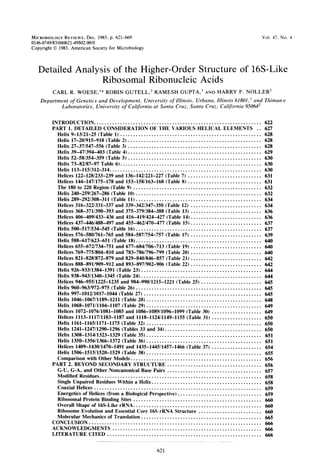
![622 WOESE ET AL.
INTRODUCTION
Translation physically links the genotype and
the phenotype and thereby defines them. The
process is a necessary precondition to the evolu-
tion of all macromolecular structure. The size of
enzymes, their complexity, and their specificity
reflect an underlying accuracy in the translation
process. Is it not true, therefore, that translation
embodies the essence of the cell?
Translation occurs in the framework of the
ribosome, an enormous and complex molecular
aggregate that comprises two ribonucleoprotein
subunits (12). The smaller of these (in bacteria)
contains about 20 separate protein components,
positioned around a large ribonucleic acid
(RNA) some 1,500 nucleotides in length. The
larger subunit comprises an RNA of about twice
that size (2,900 nucleotides), in addition to a
much smaller 5S RNA (120 nucleotides) and
about 30 distinct proteins (26, 40).
For almost three decades, biologists have
sought to understand the molecular mechanics
of translation, but with little beyond descriptive
success. In approaching translation, we have
tended to focus on the protein components as
the elements that define ribosome function-the
ribosomal RNAs (rRNAs) were seen as basically
structural. With the advent of nucleic acid se-
quencing technology, however, has come an
interest in the possible functional roles for
rRNAs. If we can transform their linear molecu-
lar sequence into a detailed and dynamic three-
dimensional form, we will almost certainly un-
derstand the all-important translation process.
The earliest speculations concerning rRNA
secondary structure were based upon a partial
(and, it turns out, incorrect) sequence from one
organism (20) and therefore need not be consid-
ered here. Studies of transfer RNA (tRNA) and
of 5S RNA (25) have forcefully demonstrated
that the only reliable way to determine second-
ary structure presently available (outside of X-
ray crystallography) is through comparative
analysis of primary structure. A comparative
approach to the 16S rRNA sequence, based
upon the Escherichia coli sequence (9, 11), the
nearly complete 16S rRNA sequence from Bac il-
lus brevis, and T1 ribonuclease (RNAse) oligo-
nucleotide catalogs from over 150 other bacte-
ria, gave us a first look at the true secondary
structure of the molecule (51, 98). The subse-
quent publication of 16S RNA sequences from
Zea mays chloroplasts (69) and mammalian mi-
tochondria (1, 21) permitted some refinement of
the original structure and provided further com-
parative proof for a number of helical elements
(54).
There now exist in the literature a number of
proposed secondary structures for 16S (and 23S)
rRNA (6-8, 30, 54, 79, 98, 105). All agree to a
first approximation (for all have used to some
extent a comparative approach). However,
there are differences in detail among them. Since
it is virtually impossible for interested biologists
to assess the relative merits of the various
models-indeed, it even requires considerable
effort to intercompare them-and since new
sequences add complexity as well as information
to the picture, it is of considerable value to
review the topic of the constraints in rRNA
sequence. The purpose of the present review are
to bring up-to-date and discuss in detail the
status of 16S rRNA secondary structure and to
present an overview on this rapidly developing
field.
The first part of the review concerns the
evidence for the individual helical elements. In
each case, extensive comparative evidence sup-
porting the double-helical stalk will be given,
and the structure will be further described in
terms of the susceptibility of various residues
therein to chemical modification and so on.
Also, the entire locale will, whenever possible,
be characterized in phylogenetic terms (conser-
vation of sequence, patterns of variability, etc.).
The data base used herein contains,the com-
plete sequences for the 16S-like rRNAs frqm E.
co/i (9), Z. mays (69) and tobacco chloroplasts
(86), and various mitochondria (1, 21, 39, 42, 74,
89: J. J. Seilhammer, G. M Olsen, and D. J.
Cummings, unpublished data); the unpublished
partial sequences from B. brevis (C. R. Woese
and H. F. Noller, unpublished data) and Bacillus
stearothermophilus (R. Gupta et al., unpub-
lished data); the 16S rRNA sequence from the
archaebacterium Halobacteriiin volcanii (33);
the 18S rRNA sequences from Sacchlaronyces
cerevisiae (64); Xenopiis laevis (65), and Dic-
tyostelium discoideum (R. McCarroll, G. J. Ol-
sen, Y. D. Stahl, C. R. Woese, and M. L. Sogin,
Biochemistry, in press) and the T, RNase cata-
logs for 16S rRNAs of over 200 organisms
(mostly eubacteria) (24 and references cited
therein; C. Woese et al., unpublished data).
Also, the susceptibility of various residues to
chemical modification has been measured by T1
and pancreatic RNase cataloging assays, using
bisulfite modification of C's (E. coli and B.
brevis 16S rRNAs), glyoxal substitution of G's
(E. coli 16S rRNA), m-chloroperbenzoic acid
modification of A's (E. coli 16S rRNA) (C. R.
Woese and L. J. Magrum, unpublished data),
and kethoxal substitution of G's (in E. coli active
[50] and inactive [36] 30S subunits and certain
ribonucleoprotein fragments), all reagents that
are known to respect secondary structure. Also
used to some extent are the relative sensitivity
of residues to enzymatic cleavage by T, and
pancreatic nucleases (which would detect un-
MICROBIOL. REV.](data:image/gif;base64,R0lGODlhAQABAIAAAAAAAP///yH5BAEAAAAALAAAAAABAAEAAAIBRAA7)