Infectitious complications in the practice of maxillofacial surgery
•
5 gefällt mir•869 views

Oral & maxillofacial infections

Empfohlen
Weitere ähnliche Inhalte
Was ist angesagt?
Was ist angesagt? (19)
Andere mochten auch
Andere mochten auch (13)
Ähnlich wie Infectitious complications in the practice of maxillofacial surgery
Ähnlich wie Infectitious complications in the practice of maxillofacial surgery (20)
Kürzlich hochgeladen
Kürzlich hochgeladen (20)
Infectitious complications in the practice of maxillofacial surgery
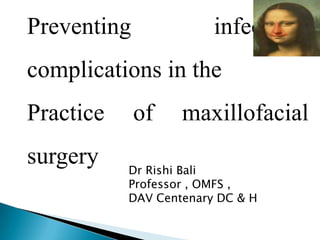
- 1. Preventing infectious complications in the Practice surgery of maxillofacial Dr Rishi Bali Professor , OMFS , DAV Centenary DC & H
- 2. Three main causes : Anaesthesia Hemorrhage Infection
- 3. Maxillofacial infections ? Considerations ? • Proximity to vital organs & siginificant mortality • Medically compromised patients • Antibiotic resistant bacteria
- 4. Fascial space infections Fungal infections Sepsis & Septic shock
- 5. Brain abscess, cavernous sinus thrombosis,Orbital cellulitis Upward spread Systemic spread SPACE INFECTIONS Septicemia,DIC Downward spread Resp. obstr ,Aspiration Pneumonia,Pneumothorax,Mediastinitis Pericarditis,Thoracic Empyema
- 6. Described in relation to the hyoid ◦ Suprahyoid ◦ Infrahyoid ◦ Entire length of the neck
- 7. Superior—skull base Inferior—hyoid Posterior—prevertebral fascia The stylopharyngeal aponeurosis of Zuckerkandel : barrier to infection from the pre to the post styloid compartment.
- 8. Carotid sheath contributions from all 3 layers of deep fascia & can be secondarily infected Airway obstruction, pneumothorax, Mediastinitis, Horner’s syndrome, IJV thrombophlebitis,LeMierre syndrome Communicates retropharyngeal space with
- 10. 39 yrs pregnant pt Lt Submandibular Pterygomand peritonsillar & Parapharyngeal infection Mild airway compromise , TLC 18,500 , BP 92/50 ,HR 120 , Temp 102, RR 21/min Focus removal - I&D Patient died ? ?
- 11. TRIAGE : Occult Obvious distress, Near total obstruction Fiberoptic intubation Tracheotomy under LA If FB/ skilled anaesthetist not available Needle decompression before airway control Avoid Blind nasal intubation
- 12. IMAGING Lateral neck film (6 at 2 & 22 at 6 ) False negative rate 33% CECT is the imaging of choice 12
- 14. Cavernous Sinus Thrombosis (CST) •Periorbital edema, headache, photophobia, proptosis,ptosis , chemosis, 3 to 6 cranial nerve palsies VI nerve earliest. •Sluggish pupillary reaction •Sensory deficits of the opth & max div of the fifth nerve . •Headache , Nuchal rigidity & Sepsis.
- 15. Fungal sinusitis • Reported with increasing frequency • Invasive form : associated morbidity & mortality with significant
- 18. No propotosis after 3 months of Voriconazole therapy
- 22. Reversal of immunocompromised state Early intervention & antifungals based on micro. Accurate Assesment of spread : aggressive Surgery Regular follow up with imaging for minimum of one year Singh V ,Bali R. Rhinocerebral mucormycosis : A diagnostic challenge & therapeutic dilemma . JOMS2012 June;70(6):1369
- 23. Facial swelling & Pain Opthalmoplegia& blurred vision
- 26. Lt interpositional arthroplasty in 7 yr male Blood loss approx. 250 ml & Uneventful extubation Post op. phase hypotension 98/58 mmhg. • Hemoglobin 10 .4gm % • Drain & surgical site were evaluated • Fast NS followed by colloids • Blood transfusion 1 unit started 10 hrs after the surgery
- 30. Infection SIRS Sepsis Severe Sepsis Ref : Bone et al. Chest. 1992;101:1644-1654.
- 33. Altered Consciousness Tachypnea PaO2 250 Liver Enzymes >2x ULN Tachycardia SBP 90 despite fluids Urine Output <0.5 mL/kg/hr despite fluids Creatinine Platelets <100,000/mm3 PTT≥60 Low pH with high lactate (lactate>2mMol) Balk RA. Crit Care Clin. 2000;16:179-192.
- 34. ◦ CRP , Blood Glucose ◦ Leukocytosis or Leukopenia ◦ Toxic granulation of neutrophils & band forms ◦ Arterial Blood gases ◦ Cultures : Blood / Pus http://www.survivingsepsis.org/campaign
- 35. Sound knowledge of pathophysiology of infectious processes ,the ability to diagnose early , anticipate complications & decisively interrupt them can prevent many morbidities & mortalities in Maxillofacial surgery
Hinweis der Redaktion
- Due to the proximity of the central nervous system and respiratory passages, timely efforts are required to diagnose and properly manage maxillofacial infections. Do not underestimate maxillofacial infections”Multiple severe complications of OI have been reported, such as airwayobstruction , mediastinitis , necrotizing fascitis, cavernous sinus thrombosis, sepsis,thoracic empyema, cerebral abscess and osteomyelitis.
- Superficial spacePrevertebral spaceRetropharyngeal spaceDanger spaceVisceral vascular space
- Posterior to pharynx and esophagusAnterior border is alar layer of deep fasciaPosterior border is prevertebral layerExtends from skull base to diaphragm and is so named because it contains loose areolar tissue and offers little resistance to the spread of infectionAnterior to alar layer of deep fasciaExtends from skull base to T1-T2
- Goal is to identify the urgencyORD :pts at risk after sedative medication or manipulation. Trismus ,reactive airway edema .Depressed airway that can not be protected .proximity of surgical site entails risk of aspiration of purulent material .
- Magnetic resonance image showing bilateral retroseptal infection with intracranial extension involving the meninges of thetemporal lobes and cavernous sinus and a left temporal space abscess.Desa and Green. Cavernous Sinus Thrombosis. J Oral MaxillofacSurg 2012.
- Dental origin -7% of all cases of CST.The infection can begin with unilateral involvement, but can develop bilaterally through the circular sinus. The cavernous sinuses and their connections are devoid of valves, consequently bidirectional spread of infection, and thrombi can occur throughout this network. Organisms may reach the cavernous sinus from the face by an anterograde route along ophthalmic veins connected to angular veins, or by a retrograde route along emissary veins connected to the pterygoid venous plexus. Contrast enhanced CT scan may reveal the primary source of infection, thickening of the superior ophthalmic vein and irregular filling defects in the cavernous sinus
- Let us see how the changes occurs in the form of proptosis during course of treatment
- Negatively stained hyphae
- If the source of immunocompromise state can not be reversed. Then the other adjuncts are almost always ineffectual.Microbiological :Culture,PCR & DNA Probes
- Zygomatic swing osteotomy