SlideShare ist ein Scribd-Unternehmen logo
1 von 49
Dr.Giridhar.A
                         Giridhar Eye Institute
                                       Cochin


Giridhar Eye Institute
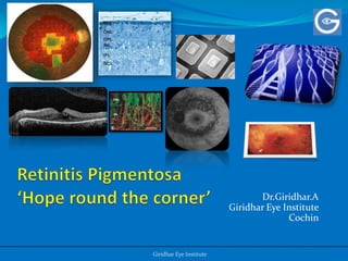
Retinitis pigmentosa
 Retinitis pigmentosa is a clinically and genetically
 heterogeneous group of hereditary disorders in
 which there is progressive loss of photoreceptor and
 pigment epithelial function.

 The prevalence of retinitis pigmentosa is between
 l/3000 and 1/5000 making it one of the most
 common causes of visual impairment in all age
 groups.
Introduction
   Retinitis pigmentosa, the most frequent cause of inherited
     retinal degeneration in patients, is caused by mutations in
     a number of retina-specific genes.

   Patients with retinitis pigmentosa typically experience
    nyctalopia in adolescence or early adulthood because of
    primary degeneration of their rods, usually beginning in
    the midperipheral retina.
   As the disease progresses, death of both rods and cones
    gives rise to a characteristic ring-shaped scotoma that
    expands with time to involve the far periphery and macula

Berson EL. Retinitis pigmentosa: unfolding its mystery. ProcnatlacadSci U S A
1996;93:4526–4528.
Introduction
 Essential diagnostic criteria include
 Bilateral involvement
 Peripheral visual field loss
 Rod-dominated photoreceptor dysfunction (documented by either an elevated
  dark adaptation threshold or electroretinogram amplitude reduction)
 Progression of the disease.

   Common eye findings are :
   Posterior subcapsular cataract
   Degeneration of the vitreous
   Waxy yellow optic disk atrophy
   Narrowed retinal vessels
   Irregular reflexes of the inner limiting membrane
   Midperipheral atrophy of the pigment epithelium
   Bone spicule pigmentation.
Macular changes
 Fishman et al”’ reported three types of macular lesions in retinitispigmentosa
  patients:

 Group 1 - 58%~ of patients had atrophy of the macular area with thinning of the
  retinal pigment epithelium and mottled transmission defects on fluorescein
  angiography.

 Group 2 - 19%’ showed cystic lesions or partial thickness holes within the macula
  with radial, inner retinal traction lines and/or various degrees of preretinal
  membranes causing a “surface wrinkling phenomenon”, among these patients the
  overall clinical severity of the retinal degeneration varied, but several had minimal
  retinal changes in the periphery.

 Group 3 - 23%) of patients had cystoid macular edema and evidence of increased
  capillary permeability on fluorescein angiography

 These patients, like those in Group 2, had minimal or no pigmentary changes in
  the midperiphery, suggesting more recent onset or less severe retinitis pigmentosa
Vitreous changes
 The great majority of retinitis pigmentosa patients have
  changes in the vitreous.

Pruett et al has classified into four groups:

 Stage I - fine colorless dust-like particles evenly distributed
  throughout the vitreous
 Stage II - posterior vitreous detachment;
 Stage Ill - vitreous condensation with the appearance of a
  matrix or reticulum of spindle-shaped condensations
  and/or cotton ball-like opacities
 Stage IV - collapse of the vitreous with greatly reduced
  volume.
Introduction
    RP can be passed on by all types of inheritance:
   20% of RP is autosomal dominant (ADRP),
   20% is autosomal recessive (ARRP)
   10% is X linked (XLRP), while the remaining 50% is found
    in patients without any known affected relatives

  Patients with RP who were at least 45 years or older found the
    following findings:
   52% had 20/40 or better vision in at least one eye,
   25% had 20/200 or worse vision
   0.5% had no light perception.


Grover S, Fishman GA, Anderson RJ, et al. Visual acuity impairment in patients with
retinitis pigmentosa at age 45 years or older. Ophthalmology. Sep 1999;106(9):1780-5
The Pathology
 Earlier histologic studies of photoreceptors in retinitis pigmentosa retinas
  demonstrated outer segment shortening and progressive loss of these critical
  cells

   Milam AH, Li Z-Y, Fariss RN. Histopathology of the human retina in retinitis pigmentosa. ProgRetin Eye Res 1998;17:175–205


 Recent studies revealed that peripheral rods in degenerating retinas with
    retinitis pigmentosa undergo a remarkable process of neurite
    sprouting, forming long axon-like processes that extend through the outer
    plexiform layer into the inner retina

   Li Z-Y, Kljavin IJ, Milam AH. Rod photoreceptor neurite sprouting in retinitis pigmentosa. J
    Neurosci1995;15:5429– 5438.
   Milam AH, Li Z-Y, Cideciyan AV, Jacobson SG. Clinicopathologic effects of the Q64ter rhodopsin mutation in
    retinitispigmentosa. InvestOphthalmol Vis Sci1996;37: 753–765.
Retinal layers


(A) Normal human retina . RPE 5 retinal pigment
                                                          (B)Retinitis pigmentosa retina. The retina is thin
epithelium; OS 5 outer segments; ONL 5 outer nuclear      because many photoreceptors have died and the
layer; OPL 5 outer plexiform layer; INL 5 inner nuclear   remaining photoreceptors lack outer segments. Some
layer; IPL 5 inner plexiform layer; GCL 5 ganglion cell   photoreceptor somata are retained in the outer
layer                                                     nuclear layer, adjacent to the retinal pigment
                                                          epithelium..The outer plexiform layer is thinned but
                                                          the inner nuclear layer, inner plexiform layer, and
                                                          ganglion cell layer appear relatively normal.
Immunolabeling of Rod Photoreceptors With Anti-opsin

 (Green) In Normal and Retinitis Pigmentosa Retinas                                              .


(C) Opsinimmunolabeling (green) is strongest in the outer segments (OS) and weak in the plasma membranes of
rod somata in the outer nuclear layer and synapses in the outer plexiform layer. (D) Retinitis pigmentosa The
surviving rods are opsin positive (green) but few retain outer segments. Rod neurites (arrows) extend past the
inner nuclear layer, and some terminate in the ganglion cell layer
Neurite sprouting
 Rods, amacrine and horizontal cells undergo
  neurite sprouting in human retinas with retinitis
  pigmentosa.
 These changes in the retinal neurons may
  contribute to the electroretinographic
  abnormalities and progressive decline in vision
  noted by patients with retinitis pigmentosa.
 These alterations may also complicate strategies
  for treatment of retinitis pigmentosa.
Immunolabeling


Retinitis pigmentosa retina .Localization of opsin (green) in rods in the outer
nuclear layer (ONL) and glialfibrillary acid protein (red) in reactive Muller
cells(red).
Investigations
 Conventional investigations:
 Electroretinography – ERG
 Dark Adaptation and Visual Sensitivity
 Visual Field
 Fundus Reflectometry
 Contrast Sensitivity
 The Electrooculogram – EOG
 The Visually Evoked Response
 Fluorescein Angiography
 Vitreous Fluorophotometry
Newer Clinical Investigations
 Spectral domain OCT
 Micro perimetry
 Fundus Auto fluorescence
 Genetic subtyping
SD OCT in RP
 The functional and morphologic data for the patients
  with classic RP and without macular complications
  were not significantly different from those for healthy
  patients.
 SD OCT enables to obtain more precise information
  about the changes in macular status.
SD OCT in RP
                                      1                        2


                                          3                    4


1. A retinitis pigmentosa (RP) patient with no abnormalities
2. Macular edema
3. Vitreomacular traction
4. Retinal thinning
Microperimetry


                 A picture of the fundus in a
                 patient with retinitis
                 pigmentosa is overlaid with
                 microperimetry (MP-1)
                 results, showing no retinal
                 sensitivity in the peripheral
                 visual field.
Fundus Autofluorescence
TheHyperautofluorescent Ring

 Progressive constriction of the hyperautofluorescent
 ring and a concordant decrease in IS/OS junction
 length are observed over time.
History of genetics in RP
 The striking pedigree by Franceschetti (1953) was
  reproduced in the book by Francois (1961).
 Boughman et al. (1980) estimated the overall frequency at
  about 1 in 3,700, whereas the incidence of the recessive
  type, with at least 2 genocopies, was estimated to be about 1
  in 4,450. No evidence of ethnic heterogeneity was found.
 Heckenlively et al. (1981) identified 43 cases of autosomal
  recessive RP among the Navajo Indians.
 In Shanghai, Hu(1982) analyzed 151 pedigrees with 209
  cases of RP. Of these cases, the proportions of autosomal
  recessive (AR), autosomal dominant (AD), X-linked
  recessive (XR), and simplex cases were 33.1, 11, 7.7 and
  48.3%, respectively.
Genetics in RP
 The greatest roadblock to molecular diagnosis of RP is the
  availability of genetic testing.

 THE GOALS AHEAD IN MOLECULAR GENETICS :

1. Most of the genes causing RP must be identified.
2. It must be possible to detect nearly all of the disease-
   causing mutations within these genes.
3. Mutation testing must become inexpensive, reliable, and
   widely available.
4. We must be able to understand, interpret, and explain the
   molecular information.
The total prevalence is 1 case per 3100
 persons (range, 1 case per 3000 persons to
 1 case per 7000 persons), or 32.2 cases per
 100 000 persons.


Haim et al. Epidemiology of retinitis pigmentosa
in Denmark. ActaOphthalmol Scand Suppl.
2002;233:1-34.
Why finding the underlying disease causing
mutation should matter to the patient or the
clinician?
 Identifying the underlying mutation(s) can establish the
  diagnosis
 Knowing the genetic cause is essential for family
  counseling and for predicting recurrence risk and
  prognosis.
 Each new mutation that is found contributes to a better
  understanding of ocular biology
 The era of gene-specific and mutation specific treatments
  for inherited retinal diseases is quickly approaching*
*Bennett J. Gene therapy for Leber congenital amaurosis. Novartis Found Symp.
2004;255:195-202.
Chader GJ. Beyond basic research for inherited and orphan retinal diseases: successes
and challenges. Retina. 2005;25:S15-S17
Number of mapped and identified retinal
disease genes from 1980 to 2006.
Genes and Mapped Loci Causing Nonsyndromic, Nonsystemic
                  Retinitis Pigmentosa
Genes and Mapped Loci Causing Nonsyndromic, Nonsystemic
                  Retinitis Pigmentosa
Genes and Mapped Loci Causing Nonsyndromic, Nonsystemic
                  Retinitis Pigmentosa
Mutations in Genes That Cause an Appreciable Fraction of Retinitis
                           Pigmentosa Cases


Perspective on Genes and Mutations Causing Retinitis Pigmentosa
Stephen et al Arch Ophthalmol. 2007;125:151-158
Genetic testing
 At least 35 different genes or loci are known to cause "nonsyndromic RP“

DNA testing is available on a clinical basis for:
 RLBP1 (autosomal recessive, Bothnia type RP)
 RP1 (autosomal dominant, RP1)
 RHO (autosomal dominant, RP4)
 RDS (autosomal dominant, RP7)
 PRPF8 (autosomal dominant, RP13)
 PRPF3 (autosomal dominant, RP18)
 CRB1 (autosomal recessive, RP12)
 ABCA4 (autosomal recessive, RP19)
 RPE65 (autosomal recessive, RP20)

For all other genes, molecular genetic testing is available on a research basis only
RP2 Gene in X linked inheritance
 Researchers emphasis the screening of the RP2 gene in
  patients younger than 16 years characterized by X-
  linked inheritance, decreased best-corrected visual
  acuity, high myopia, and early-onset macular atrophy
 Patients exhibiting a choroideremia like fundus
  without choroideremia gene mutations should also be
  screened for RP2 mutations.


RP2 Phenotype and Pathogenetic Correlations in X-Linked Retinitis
Pigmentosa Thiran et al Arch Ophthalmol. 2010;128(7):915-923
Management of RP (Current perspective )
 Vitamin A/beta-carotene Antioxidants may be useful
  in treating patients with RP.
 A recent comprehensive epidemiologic study
  concluded that very high daily doses of vitamin A
  palmitate (15,000 U/d) slow the progress of RP by
  about 2% per year.

 The effects are modest; therefore, this treatment must
 be weighed against the uncertain risk of long-term
 adverse effects from large chronic doses of vitamin A.
Lutein supplement
  Recent studies shows Lutein supplementation of 12
    mg/d slowed loss of midperipheralvisualfield on
    average among non smoking adults with retinitis
    pigmentosa taking vitamin A.


Clinical Trial of Lutein in Patients With Retinitis Pigmentosa Receiving
Vitamin A Eliot et al. Arch Ophthalmol. 2010;128(4):403-411
Management of RP (Current perspective )

 DHA is an omega-3 polyunsaturated fatty acid and
  antioxidant.
 Studies reported trends of less ERG change in patients
  with higher levels of DHA.

 However, a recent study compared DHA plus vitamin
 A to vitamin A alone in patients with RP over 4 years.
 Benefit of DHA was not seen. Further clinical trials
 must be done to determine DHA benefit.
Management of RP (Current perspective )
Acetazolamide
 Oral acetazolamide has shown the most encouraging
  results with some improvement in visual function.
 Studies by Fishman et al and Cox et al have
  demonstrated improvement in Snelling visual acuity
  with oral acetazolamide for patients who have RP with
  macular edema*.
  Adverse effects, including fatigue, renal stones, loss of
  appetite, hand tingling, and anemia, may limit its use.


*Fishman GA, Gilbert LD, Fiscella RG, Kimura AE, Jampol LM. Acetazolamide for treatment of chronic
macular edema in retinitis pigmentosa. Arch Ophthalmol. Oct 1989;107(10):1445-52
Management of RP (Current perspective )

 Ascorbic acid 1000 mg/d
 Intra vitreal Triamcinolone for macular edema in RP*
 Rehabilitation with Low vision aids


*Treatment of Cystoid Macular Edema in Retinitis Pigmentosa With
IntravitrealTriamcinolone. Lucia et al Arch Ophthalmol. 2007;125:759-764
Recent advances
     Transplant of Rods in Mice Retina
                                   Mice receiving rod-rich transplants
                                   demonstrated statistically significant greater
                                   cone numbers, with rescue of 40% of host
                                   cones normally destined to die during this
                                   period.


                                   Such findings indicate that transplantation
                                   of rods could limit loss of cones, thus
                                   preserving useful vision in human retinitis
                                   pigmentosa.


Selective Transplantation of Rods Delays Cone Loss in a Retinitis Pigmentosa Model.
SaddekMohand-Said, MD Arch Ophthalmol. 2000;118:807-811
Recent advances
 Sheet Transplant of Fetal Retina With Retinal Pigment
   Epithelium in Retinitis Pigmentosa:

 Norman D. Radtke and co workers transplanted a
   sheet of fetal neural retina with its retinal pigment
   epithelium into the subretinal space under the fovea
   unilaterally in a patient with Retinitis Pigmentosa with
   visual acuity of 20/800 in the treated eye.


Vision Change After Sheet Transplant of Fetal Retina With Retinal Pigment
Epithelium to a Patient With Retinitis Pigmentosa Norman et al Arch Ophthalmol.
2004;122:1159-1165
Fetal Retinal Transplant


Fundus images: Arrows indicate the same blood vessel landmark in all images. A, Three weeks prior to
transplantation. B, Two weeks after transplantation, showing heavy pigmentation of the transplant. The
transplant area is outlined by white dots. C, Six months after transplantation, there is a loss of pigment. A
white scar is recognizable at the retinotomy site. White dots outline the same area as in B. D, Twelve months
after transplantation, there is no change compared with 6 months.
The Hope
 A change in visual acuity from 20/800 to 20/400 (7
 months), 20/250 (9 months), and 20/160 (1 year) was
 observed by ETDRS visual acuity testing.

 At 2 years 2 months after the surgery, the patient noted
  that she could definitely see better with the eye that
  was operated on.
 She could also read the large-print Reader’s Digest and
  print on the computer with the eye that was operated
  on that she could not read with the other eye.
Artificial Silicon Retina Microchip
   The ASR microchip is a 2-mm-diameter silicon-based
    device that contains approximately 5000
    microelectrode- tipped microphotodiodes and is
    powered by incident light.

   The right eyes of 6 patients with retinitis pigmentosa
    were implanted with the ASR microchip while the left
    eyes served as controls. Safety and visual function
    information was collected.


The Artificial Silicon Retina Microchip for the Treatment of Vision Loss
From Retinitis Pigmentosa Alan Y. Chow, MD Arch Ophthalmol. 2004;122:460-469
ASR MICROCHIP


                                         B -The ASR microchip
A -The ASR’s size relative to a penny.   (original magnification ×36).


C-The ASR pixels                         D -Subretinal location of the implanted ASR
(original magnification 1400).           microchip.
ASR Implant

              Fundus
              photographs and
              fluorescein
              angiograms of an
              implanted
              artificial silicon
              retina microchip
              in the superior
              temporal retina.
The Hope
 No significant safety-related adverse effects were observed.
 Subjective improvements included improved perception of
  brightness, contrast, color, movement, shape, resolution, a
  nd visual field size

 The observation of retinal visual improvement in areas far
  from the implant site suggests a possible generalized
  neurotrophic-type rescue effect on the damaged retina
  caused by the presence of the ASR.
 A larger clinical trial is indicated to further evaluate the
  safety and efficacy of a subretinally implanted ASR.
Stem Cells
   UK Researchers working with mice, transplanted
    mouse stem cells which were at an advanced stage of
    development, and already programmed to develop
    into photoreceptor cells, into mice that had been
    genetically induced to mimic the human conditions of
    retinitis pigmentosa.
   These transplanted cells integrate, differentiate into
    rod photoreceptors, form synaptic connections and
    improve visual function.
   This research may in the future lead to using
    transplants in humans to relieve blindness.


Retinal repair by transplantation of photoreceptor precursors
R. E. MacLaren. Nature 444, 203-207 2006
“Pikachurin” The sight saving protein ?

    Pikachurin is a dystroglycan-interacting
      polysaccharide which has an essential role in the
      precise interactions between the photoreceptor ribbon
      synapse and the bipolar dendrites


    The protein localizes to the synaptic cleft in the photoreceptor ribbon
      synapse, which transmits signals from visual cells to the brain,
"Pikachurin, a dystroglycan ligand, is essential for photoreceptor ribbon synapse formation".   Sato (2008). Nat.
Neurosci. 11 (8): 923–31
Gene Therapy
 Expression of archaebacterialhalorhodopsin in
    light-insensitive cones can substitute for the
    native phototransduction cascade and restore
    their light sensitivity in mouse models of retinitis
    pigmentosa.

 Using human ex vivo retinas, researchers shows
    that halorhodopsin can reactivate light-
    insensitive human photoreceptors.


Genetic Reactivation of Cone Photoreceptors Restores Visual Responses in Retinitis pigmentosa
Volker et al Science 23 July 2010
0.15% UF-021 (unoprostone isopropyl)
(OCUSEVA)
 Phase 2 Clinical Trial Data of UF-021 in Retinitis
    Pigmentosa Patients Presented at ARVO 2011
   The purpose of the phase 2 study was to test the effects of
    unoprostone isopropyl in protecting and improving the
    central vision in mid-stage to late-stage RP patients
   The primary efficacy endpoint was the change seen on the
    visual field analysis as checked by an instrument called the
    MP-1 microperimeter
   The results show promise, specifically with the advantage
    that it only requires instillation of drops, rather than a
    completed surgical intervention.
   BUT the group that was instilled with placebo drops still
    showed improvement in about 17 of the 112 patients in the
    trial (15.2%).
Tauroursodeoxycholic acid (TUDCA)

   Tauroursodeoxycholic acid (TUDCA) is an
    ambiphilic bile acid.
   Research had positive affects on the health of
    the mouse retina, including a reduced accumulation of
    superoxide radicals, rod cell death, and disruption of
    cone inner and outer segments. The findings of the
    study are elucidating optimized conditions for RP
    treatment


Phillips et al"Tauroursodeoxycholic acid preservation of photoreceptor structure and function in the rd10 mouse
through postnatal day 30". Invest Ophthalmol Vis Sci. 2008 May;49(5):2148-55
Is there hope?

Weitere ähnliche Inhalte

Was ist angesagt?

hereditary macular and choroidal dystrophies
hereditary macular and choroidal dystrophies hereditary macular and choroidal dystrophies
hereditary macular and choroidal dystrophies Priyanka Choudhary
 
Retinitis pigmentosa ppt
Retinitis pigmentosa ppt Retinitis pigmentosa ppt
Retinitis pigmentosa ppt Mehedi Hasan
 
Retinitis pigmentosa and allied disorders
Retinitis pigmentosa and allied disordersRetinitis pigmentosa and allied disorders
Retinitis pigmentosa and allied disordersShruti Laddha
 
Retinitis pigmentosa (opthalmology)
Retinitis pigmentosa (opthalmology)Retinitis pigmentosa (opthalmology)
Retinitis pigmentosa (opthalmology)Ma Wady
 
Retinitis pigmentosa
Retinitis pigmentosaRetinitis pigmentosa
Retinitis pigmentosaJananieIyer
 
Retinal dystrophy
Retinal dystrophyRetinal dystrophy
Retinal dystrophydipusarkar2
 
XERODERMA PIGMENTOSUM.pptx
XERODERMA PIGMENTOSUM.pptxXERODERMA PIGMENTOSUM.pptx
XERODERMA PIGMENTOSUM.pptxNikhil Das
 
Retinitis Pigmentosa
Retinitis PigmentosaRetinitis Pigmentosa
Retinitis PigmentosaInsha Aleena
 
Hereditary vitreoretinal degenerations
Hereditary vitreoretinal degenerationsHereditary vitreoretinal degenerations
Hereditary vitreoretinal degenerationsShruti Laddha
 
Discuss Night Blindness!
Discuss Night Blindness!Discuss Night Blindness!
Discuss Night Blindness!personalp
 
Hereditary macular dystrophies
Hereditary macular  dystrophiesHereditary macular  dystrophies
Hereditary macular dystrophiesShruti Laddha
 
Pediatric Eye Conditions In Wisconsins Preschool Population
Pediatric Eye Conditions In Wisconsins Preschool PopulationPediatric Eye Conditions In Wisconsins Preschool Population
Pediatric Eye Conditions In Wisconsins Preschool Populationguest366b411
 
CENTRAL SEROUS CHORIOETINOPATHY
CENTRAL SEROUS CHORIOETINOPATHYCENTRAL SEROUS CHORIOETINOPATHY
CENTRAL SEROUS CHORIOETINOPATHYSivateja Challa
 

Was ist angesagt? (20)

Cone and Rod Dystrophy
Cone and Rod DystrophyCone and Rod Dystrophy
Cone and Rod Dystrophy
 
Retinitis pigmentosa
Retinitis pigmentosaRetinitis pigmentosa
Retinitis pigmentosa
 
hereditary macular and choroidal dystrophies
hereditary macular and choroidal dystrophies hereditary macular and choroidal dystrophies
hereditary macular and choroidal dystrophies
 
Retinitis pigmentosa ppt
Retinitis pigmentosa ppt Retinitis pigmentosa ppt
Retinitis pigmentosa ppt
 
Retinitis pigmentosa and allied disorders
Retinitis pigmentosa and allied disordersRetinitis pigmentosa and allied disorders
Retinitis pigmentosa and allied disorders
 
Retinitis pigmentosa (opthalmology)
Retinitis pigmentosa (opthalmology)Retinitis pigmentosa (opthalmology)
Retinitis pigmentosa (opthalmology)
 
Retinitis pigmentosa
Retinitis pigmentosaRetinitis pigmentosa
Retinitis pigmentosa
 
Retinal dystrophy
Retinal dystrophyRetinal dystrophy
Retinal dystrophy
 
Lecture pspos
Lecture   psposLecture   pspos
Lecture pspos
 
XERODERMA PIGMENTOSUM.pptx
XERODERMA PIGMENTOSUM.pptxXERODERMA PIGMENTOSUM.pptx
XERODERMA PIGMENTOSUM.pptx
 
Retinitis Pigmentosa
Retinitis PigmentosaRetinitis Pigmentosa
Retinitis Pigmentosa
 
Macular disorders best disease
Macular disorders best diseaseMacular disorders best disease
Macular disorders best disease
 
Hereditary vitreoretinal degenerations
Hereditary vitreoretinal degenerationsHereditary vitreoretinal degenerations
Hereditary vitreoretinal degenerations
 
Autosomal dominant optic atrophy
Autosomal dominant optic atrophy Autosomal dominant optic atrophy
Autosomal dominant optic atrophy
 
Retinitis pigmentosa 2
Retinitis pigmentosa 2Retinitis pigmentosa 2
Retinitis pigmentosa 2
 
Retinitis pigmentosa 3
Retinitis pigmentosa 3Retinitis pigmentosa 3
Retinitis pigmentosa 3
 
Discuss Night Blindness!
Discuss Night Blindness!Discuss Night Blindness!
Discuss Night Blindness!
 
Hereditary macular dystrophies
Hereditary macular  dystrophiesHereditary macular  dystrophies
Hereditary macular dystrophies
 
Pediatric Eye Conditions In Wisconsins Preschool Population
Pediatric Eye Conditions In Wisconsins Preschool PopulationPediatric Eye Conditions In Wisconsins Preschool Population
Pediatric Eye Conditions In Wisconsins Preschool Population
 
CENTRAL SEROUS CHORIOETINOPATHY
CENTRAL SEROUS CHORIOETINOPATHYCENTRAL SEROUS CHORIOETINOPATHY
CENTRAL SEROUS CHORIOETINOPATHY
 

Andere mochten auch

Retinitis pigmentosa
Retinitis pigmentosaRetinitis pigmentosa
Retinitis pigmentosaKenny Correa
 
that's what I do 2013
that's what I do 2013that's what I do 2013
that's what I do 2013Anna Huguet
 
Dr Gerry Chader - AGM 2011 - Research Advancements
Dr Gerry Chader - AGM 2011 - Research AdvancementsDr Gerry Chader - AGM 2011 - Research Advancements
Dr Gerry Chader - AGM 2011 - Research AdvancementsFighting Blindness
 
Transgenic organisms powerpoint
Transgenic organisms powerpointTransgenic organisms powerpoint
Transgenic organisms powerpoint14wilsst
 
Neuropatía Optica Hereditaria de Leber
Neuropatía Optica Hereditaria de LeberNeuropatía Optica Hereditaria de Leber
Neuropatía Optica Hereditaria de LeberEdgar Gtz
 
ophthalmology.squint.(dr.khald)
ophthalmology.squint.(dr.khald)ophthalmology.squint.(dr.khald)
ophthalmology.squint.(dr.khald)student
 
Heterophoria n tropia
Heterophoria n tropiaHeterophoria n tropia
Heterophoria n tropiaJunu Shrestha
 
9. retinitis pigmentosa y otras enfermedades retinianas hereditarias
9. retinitis pigmentosa y otras enfermedades retinianas hereditarias9. retinitis pigmentosa y otras enfermedades retinianas hereditarias
9. retinitis pigmentosa y otras enfermedades retinianas hereditariasMarvin Barahona
 
Retinosis pigmentaria
Retinosis pigmentariaRetinosis pigmentaria
Retinosis pigmentariaAbril Santos
 
Retinitis Pigmentaria
Retinitis Pigmentaria  Retinitis Pigmentaria
Retinitis Pigmentaria Felipe Godoy
 
Transgenic animals new
Transgenic animals newTransgenic animals new
Transgenic animals newKarthi Kumar
 
KnockOut mouse technology By Bikash karki
KnockOut mouse technology By Bikash karkiKnockOut mouse technology By Bikash karki
KnockOut mouse technology By Bikash karkiBikash Karki
 

Andere mochten auch (20)

Retinitis pigmentosa
Retinitis pigmentosaRetinitis pigmentosa
Retinitis pigmentosa
 
that's what I do 2013
that's what I do 2013that's what I do 2013
that's what I do 2013
 
Dr Gerry Chader - AGM 2011 - Research Advancements
Dr Gerry Chader - AGM 2011 - Research AdvancementsDr Gerry Chader - AGM 2011 - Research Advancements
Dr Gerry Chader - AGM 2011 - Research Advancements
 
Transgenic organisms powerpoint
Transgenic organisms powerpointTransgenic organisms powerpoint
Transgenic organisms powerpoint
 
Neuropatía Optica Hereditaria de Leber
Neuropatía Optica Hereditaria de LeberNeuropatía Optica Hereditaria de Leber
Neuropatía Optica Hereditaria de Leber
 
ophthalmology.squint.(dr.khald)
ophthalmology.squint.(dr.khald)ophthalmology.squint.(dr.khald)
ophthalmology.squint.(dr.khald)
 
Heterophoria n tropia
Heterophoria n tropiaHeterophoria n tropia
Heterophoria n tropia
 
9. retinitis pigmentosa y otras enfermedades retinianas hereditarias
9. retinitis pigmentosa y otras enfermedades retinianas hereditarias9. retinitis pigmentosa y otras enfermedades retinianas hereditarias
9. retinitis pigmentosa y otras enfermedades retinianas hereditarias
 
Retinosis pigmentaria
Retinosis pigmentariaRetinosis pigmentaria
Retinosis pigmentaria
 
Case presentation-Retinitis Pigmentosa Albecanse
Case presentation-Retinitis Pigmentosa AlbecanseCase presentation-Retinitis Pigmentosa Albecanse
Case presentation-Retinitis Pigmentosa Albecanse
 
Retinitis Pigmentaria
Retinitis Pigmentaria  Retinitis Pigmentaria
Retinitis Pigmentaria
 
Transgenic animals new
Transgenic animals newTransgenic animals new
Transgenic animals new
 
Animal models
Animal modelsAnimal models
Animal models
 
Iridocyclitis
Iridocyclitis Iridocyclitis
Iridocyclitis
 
RETINITIS PIGMENTOSA
RETINITIS PIGMENTOSARETINITIS PIGMENTOSA
RETINITIS PIGMENTOSA
 
Knock out mice
Knock out miceKnock out mice
Knock out mice
 
Standards of psy..
Standards of psy..Standards of psy..
Standards of psy..
 
KnockOut mouse technology By Bikash karki
KnockOut mouse technology By Bikash karkiKnockOut mouse technology By Bikash karki
KnockOut mouse technology By Bikash karki
 
ppt on Eye anatomy
ppt on Eye anatomy ppt on Eye anatomy
ppt on Eye anatomy
 
Knock out technology (final)
Knock out technology (final)Knock out technology (final)
Knock out technology (final)
 

Ähnlich wie Dr Giridhar on RO - Ray of Hope?

Retinal Dystrophy 1. Farhad.pptx
Retinal Dystrophy 1. Farhad.pptxRetinal Dystrophy 1. Farhad.pptx
Retinal Dystrophy 1. Farhad.pptxFarhad Ahmed Dipu
 
Retinitis Pigmetosa_Xerophthalmia_Dr. Pradeep Bastola.pptx
Retinitis Pigmetosa_Xerophthalmia_Dr. Pradeep Bastola.pptxRetinitis Pigmetosa_Xerophthalmia_Dr. Pradeep Bastola.pptx
Retinitis Pigmetosa_Xerophthalmia_Dr. Pradeep Bastola.pptxDr. Pradeep Bastola
 
Pathological myopia 01.03.2014
Pathological myopia 01.03.2014Pathological myopia 01.03.2014
Pathological myopia 01.03.2014Mohammad Bawtag
 
Pigmentary glaucoma - Dr Shylesh B Dabke
Pigmentary glaucoma - Dr Shylesh B DabkePigmentary glaucoma - Dr Shylesh B Dabke
Pigmentary glaucoma - Dr Shylesh B DabkeShylesh Dabke
 
Pathological Myopia.pptx
Pathological Myopia.pptxPathological Myopia.pptx
Pathological Myopia.pptxMohammad Bawtag
 
Cure of leber congenital amaurosis
Cure of leber congenital amaurosisCure of leber congenital amaurosis
Cure of leber congenital amaurosisVibha Sharma
 
Optic coherence tomography
Optic coherence tomographyOptic coherence tomography
Optic coherence tomographyNeurologyKota
 
Retinal Dystrophy.pptx vvvvvvvvvvvvvvvvv
Retinal Dystrophy.pptx vvvvvvvvvvvvvvvvvRetinal Dystrophy.pptx vvvvvvvvvvvvvvvvv
Retinal Dystrophy.pptx vvvvvvvvvvvvvvvvvHarshika Malik
 
Mucopolysccharidoses : A Rare Cause For Bilateral Cloudy Cornea.
Mucopolysccharidoses : A Rare Cause For Bilateral Cloudy Cornea.Mucopolysccharidoses : A Rare Cause For Bilateral Cloudy Cornea.
Mucopolysccharidoses : A Rare Cause For Bilateral Cloudy Cornea.Dr. Jagannath Boramani
 
Retinoblastoma
RetinoblastomaRetinoblastoma
RetinoblastomaSSSIHMS-PG
 
Retinal Dystrophy. Farhad - Copy.pptx
Retinal Dystrophy. Farhad - Copy.pptxRetinal Dystrophy. Farhad - Copy.pptx
Retinal Dystrophy. Farhad - Copy.pptxFarhad Ahmed Dipu
 
A Variety of Anatomical and Clinical Manifestations of Myelinated Retinal Ner...
A Variety of Anatomical and Clinical Manifestations of Myelinated Retinal Ner...A Variety of Anatomical and Clinical Manifestations of Myelinated Retinal Ner...
A Variety of Anatomical and Clinical Manifestations of Myelinated Retinal Ner...inventionjournals
 

Ähnlich wie Dr Giridhar on RO - Ray of Hope? (20)

Retinal Dystrophy 1. Farhad.pptx
Retinal Dystrophy 1. Farhad.pptxRetinal Dystrophy 1. Farhad.pptx
Retinal Dystrophy 1. Farhad.pptx
 
Retinitis Pigmetosa_Xerophthalmia_Dr. Pradeep Bastola.pptx
Retinitis Pigmetosa_Xerophthalmia_Dr. Pradeep Bastola.pptxRetinitis Pigmetosa_Xerophthalmia_Dr. Pradeep Bastola.pptx
Retinitis Pigmetosa_Xerophthalmia_Dr. Pradeep Bastola.pptx
 
MACULAR DYSTROPHY
MACULAR DYSTROPHYMACULAR DYSTROPHY
MACULAR DYSTROPHY
 
Pathological myopia 01.03.2014
Pathological myopia 01.03.2014Pathological myopia 01.03.2014
Pathological myopia 01.03.2014
 
Pigmentary glaucoma - Dr Shylesh B Dabke
Pigmentary glaucoma - Dr Shylesh B DabkePigmentary glaucoma - Dr Shylesh B Dabke
Pigmentary glaucoma - Dr Shylesh B Dabke
 
Pathological Myopia.pptx
Pathological Myopia.pptxPathological Myopia.pptx
Pathological Myopia.pptx
 
Cure of leber congenital amaurosis
Cure of leber congenital amaurosisCure of leber congenital amaurosis
Cure of leber congenital amaurosis
 
Optic coherence tomography
Optic coherence tomographyOptic coherence tomography
Optic coherence tomography
 
Radiation
RadiationRadiation
Radiation
 
Retinal Dystrophy.pptx vvvvvvvvvvvvvvvvv
Retinal Dystrophy.pptx vvvvvvvvvvvvvvvvvRetinal Dystrophy.pptx vvvvvvvvvvvvvvvvv
Retinal Dystrophy.pptx vvvvvvvvvvvvvvvvv
 
Mucopolysccharidoses : A Rare Cause For Bilateral Cloudy Cornea.
Mucopolysccharidoses : A Rare Cause For Bilateral Cloudy Cornea.Mucopolysccharidoses : A Rare Cause For Bilateral Cloudy Cornea.
Mucopolysccharidoses : A Rare Cause For Bilateral Cloudy Cornea.
 
pseudoexfoliative glaucoma
pseudoexfoliative glaucomapseudoexfoliative glaucoma
pseudoexfoliative glaucoma
 
Optic neuritis
Optic neuritisOptic neuritis
Optic neuritis
 
Retinoblastoma
RetinoblastomaRetinoblastoma
Retinoblastoma
 
Retinitis pigmentosa
Retinitis pigmentosaRetinitis pigmentosa
Retinitis pigmentosa
 
Hereditary Retinal disease .pptx
Hereditary Retinal disease .pptxHereditary Retinal disease .pptx
Hereditary Retinal disease .pptx
 
Retinoblastoma
RetinoblastomaRetinoblastoma
Retinoblastoma
 
Retinal Dystrophy. Farhad - Copy.pptx
Retinal Dystrophy. Farhad - Copy.pptxRetinal Dystrophy. Farhad - Copy.pptx
Retinal Dystrophy. Farhad - Copy.pptx
 
A Variety of Anatomical and Clinical Manifestations of Myelinated Retinal Ner...
A Variety of Anatomical and Clinical Manifestations of Myelinated Retinal Ner...A Variety of Anatomical and Clinical Manifestations of Myelinated Retinal Ner...
A Variety of Anatomical and Clinical Manifestations of Myelinated Retinal Ner...
 
Retinoblastoma
RetinoblastomaRetinoblastoma
Retinoblastoma
 

Kürzlich hochgeladen

Asthma Review - GINA guidelines summary 2024
Asthma Review - GINA guidelines summary 2024Asthma Review - GINA guidelines summary 2024
Asthma Review - GINA guidelines summary 2024Gabriel Guevara MD
 
call girls in munirka DELHI 🔝 >༒9540349809 🔝 genuine Escort Service 🔝✔️✔️
call girls in munirka  DELHI 🔝 >༒9540349809 🔝 genuine Escort Service 🔝✔️✔️call girls in munirka  DELHI 🔝 >༒9540349809 🔝 genuine Escort Service 🔝✔️✔️
call girls in munirka DELHI 🔝 >༒9540349809 🔝 genuine Escort Service 🔝✔️✔️saminamagar
 
Russian Call Girls in Pune Riya 9907093804 Short 1500 Night 6000 Best call gi...
Russian Call Girls in Pune Riya 9907093804 Short 1500 Night 6000 Best call gi...Russian Call Girls in Pune Riya 9907093804 Short 1500 Night 6000 Best call gi...
Russian Call Girls in Pune Riya 9907093804 Short 1500 Night 6000 Best call gi...Miss joya
 
Book Call Girls in Kasavanahalli - 7001305949 with real photos and phone numbers
Book Call Girls in Kasavanahalli - 7001305949 with real photos and phone numbersBook Call Girls in Kasavanahalli - 7001305949 with real photos and phone numbers
Book Call Girls in Kasavanahalli - 7001305949 with real photos and phone numbersnarwatsonia7
 
Call Girls Service Chennai Jiya 7001305949 Independent Escort Service Chennai
Call Girls Service Chennai Jiya 7001305949 Independent Escort Service ChennaiCall Girls Service Chennai Jiya 7001305949 Independent Escort Service Chennai
Call Girls Service Chennai Jiya 7001305949 Independent Escort Service ChennaiNehru place Escorts
 
See the 2,456 pharmacies on the National E-Pharmacy Platform
See the 2,456 pharmacies on the National E-Pharmacy PlatformSee the 2,456 pharmacies on the National E-Pharmacy Platform
See the 2,456 pharmacies on the National E-Pharmacy PlatformKweku Zurek
 
Call Girls ITPL Just Call 7001305949 Top Class Call Girl Service Available
Call Girls ITPL Just Call 7001305949 Top Class Call Girl Service AvailableCall Girls ITPL Just Call 7001305949 Top Class Call Girl Service Available
Call Girls ITPL Just Call 7001305949 Top Class Call Girl Service Availablenarwatsonia7
 
Call Girls Hosur Just Call 7001305949 Top Class Call Girl Service Available
Call Girls Hosur Just Call 7001305949 Top Class Call Girl Service AvailableCall Girls Hosur Just Call 7001305949 Top Class Call Girl Service Available
Call Girls Hosur Just Call 7001305949 Top Class Call Girl Service Availablenarwatsonia7
 
Call Girls Service In Shyam Nagar Whatsapp 8445551418 Independent Escort Service
Call Girls Service In Shyam Nagar Whatsapp 8445551418 Independent Escort ServiceCall Girls Service In Shyam Nagar Whatsapp 8445551418 Independent Escort Service
Call Girls Service In Shyam Nagar Whatsapp 8445551418 Independent Escort Serviceparulsinha
 
Call Girl Koramangala | 7001305949 At Low Cost Cash Payment Booking
Call Girl Koramangala | 7001305949 At Low Cost Cash Payment BookingCall Girl Koramangala | 7001305949 At Low Cost Cash Payment Booking
Call Girl Koramangala | 7001305949 At Low Cost Cash Payment Bookingnarwatsonia7
 
Call Girls Hsr Layout Just Call 7001305949 Top Class Call Girl Service Available
Call Girls Hsr Layout Just Call 7001305949 Top Class Call Girl Service AvailableCall Girls Hsr Layout Just Call 7001305949 Top Class Call Girl Service Available
Call Girls Hsr Layout Just Call 7001305949 Top Class Call Girl Service Availablenarwatsonia7
 
call girls in green park DELHI 🔝 >༒9540349809 🔝 genuine Escort Service 🔝✔️✔️
call girls in green park  DELHI 🔝 >༒9540349809 🔝 genuine Escort Service 🔝✔️✔️call girls in green park  DELHI 🔝 >༒9540349809 🔝 genuine Escort Service 🔝✔️✔️
call girls in green park DELHI 🔝 >༒9540349809 🔝 genuine Escort Service 🔝✔️✔️saminamagar
 
Call Girls Kanakapura Road Just Call 7001305949 Top Class Call Girl Service A...
Call Girls Kanakapura Road Just Call 7001305949 Top Class Call Girl Service A...Call Girls Kanakapura Road Just Call 7001305949 Top Class Call Girl Service A...
Call Girls Kanakapura Road Just Call 7001305949 Top Class Call Girl Service A...narwatsonia7
 
Call Girls Whitefield Just Call 7001305949 Top Class Call Girl Service Available
Call Girls Whitefield Just Call 7001305949 Top Class Call Girl Service AvailableCall Girls Whitefield Just Call 7001305949 Top Class Call Girl Service Available
Call Girls Whitefield Just Call 7001305949 Top Class Call Girl Service Availablenarwatsonia7
 
call girls in Connaught Place DELHI 🔝 >༒9540349809 🔝 genuine Escort Service ...
call girls in Connaught Place  DELHI 🔝 >༒9540349809 🔝 genuine Escort Service ...call girls in Connaught Place  DELHI 🔝 >༒9540349809 🔝 genuine Escort Service ...
call girls in Connaught Place DELHI 🔝 >༒9540349809 🔝 genuine Escort Service ...saminamagar
 
Call Girls In Andheri East Call 9920874524 Book Hot And Sexy Girls
Call Girls In Andheri East Call 9920874524 Book Hot And Sexy GirlsCall Girls In Andheri East Call 9920874524 Book Hot And Sexy Girls
Call Girls In Andheri East Call 9920874524 Book Hot And Sexy Girlsnehamumbai
 
Call Girls Service Nandiambakkam | 7001305949 At Low Cost Cash Payment Booking
Call Girls Service Nandiambakkam | 7001305949 At Low Cost Cash Payment BookingCall Girls Service Nandiambakkam | 7001305949 At Low Cost Cash Payment Booking
Call Girls Service Nandiambakkam | 7001305949 At Low Cost Cash Payment BookingNehru place Escorts
 
Kolkata Call Girls Services 9907093804 @24x7 High Class Babes Here Call Now
Kolkata Call Girls Services 9907093804 @24x7 High Class Babes Here Call NowKolkata Call Girls Services 9907093804 @24x7 High Class Babes Here Call Now
Kolkata Call Girls Services 9907093804 @24x7 High Class Babes Here Call NowNehru place Escorts
 
Russian Call Girls Chickpet - 7001305949 Booking and charges genuine rate for...
Russian Call Girls Chickpet - 7001305949 Booking and charges genuine rate for...Russian Call Girls Chickpet - 7001305949 Booking and charges genuine rate for...
Russian Call Girls Chickpet - 7001305949 Booking and charges genuine rate for...narwatsonia7
 
Call Girls Thane Just Call 9910780858 Get High Class Call Girls Service
Call Girls Thane Just Call 9910780858 Get High Class Call Girls ServiceCall Girls Thane Just Call 9910780858 Get High Class Call Girls Service
Call Girls Thane Just Call 9910780858 Get High Class Call Girls Servicesonalikaur4
 

Kürzlich hochgeladen (20)

Asthma Review - GINA guidelines summary 2024
Asthma Review - GINA guidelines summary 2024Asthma Review - GINA guidelines summary 2024
Asthma Review - GINA guidelines summary 2024
 
call girls in munirka DELHI 🔝 >༒9540349809 🔝 genuine Escort Service 🔝✔️✔️
call girls in munirka  DELHI 🔝 >༒9540349809 🔝 genuine Escort Service 🔝✔️✔️call girls in munirka  DELHI 🔝 >༒9540349809 🔝 genuine Escort Service 🔝✔️✔️
call girls in munirka DELHI 🔝 >༒9540349809 🔝 genuine Escort Service 🔝✔️✔️
 
Russian Call Girls in Pune Riya 9907093804 Short 1500 Night 6000 Best call gi...
Russian Call Girls in Pune Riya 9907093804 Short 1500 Night 6000 Best call gi...Russian Call Girls in Pune Riya 9907093804 Short 1500 Night 6000 Best call gi...
Russian Call Girls in Pune Riya 9907093804 Short 1500 Night 6000 Best call gi...
 
Book Call Girls in Kasavanahalli - 7001305949 with real photos and phone numbers
Book Call Girls in Kasavanahalli - 7001305949 with real photos and phone numbersBook Call Girls in Kasavanahalli - 7001305949 with real photos and phone numbers
Book Call Girls in Kasavanahalli - 7001305949 with real photos and phone numbers
 
Call Girls Service Chennai Jiya 7001305949 Independent Escort Service Chennai
Call Girls Service Chennai Jiya 7001305949 Independent Escort Service ChennaiCall Girls Service Chennai Jiya 7001305949 Independent Escort Service Chennai
Call Girls Service Chennai Jiya 7001305949 Independent Escort Service Chennai
 
See the 2,456 pharmacies on the National E-Pharmacy Platform
See the 2,456 pharmacies on the National E-Pharmacy PlatformSee the 2,456 pharmacies on the National E-Pharmacy Platform
See the 2,456 pharmacies on the National E-Pharmacy Platform
 
Call Girls ITPL Just Call 7001305949 Top Class Call Girl Service Available
Call Girls ITPL Just Call 7001305949 Top Class Call Girl Service AvailableCall Girls ITPL Just Call 7001305949 Top Class Call Girl Service Available
Call Girls ITPL Just Call 7001305949 Top Class Call Girl Service Available
 
Call Girls Hosur Just Call 7001305949 Top Class Call Girl Service Available
Call Girls Hosur Just Call 7001305949 Top Class Call Girl Service AvailableCall Girls Hosur Just Call 7001305949 Top Class Call Girl Service Available
Call Girls Hosur Just Call 7001305949 Top Class Call Girl Service Available
 
Call Girls Service In Shyam Nagar Whatsapp 8445551418 Independent Escort Service
Call Girls Service In Shyam Nagar Whatsapp 8445551418 Independent Escort ServiceCall Girls Service In Shyam Nagar Whatsapp 8445551418 Independent Escort Service
Call Girls Service In Shyam Nagar Whatsapp 8445551418 Independent Escort Service
 
Call Girl Koramangala | 7001305949 At Low Cost Cash Payment Booking
Call Girl Koramangala | 7001305949 At Low Cost Cash Payment BookingCall Girl Koramangala | 7001305949 At Low Cost Cash Payment Booking
Call Girl Koramangala | 7001305949 At Low Cost Cash Payment Booking
 
Call Girls Hsr Layout Just Call 7001305949 Top Class Call Girl Service Available
Call Girls Hsr Layout Just Call 7001305949 Top Class Call Girl Service AvailableCall Girls Hsr Layout Just Call 7001305949 Top Class Call Girl Service Available
Call Girls Hsr Layout Just Call 7001305949 Top Class Call Girl Service Available
 
call girls in green park DELHI 🔝 >༒9540349809 🔝 genuine Escort Service 🔝✔️✔️
call girls in green park  DELHI 🔝 >༒9540349809 🔝 genuine Escort Service 🔝✔️✔️call girls in green park  DELHI 🔝 >༒9540349809 🔝 genuine Escort Service 🔝✔️✔️
call girls in green park DELHI 🔝 >༒9540349809 🔝 genuine Escort Service 🔝✔️✔️
 
Call Girls Kanakapura Road Just Call 7001305949 Top Class Call Girl Service A...
Call Girls Kanakapura Road Just Call 7001305949 Top Class Call Girl Service A...Call Girls Kanakapura Road Just Call 7001305949 Top Class Call Girl Service A...
Call Girls Kanakapura Road Just Call 7001305949 Top Class Call Girl Service A...
 
Call Girls Whitefield Just Call 7001305949 Top Class Call Girl Service Available
Call Girls Whitefield Just Call 7001305949 Top Class Call Girl Service AvailableCall Girls Whitefield Just Call 7001305949 Top Class Call Girl Service Available
Call Girls Whitefield Just Call 7001305949 Top Class Call Girl Service Available
 
call girls in Connaught Place DELHI 🔝 >༒9540349809 🔝 genuine Escort Service ...
call girls in Connaught Place  DELHI 🔝 >༒9540349809 🔝 genuine Escort Service ...call girls in Connaught Place  DELHI 🔝 >༒9540349809 🔝 genuine Escort Service ...
call girls in Connaught Place DELHI 🔝 >༒9540349809 🔝 genuine Escort Service ...
 
Call Girls In Andheri East Call 9920874524 Book Hot And Sexy Girls
Call Girls In Andheri East Call 9920874524 Book Hot And Sexy GirlsCall Girls In Andheri East Call 9920874524 Book Hot And Sexy Girls
Call Girls In Andheri East Call 9920874524 Book Hot And Sexy Girls
 
Call Girls Service Nandiambakkam | 7001305949 At Low Cost Cash Payment Booking
Call Girls Service Nandiambakkam | 7001305949 At Low Cost Cash Payment BookingCall Girls Service Nandiambakkam | 7001305949 At Low Cost Cash Payment Booking
Call Girls Service Nandiambakkam | 7001305949 At Low Cost Cash Payment Booking
 
Kolkata Call Girls Services 9907093804 @24x7 High Class Babes Here Call Now
Kolkata Call Girls Services 9907093804 @24x7 High Class Babes Here Call NowKolkata Call Girls Services 9907093804 @24x7 High Class Babes Here Call Now
Kolkata Call Girls Services 9907093804 @24x7 High Class Babes Here Call Now
 
Russian Call Girls Chickpet - 7001305949 Booking and charges genuine rate for...
Russian Call Girls Chickpet - 7001305949 Booking and charges genuine rate for...Russian Call Girls Chickpet - 7001305949 Booking and charges genuine rate for...
Russian Call Girls Chickpet - 7001305949 Booking and charges genuine rate for...
 
Call Girls Thane Just Call 9910780858 Get High Class Call Girls Service
Call Girls Thane Just Call 9910780858 Get High Class Call Girls ServiceCall Girls Thane Just Call 9910780858 Get High Class Call Girls Service
Call Girls Thane Just Call 9910780858 Get High Class Call Girls Service
 

Dr Giridhar on RO - Ray of Hope?

  • 1. Dr.Giridhar.A Giridhar Eye Institute Cochin Giridhar Eye Institute
  • 2. Retinitis pigmentosa  Retinitis pigmentosa is a clinically and genetically heterogeneous group of hereditary disorders in which there is progressive loss of photoreceptor and pigment epithelial function.  The prevalence of retinitis pigmentosa is between l/3000 and 1/5000 making it one of the most common causes of visual impairment in all age groups.
  • 3. Introduction  Retinitis pigmentosa, the most frequent cause of inherited retinal degeneration in patients, is caused by mutations in a number of retina-specific genes.  Patients with retinitis pigmentosa typically experience nyctalopia in adolescence or early adulthood because of primary degeneration of their rods, usually beginning in the midperipheral retina.  As the disease progresses, death of both rods and cones gives rise to a characteristic ring-shaped scotoma that expands with time to involve the far periphery and macula Berson EL. Retinitis pigmentosa: unfolding its mystery. ProcnatlacadSci U S A 1996;93:4526–4528.
  • 4. Introduction  Essential diagnostic criteria include  Bilateral involvement  Peripheral visual field loss  Rod-dominated photoreceptor dysfunction (documented by either an elevated dark adaptation threshold or electroretinogram amplitude reduction)  Progression of the disease.  Common eye findings are :  Posterior subcapsular cataract  Degeneration of the vitreous  Waxy yellow optic disk atrophy  Narrowed retinal vessels  Irregular reflexes of the inner limiting membrane  Midperipheral atrophy of the pigment epithelium  Bone spicule pigmentation.
  • 5. Macular changes  Fishman et al”’ reported three types of macular lesions in retinitispigmentosa patients:  Group 1 - 58%~ of patients had atrophy of the macular area with thinning of the retinal pigment epithelium and mottled transmission defects on fluorescein angiography.  Group 2 - 19%’ showed cystic lesions or partial thickness holes within the macula with radial, inner retinal traction lines and/or various degrees of preretinal membranes causing a “surface wrinkling phenomenon”, among these patients the overall clinical severity of the retinal degeneration varied, but several had minimal retinal changes in the periphery.  Group 3 - 23%) of patients had cystoid macular edema and evidence of increased capillary permeability on fluorescein angiography  These patients, like those in Group 2, had minimal or no pigmentary changes in the midperiphery, suggesting more recent onset or less severe retinitis pigmentosa
  • 6. Vitreous changes  The great majority of retinitis pigmentosa patients have changes in the vitreous. Pruett et al has classified into four groups:  Stage I - fine colorless dust-like particles evenly distributed throughout the vitreous  Stage II - posterior vitreous detachment;  Stage Ill - vitreous condensation with the appearance of a matrix or reticulum of spindle-shaped condensations and/or cotton ball-like opacities  Stage IV - collapse of the vitreous with greatly reduced volume.
  • 7. Introduction RP can be passed on by all types of inheritance:  20% of RP is autosomal dominant (ADRP),  20% is autosomal recessive (ARRP)  10% is X linked (XLRP), while the remaining 50% is found in patients without any known affected relatives Patients with RP who were at least 45 years or older found the following findings:  52% had 20/40 or better vision in at least one eye,  25% had 20/200 or worse vision  0.5% had no light perception. Grover S, Fishman GA, Anderson RJ, et al. Visual acuity impairment in patients with retinitis pigmentosa at age 45 years or older. Ophthalmology. Sep 1999;106(9):1780-5
  • 8. The Pathology  Earlier histologic studies of photoreceptors in retinitis pigmentosa retinas demonstrated outer segment shortening and progressive loss of these critical cells  Milam AH, Li Z-Y, Fariss RN. Histopathology of the human retina in retinitis pigmentosa. ProgRetin Eye Res 1998;17:175–205  Recent studies revealed that peripheral rods in degenerating retinas with retinitis pigmentosa undergo a remarkable process of neurite sprouting, forming long axon-like processes that extend through the outer plexiform layer into the inner retina  Li Z-Y, Kljavin IJ, Milam AH. Rod photoreceptor neurite sprouting in retinitis pigmentosa. J Neurosci1995;15:5429– 5438.  Milam AH, Li Z-Y, Cideciyan AV, Jacobson SG. Clinicopathologic effects of the Q64ter rhodopsin mutation in retinitispigmentosa. InvestOphthalmol Vis Sci1996;37: 753–765.
  • 9. Retinal layers (A) Normal human retina . RPE 5 retinal pigment (B)Retinitis pigmentosa retina. The retina is thin epithelium; OS 5 outer segments; ONL 5 outer nuclear because many photoreceptors have died and the layer; OPL 5 outer plexiform layer; INL 5 inner nuclear remaining photoreceptors lack outer segments. Some layer; IPL 5 inner plexiform layer; GCL 5 ganglion cell photoreceptor somata are retained in the outer layer nuclear layer, adjacent to the retinal pigment epithelium..The outer plexiform layer is thinned but the inner nuclear layer, inner plexiform layer, and ganglion cell layer appear relatively normal.
  • 10. Immunolabeling of Rod Photoreceptors With Anti-opsin (Green) In Normal and Retinitis Pigmentosa Retinas . (C) Opsinimmunolabeling (green) is strongest in the outer segments (OS) and weak in the plasma membranes of rod somata in the outer nuclear layer and synapses in the outer plexiform layer. (D) Retinitis pigmentosa The surviving rods are opsin positive (green) but few retain outer segments. Rod neurites (arrows) extend past the inner nuclear layer, and some terminate in the ganglion cell layer
  • 11. Neurite sprouting  Rods, amacrine and horizontal cells undergo neurite sprouting in human retinas with retinitis pigmentosa.  These changes in the retinal neurons may contribute to the electroretinographic abnormalities and progressive decline in vision noted by patients with retinitis pigmentosa.  These alterations may also complicate strategies for treatment of retinitis pigmentosa.
  • 12. Immunolabeling Retinitis pigmentosa retina .Localization of opsin (green) in rods in the outer nuclear layer (ONL) and glialfibrillary acid protein (red) in reactive Muller cells(red).
  • 13. Investigations  Conventional investigations:  Electroretinography – ERG  Dark Adaptation and Visual Sensitivity  Visual Field  Fundus Reflectometry  Contrast Sensitivity  The Electrooculogram – EOG  The Visually Evoked Response  Fluorescein Angiography  Vitreous Fluorophotometry
  • 14. Newer Clinical Investigations  Spectral domain OCT  Micro perimetry  Fundus Auto fluorescence  Genetic subtyping
  • 15. SD OCT in RP  The functional and morphologic data for the patients with classic RP and without macular complications were not significantly different from those for healthy patients.  SD OCT enables to obtain more precise information about the changes in macular status.
  • 16. SD OCT in RP 1 2 3 4 1. A retinitis pigmentosa (RP) patient with no abnormalities 2. Macular edema 3. Vitreomacular traction 4. Retinal thinning
  • 17. Microperimetry A picture of the fundus in a patient with retinitis pigmentosa is overlaid with microperimetry (MP-1) results, showing no retinal sensitivity in the peripheral visual field.
  • 19. TheHyperautofluorescent Ring  Progressive constriction of the hyperautofluorescent ring and a concordant decrease in IS/OS junction length are observed over time.
  • 20. History of genetics in RP  The striking pedigree by Franceschetti (1953) was reproduced in the book by Francois (1961).  Boughman et al. (1980) estimated the overall frequency at about 1 in 3,700, whereas the incidence of the recessive type, with at least 2 genocopies, was estimated to be about 1 in 4,450. No evidence of ethnic heterogeneity was found.  Heckenlively et al. (1981) identified 43 cases of autosomal recessive RP among the Navajo Indians.  In Shanghai, Hu(1982) analyzed 151 pedigrees with 209 cases of RP. Of these cases, the proportions of autosomal recessive (AR), autosomal dominant (AD), X-linked recessive (XR), and simplex cases were 33.1, 11, 7.7 and 48.3%, respectively.
  • 21. Genetics in RP  The greatest roadblock to molecular diagnosis of RP is the availability of genetic testing.  THE GOALS AHEAD IN MOLECULAR GENETICS : 1. Most of the genes causing RP must be identified. 2. It must be possible to detect nearly all of the disease- causing mutations within these genes. 3. Mutation testing must become inexpensive, reliable, and widely available. 4. We must be able to understand, interpret, and explain the molecular information.
  • 22. The total prevalence is 1 case per 3100 persons (range, 1 case per 3000 persons to 1 case per 7000 persons), or 32.2 cases per 100 000 persons. Haim et al. Epidemiology of retinitis pigmentosa in Denmark. ActaOphthalmol Scand Suppl. 2002;233:1-34.
  • 23. Why finding the underlying disease causing mutation should matter to the patient or the clinician?  Identifying the underlying mutation(s) can establish the diagnosis  Knowing the genetic cause is essential for family counseling and for predicting recurrence risk and prognosis.  Each new mutation that is found contributes to a better understanding of ocular biology  The era of gene-specific and mutation specific treatments for inherited retinal diseases is quickly approaching* *Bennett J. Gene therapy for Leber congenital amaurosis. Novartis Found Symp. 2004;255:195-202. Chader GJ. Beyond basic research for inherited and orphan retinal diseases: successes and challenges. Retina. 2005;25:S15-S17
  • 24. Number of mapped and identified retinal disease genes from 1980 to 2006.
  • 25. Genes and Mapped Loci Causing Nonsyndromic, Nonsystemic Retinitis Pigmentosa
  • 26. Genes and Mapped Loci Causing Nonsyndromic, Nonsystemic Retinitis Pigmentosa
  • 27. Genes and Mapped Loci Causing Nonsyndromic, Nonsystemic Retinitis Pigmentosa
  • 28. Mutations in Genes That Cause an Appreciable Fraction of Retinitis Pigmentosa Cases Perspective on Genes and Mutations Causing Retinitis Pigmentosa Stephen et al Arch Ophthalmol. 2007;125:151-158
  • 29. Genetic testing  At least 35 different genes or loci are known to cause "nonsyndromic RP“ DNA testing is available on a clinical basis for:  RLBP1 (autosomal recessive, Bothnia type RP)  RP1 (autosomal dominant, RP1)  RHO (autosomal dominant, RP4)  RDS (autosomal dominant, RP7)  PRPF8 (autosomal dominant, RP13)  PRPF3 (autosomal dominant, RP18)  CRB1 (autosomal recessive, RP12)  ABCA4 (autosomal recessive, RP19)  RPE65 (autosomal recessive, RP20) For all other genes, molecular genetic testing is available on a research basis only
  • 30. RP2 Gene in X linked inheritance  Researchers emphasis the screening of the RP2 gene in patients younger than 16 years characterized by X- linked inheritance, decreased best-corrected visual acuity, high myopia, and early-onset macular atrophy  Patients exhibiting a choroideremia like fundus without choroideremia gene mutations should also be screened for RP2 mutations. RP2 Phenotype and Pathogenetic Correlations in X-Linked Retinitis Pigmentosa Thiran et al Arch Ophthalmol. 2010;128(7):915-923
  • 31. Management of RP (Current perspective )  Vitamin A/beta-carotene Antioxidants may be useful in treating patients with RP.  A recent comprehensive epidemiologic study concluded that very high daily doses of vitamin A palmitate (15,000 U/d) slow the progress of RP by about 2% per year.  The effects are modest; therefore, this treatment must be weighed against the uncertain risk of long-term adverse effects from large chronic doses of vitamin A.
  • 32. Lutein supplement  Recent studies shows Lutein supplementation of 12 mg/d slowed loss of midperipheralvisualfield on average among non smoking adults with retinitis pigmentosa taking vitamin A. Clinical Trial of Lutein in Patients With Retinitis Pigmentosa Receiving Vitamin A Eliot et al. Arch Ophthalmol. 2010;128(4):403-411
  • 33. Management of RP (Current perspective )  DHA is an omega-3 polyunsaturated fatty acid and antioxidant.  Studies reported trends of less ERG change in patients with higher levels of DHA.  However, a recent study compared DHA plus vitamin A to vitamin A alone in patients with RP over 4 years. Benefit of DHA was not seen. Further clinical trials must be done to determine DHA benefit.
  • 34. Management of RP (Current perspective ) Acetazolamide  Oral acetazolamide has shown the most encouraging results with some improvement in visual function.  Studies by Fishman et al and Cox et al have demonstrated improvement in Snelling visual acuity with oral acetazolamide for patients who have RP with macular edema*. Adverse effects, including fatigue, renal stones, loss of appetite, hand tingling, and anemia, may limit its use. *Fishman GA, Gilbert LD, Fiscella RG, Kimura AE, Jampol LM. Acetazolamide for treatment of chronic macular edema in retinitis pigmentosa. Arch Ophthalmol. Oct 1989;107(10):1445-52
  • 35. Management of RP (Current perspective )  Ascorbic acid 1000 mg/d  Intra vitreal Triamcinolone for macular edema in RP*  Rehabilitation with Low vision aids *Treatment of Cystoid Macular Edema in Retinitis Pigmentosa With IntravitrealTriamcinolone. Lucia et al Arch Ophthalmol. 2007;125:759-764
  • 36. Recent advances Transplant of Rods in Mice Retina Mice receiving rod-rich transplants demonstrated statistically significant greater cone numbers, with rescue of 40% of host cones normally destined to die during this period. Such findings indicate that transplantation of rods could limit loss of cones, thus preserving useful vision in human retinitis pigmentosa. Selective Transplantation of Rods Delays Cone Loss in a Retinitis Pigmentosa Model. SaddekMohand-Said, MD Arch Ophthalmol. 2000;118:807-811
  • 37. Recent advances  Sheet Transplant of Fetal Retina With Retinal Pigment Epithelium in Retinitis Pigmentosa:  Norman D. Radtke and co workers transplanted a sheet of fetal neural retina with its retinal pigment epithelium into the subretinal space under the fovea unilaterally in a patient with Retinitis Pigmentosa with visual acuity of 20/800 in the treated eye. Vision Change After Sheet Transplant of Fetal Retina With Retinal Pigment Epithelium to a Patient With Retinitis Pigmentosa Norman et al Arch Ophthalmol. 2004;122:1159-1165
  • 38. Fetal Retinal Transplant Fundus images: Arrows indicate the same blood vessel landmark in all images. A, Three weeks prior to transplantation. B, Two weeks after transplantation, showing heavy pigmentation of the transplant. The transplant area is outlined by white dots. C, Six months after transplantation, there is a loss of pigment. A white scar is recognizable at the retinotomy site. White dots outline the same area as in B. D, Twelve months after transplantation, there is no change compared with 6 months.
  • 39. The Hope  A change in visual acuity from 20/800 to 20/400 (7 months), 20/250 (9 months), and 20/160 (1 year) was observed by ETDRS visual acuity testing.  At 2 years 2 months after the surgery, the patient noted that she could definitely see better with the eye that was operated on.  She could also read the large-print Reader’s Digest and print on the computer with the eye that was operated on that she could not read with the other eye.
  • 40. Artificial Silicon Retina Microchip  The ASR microchip is a 2-mm-diameter silicon-based device that contains approximately 5000 microelectrode- tipped microphotodiodes and is powered by incident light.  The right eyes of 6 patients with retinitis pigmentosa were implanted with the ASR microchip while the left eyes served as controls. Safety and visual function information was collected. The Artificial Silicon Retina Microchip for the Treatment of Vision Loss From Retinitis Pigmentosa Alan Y. Chow, MD Arch Ophthalmol. 2004;122:460-469
  • 41. ASR MICROCHIP B -The ASR microchip A -The ASR’s size relative to a penny. (original magnification ×36). C-The ASR pixels D -Subretinal location of the implanted ASR (original magnification 1400). microchip.
  • 42. ASR Implant Fundus photographs and fluorescein angiograms of an implanted artificial silicon retina microchip in the superior temporal retina.
  • 43. The Hope  No significant safety-related adverse effects were observed.  Subjective improvements included improved perception of brightness, contrast, color, movement, shape, resolution, a nd visual field size  The observation of retinal visual improvement in areas far from the implant site suggests a possible generalized neurotrophic-type rescue effect on the damaged retina caused by the presence of the ASR.  A larger clinical trial is indicated to further evaluate the safety and efficacy of a subretinally implanted ASR.
  • 44. Stem Cells  UK Researchers working with mice, transplanted mouse stem cells which were at an advanced stage of development, and already programmed to develop into photoreceptor cells, into mice that had been genetically induced to mimic the human conditions of retinitis pigmentosa.  These transplanted cells integrate, differentiate into rod photoreceptors, form synaptic connections and improve visual function.  This research may in the future lead to using transplants in humans to relieve blindness. Retinal repair by transplantation of photoreceptor precursors R. E. MacLaren. Nature 444, 203-207 2006
  • 45. “Pikachurin” The sight saving protein ?  Pikachurin is a dystroglycan-interacting polysaccharide which has an essential role in the precise interactions between the photoreceptor ribbon synapse and the bipolar dendrites  The protein localizes to the synaptic cleft in the photoreceptor ribbon synapse, which transmits signals from visual cells to the brain, "Pikachurin, a dystroglycan ligand, is essential for photoreceptor ribbon synapse formation". Sato (2008). Nat. Neurosci. 11 (8): 923–31
  • 46. Gene Therapy  Expression of archaebacterialhalorhodopsin in light-insensitive cones can substitute for the native phototransduction cascade and restore their light sensitivity in mouse models of retinitis pigmentosa.  Using human ex vivo retinas, researchers shows that halorhodopsin can reactivate light- insensitive human photoreceptors. Genetic Reactivation of Cone Photoreceptors Restores Visual Responses in Retinitis pigmentosa Volker et al Science 23 July 2010
  • 47. 0.15% UF-021 (unoprostone isopropyl) (OCUSEVA)  Phase 2 Clinical Trial Data of UF-021 in Retinitis Pigmentosa Patients Presented at ARVO 2011  The purpose of the phase 2 study was to test the effects of unoprostone isopropyl in protecting and improving the central vision in mid-stage to late-stage RP patients  The primary efficacy endpoint was the change seen on the visual field analysis as checked by an instrument called the MP-1 microperimeter  The results show promise, specifically with the advantage that it only requires instillation of drops, rather than a completed surgical intervention.  BUT the group that was instilled with placebo drops still showed improvement in about 17 of the 112 patients in the trial (15.2%).
  • 48. Tauroursodeoxycholic acid (TUDCA)  Tauroursodeoxycholic acid (TUDCA) is an ambiphilic bile acid.  Research had positive affects on the health of the mouse retina, including a reduced accumulation of superoxide radicals, rod cell death, and disruption of cone inner and outer segments. The findings of the study are elucidating optimized conditions for RP treatment Phillips et al"Tauroursodeoxycholic acid preservation of photoreceptor structure and function in the rd10 mouse through postnatal day 30". Invest Ophthalmol Vis Sci. 2008 May;49(5):2148-55