Mammary glands power point by Dr. Rekha Pathak senior scientist IVRI
•Als PPT, PDF herunterladen•
18 gefällt mir•9,055 views

the power point has been made with illustrations and videos to make it more explicable

Empfohlen
Weitere ähnliche Inhalte
Was ist angesagt?
Was ist angesagt? (20)
Andere mochten auch
Andere mochten auch (20)
Ähnlich wie Mammary glands power point by Dr. Rekha Pathak senior scientist IVRI
Ähnlich wie Mammary glands power point by Dr. Rekha Pathak senior scientist IVRI (20)
Mehr von Rekha Pathak
Mehr von Rekha Pathak (20)
Mammary glands power point by Dr. Rekha Pathak senior scientist IVRI
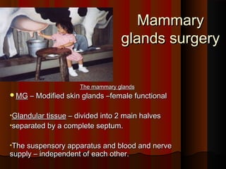
- 1. Mammary glands surgery The mammary glands MG – Modified skin glands –female functional •Glandular tissue – divided into 2 main halves •separated by a complete septum. •The suspensory apparatus and blood and nerve supply – independent of each other.
- 3. Section of mammary glands
- 5. Nervous stimulation on udder
- 7. Anatomy So one half can be easily removed in diseased condition without affecting the other. Each half---------- Cranial quarter ---------- Caudal quarter
- 8. Anatomy of mammary glands 2 quarters of each half- independent glandular tissue but common blood and nerve supply and lymph drainage.( B.S.-ext. pudic and perineal)- inguinal nerve
- 9. Structure of MG From outside to inside – teat consist of 1.Skin(E,D) 2.Muscular layer-M- (outer longitudinal and inner circular which extends distally – spincter of the teat canal-S.)
- 10. 3. fibrous layer- binding layer for muscle with mucosa.- C 4. mucus mem- longitudinal and transverse folds- intersect – form pockets or recess-Mc Bacteria resides
- 11. Structure of MG
- 12. At distal- mucosa-rose flower like folds pattern- rosette of Furstenberg.----- R Duct system – 2 parts 1.teat sinus/cistern 2. streak canal.(pappilary duct)
- 13. Structure of MG Teat cistern – separated from gland cistern – annular fold-A Ventrally the rostte of Furstenberg separates teat cistern from the streak canal. Closing mechanism – rostte of Furstenberg- sphincter muscle- prevents milk leakage and entry of microbes.
- 14. Anaesthesia Surgery of MG- ring block- 10-12 ml lignocaine 2% Posterior epidural block. Spinal anaesthetics
- 16. Supernumery teats Supernumery teats – teats in between normal teats Removed for – cosmetic- interfere with milking procedure. - unfit character for breeding 2 elliptical incisions- close with non-absorbable. FUSED TEATS- skin are fused- without involving teat canal or muscles. Divided surgically and cutaneous wound sutured
- 17. Teat laceration Teat lacerations Higher in goats(pendulous udder and long teats) Etio-Direct injury Superficial wounds – general principles
- 18. Teat laceration Large wounds – involving skin and muscularis but not mucosa. suture Deep lacerations – involve mucosa, a complete longitudinal tearing.
- 19. Teat laceration Ring block – tourniquet – check haemorhage and milk inflow into cistern. Teat siphon inserted – debridement is properly done Close the mucosa – simple continuous- atraumatic needle. Finally skin
- 20. Teat lacerations Check leakage to ensure a proper sealing –fistula may form later. Antibiotic preparation into teat. Polyethy- catheter – mastitis.
- 22. Teat Fistula Teat cistern and teat surface- milk flows in lactating animals. Aquired and rarely congenital. Best treated during dry period.
- 23. Teat Fistula If very small- mild chemical cauterization. If large- reconstructive surgery. If inflamed delay the operation since chance of recurrence. Repair-2 elliptical incisions – debridement and undermining- close.
- 24. Papilloma/warts Papilloma/warts- finger-like Isolated or multiple projections Ligate at the base – drops off.-if not surgical.
- 25. LACTOLITHS: LACTOLITHS: Teat cistern liths due to mineral deposits. Concretions and rarely as organized calculi.- obstruction to milking. Lodged at teat orifice. If small removed by teat orifice by milking. Mosquito forceps if large.or use teat bistoury to slit the contracted sphincter.
- 26. Polyp Polyp: Pea sized growth- attached to wall of teat cistern-clamped and removed by alligator forceps.
- 27. Teat spider :(memberanous obstruction) Congenital Aquired Teat spider : Improper Injury, (memberanous development tumour or obstruction) of teat infection Congenital or aquired Milk pocket present usually not Symptom: present Obstruction to milk Treatment Rewarding flow not and rewarding prognosis is good
- 28. Teat spider :(memberanous obstruction) Milk pocket-fluctuating milk above the obstruction. In congenital- milk pocket is absent. Treatment is not rewarding. If the milk pocket is palpated prognosis is good. Hudson ‘s teat spiral is introduced with 3-4 revolutions. Milk also prevents the stricture formation.do not milk it completely.
- 29. Reference Reference: RPS Tyagi, Ruminant surgery
- 30. Questions???