Basic anthropometry ppt.
•Als PPT, PDF herunterladen•
175 gefällt mir•113,792 views

Basic Anthropometry lecture for medical students..

Empfohlen
Weitere ähnliche Inhalte
Was ist angesagt?
Was ist angesagt? (20)
Ähnlich wie Basic anthropometry ppt.
Ähnlich wie Basic anthropometry ppt. (20)
Mehr von Reina Ramesh
Mehr von Reina Ramesh (13)
Basic anthropometry ppt.
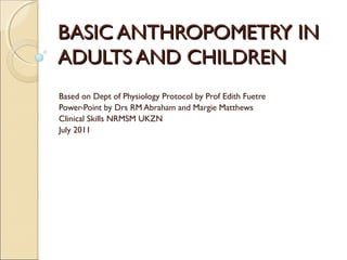
- 1. BASIC ANTHROPOMETRY IN ADULTS AND CHILDREN Based on Dept of Physiology Protocol by Prof Edith Fuetre Power-Point by Drs RM Abraham and Margie Matthews Clinical Skills NRMSM UKZN July 2011
- 2. Anthropometry: Introduction A branch of anthropology that involves the quantitative measurement of the human body. It is the single most portable, universally applicable, inexpensive and non-invasive technique for assessing the size, proportions and composition of the human body. Appropriate use and interpretation from infancy to old age is a valuable tool for guiding the health and nutritional status of individuals and populations. Paediatricians have long used child growth as an important parameter to evaluate the health and well-being of children.
- 3. Anthropometric Parameters Basic measurements Some measurements Height (length) used for nutritional Weight (mass) assessment include: Height Circumference eg OFC (infant up to age Mass 2years only) BMI Skin-fold thickness Triceps skin-fold Derived measurements Waist, Hip and Mid- of body composition arm circumference and interrelationships (MAC) and derived (e.g BMI, waist-hip measurements/ ratios ratio)
- 4. Height/Stature Measurement Technique The subject places his/her heels The subject must be barefoot, together, with both heels touching wearing as little clothing as possible the base of the vertical board The subject stands on a flat surface, The medial borders of the feet are at at a right angle to the vertical board an angle of about 60° of the stadiometer The scapulae and buttocks must also His/her weight is distributed evenly be in contact with the vertical board over both feet, with the head The subject must inhale deeply and positioned in the Frankfurt maintain a fully erect position Horizontal Plane (in this position, the without altering the load on the heels most inferior point on the left orbital The movable head board is brought margin is at the same horizontal level onto the most superior point on the as the left tragion – the line of vision head with sufficient pressure to is approximately horizontal ) compress the hair The arms hang freely by the sides of The measurement is taken to the the trunk, with palms facing the nearest 1 mm thighs
- 6. Mass/Weight Measurement Technique Subject must be barefoot and wear as little clothing as possible. Subject stands on the platform of the scale with his/her weight distributed evenly over both feet. The arms hang by the sides of the trunk, with palms facing the thighs . The subject is instructed to maintain a stable position while the measurement is taken. The measurement is taken to the nearest 0.1 kg .
- 7. Body Mass Index This ratio is expressed in Kg/m2 and provides a rough estimation of the body mass status of the individual in relation to his/her height.
- 8. BMI Ranges and Co-morbidity Risk ANTHROPOMETRY QUANTIFYING OBESITY WITH BODY MASS INDEX (WEIGHT/HEIGHT²) BMI (kg/m²) CLASSIFICATION* RISK OF OBESITY COMORBIDITY 18.5 - 24.9 Normal range Negligible 25.0 - 29.9 Overweight Mildly increased >30 Obese 30.0 - 34.9 Class I Moderate 35.0 - 39.9 Class II Severe > 40.0 Class III Very severe * Classification of the World Health Organisation (WHO) and International Obesity Task Force
- 9. Waist-to-Hip Circumference Ratio An indicator of the Men generally have a pattern of distribution higher ratio than of subcutaneous women adipose tissue. Women 0.85-1.7 (high Distribution of fat is an risk)and <0.85 (Low important indicator of risk) CHD (coronary heart Men 0.95-1.9 (high disease) risk) and <0.95 (Low More fat in the risk) abdominal area - increases risk of CHD.
- 10. Waist-to-Hip Circumference Ratio Measurement Technique Waist circumference A good quality non- stretchable measuring tape should be used. View the patient from the front. Locate the narrowest point between ribs and iliac crests. Ensure that the tape measure is at the same height around the waist. Measure and state the measurement correctly to the nearest centimetre.
- 11. Waist-to-hip circumference ratio Hip circumference View the patient from the front. Locate the greater trochanter. Hip measurement is taken at the widest lateral extension of the hips. Ensure that the tape measure is horizontal. Measure and state the measurement correctly to the nearest centimetre. Calculate Waist/Hip Ratio to 2 decimal places.
- 12. Measures of body composition Weight loss, per se, does not provide the nutritionist with an indication of type of tissue lost (i.e. weight loss due to loss of adipose tissue or loss of muscle tissue). Measurements of skin-folds, mid-arm circumference and mid-arm muscle circumference therefore provide a more comprehensive picture of body composition/ changes.
- 13. Mid-arm circumference (MAC) Locate the midpoint of the arm. Non-dominant arm elbow flexed at 90deg with palm facing upwards Measurer stands behind the subject & locates the lateral tip of the acromion and the most distal point on the olecranon process Place a tape measure so that it passes between these 2 landmarks and mark the midpoint Measure the midarm circumference The subject stands erect with arms hanging freely at the sides and the palms facing the thighs Place the tape measure perpendicular to the long axis of the arm at the marked midpoint & measure the circumference to the nearest mm. (e.g. 18.1 cm) Provide the actual MAC in cm.
- 14. Skin-fold measurements Approximately half of the In general, when measuring skin- fold thickness, total amount of fat tissue in the human body is located The assessor, using the forefinger and below the surface of the skin. the thumb, grasps and lifts the subcut. tissue and skin from the underlying muscle. This makes it possible to Places the pincers of the skin-fold predict total body fat from caliper, applying a constant pressure, skin-fold thicknesses with a 2cm below the fingers at a depth of 1cm. relative high degree of accuracy using a simple two- Holds this position for 3-4seconds. compartmental method. Takes three measurements for accuracy. This accuracy is confirmed by CT scan as well as ultrasonic Provides the actual skin-fold thickness in mm. and radiographic techniques used to measure subcut.fat.
- 15. Triceps skin-fold (TSF) A measure of subcutaneous fat stores taken at the midpoint of the posterior aspect of the humerus. Correlates closely with percentage of body fat and with total body fat. Triceps skin-fold thickness varies between 6 -12mm in lean individuals and between 40 - 50mm in obese individuals.
- 16. Triceps skin-fold measurement technique Subject should be standing with arms hanging loosely at the sides. Assessor to be positioned behind the subject. To locate the triceps skin-fold site, locate the site previously marked for the midarm circumference measurement (MAC). The triceps skin-fold site is on the posterior surface of the arm, midway between the shoulder and the elbow. Using the forefinger and the thumb the assessor grasps and lifts the subcut. tissue and skin 2cm above TSF site. Place the pincers of the skin-fold caliper at the TSF point at a depth of 1cm. Hold this position for 3-4seconds. Take three measurements for accuracy. Provide the actual skin-fold thickness in mm.
- 17. Mid-arm muscle circumference (MAMC) TSF is preferably used in conjunction Standard adult values with subscapular, biceps and supra- (helps interpret the above body iliac skin-fold measurements to compositional measurements) determine actual percentage body fat from set equations or in conjunction with MAC to determine mid-arm Triceps skin-fold (mm) muscle circumference. Male 12.5 Female 16.5 MAMC provides an index of muscle mass. Mid-arm circumference (cm) Male 29.3 MAMC (cm)= Female 28.5 MAC (cm) - [3.14 x TSF (cm)] Mid-arm muscle circumf. (cm) Male 25.3 Female 23.2
- 18. Other skin-folds measured Besides the most commonly used triceps skin-fold, other commonly measured skin-folds include the following: Biceps skin-fold Subscapular skin-fold Supra-iliac skin-fold
- 19. Biceps skin-fold measurement technique Locate the biceps skin-fold site: The assessor positioned in front of the subject. Subject should be standing erect with arms hanging loosely at their sides. To locate the biceps skin-fold site, locate the level previously marked for the mid-arm circumference measurement. The biceps skin-fold site is on the anterior surface of the arm, midway between the shoulder and elbow. Measuring skin-fold thickness Using forefinger and thumb, grasp and lift the subcutaneous tissue and skin 2cm above the midpoint . Place the pincers of the skin-fold caliper at the midpoint at a depth of 1cm. Hold this position for 3 to 4 seconds. Take three measurements for accuracy (answer in mm). Provide the actual skin-fold thickness in mm.
- 20. Subscapular skin-fold measurement technique The assessor is positioned behind the subject. The subscapular skin-fold site is located 1cm below the inferior angle of the scapula. The assessor grasps and lifts the subcut. tissue and skin at a downward angle of approximately 45° towards the lateral aspect of the body. Place the pincers of the skin-fold caliper at a depth of 1cm. Hold this position for 3 to 4 seconds. Take three measurements for accuracy (answer in mm). Provide the actual skin-fold thickness in mm.
- 21. Supra-iliac skin-fold measurement technique The assessor to be positioned in front of the subject. The supra-iliac site is located 5cm above the anterior superior iliac spine. The assessor grasps and lifts the subcut. tissue and skin at a downward angle of 45° towards the medial aspect of the body. Place the pincers of the skin-fold caliper at a depth of 1cm. Hold this position for 3 to 4 seconds. Take three measurements for accuracy (answer in mm). Provide the actual skin-fold thickness in mm.
- 22. Child Anthropometry Basic measurements in children include: Weight Clothing to be removed. Baby weighed on clean calibrated scale. The measurement is taken to at least 2 decimal places for accuracy in kg. Height (Length) An infantometer is used. The baby is placed supine with head against appropriate surface. The baby is held in a fully extended position with the heels at a 90º position. The measurement is taken to the nearest 0.1cm. OFC (Occipitofrontal circumference) The OFC of the baby is measured to the nearest 0.1cm with a firm tape measure placed appropriately.
- 23. Road-to-Health Chart A simple, cheap, practical and convenient method of monitoring child health. Growth monitoring is the most useful tool available in child health as it assists with early identification of nutritional problems, disease, and developmental problems. The most sensitive indicator of a child's growth is weight.
- 24. Growth chart Graph records child's growth progress. 1)Vertical axis is the weight axis (represented in kgs both on the right and left margin of each year starting from 0) 2)Horizontal axis is the age axis-one space per month – goes up to 5 years
- 25. Standards and reference curves on the Road-to-Health Chart If the weights of 100 healthy children according to age groups are plotted on a graph, the average weight is represented by the 50th centile reference curve (bold curve on the graph) The weights will be scattered around this 50th centile with more weights near to it rather than far above or below it. To obtain a normal range of weights, an upper and lower reference curve is also plotted, referred to as the 97th and 3rd centile reference curves. This means that the weights of 3 healthy children will fall above the 97 th centile and the weights of 3 healthy children will fall below the 3 rd centile. In statistics, a centile (or percentile) is the value of a variable below which a certain percent of observations fall. For example, the 50th percentile is the value (or score) below which 50 percent of the observations fall. It is extremely important to plot the weight in a serial fashion in order to evaluate the growth trend. (term “failure to thrive”) Note 60% of standard weight or 50th centile
- 26. Nutritional assessment Malnutrition may be acute/ chronic or a combination, with the acute form manifesting with weight loss/ failure to gain weight, and the chronic form resulting in stunting (child is shorter than normal). Normal Wasted Stunted Weight/age % 100 70 70 Weight/height % 100 70 100 Height/age % 100 100 84
- 27. Nutritional assessment Thus, the various anthropometric indices in children are used to measure the presence and severity of the various forms of malnutrition 1) Weight-for-height (decreased) indicates acute malnutrition (wasting) 2) Height-for-age (decreased) indicates chronic malnutrition (stunting) 3) Weight-for-age (decreased) in any protein-energy malnutrition (underweight)
- 28. Types of Malnutrition Malnutrition is a group of conditions in children and adults generally related to poor quality or insufficient quantity of nutrient intake, absorption, or utilization There are two major types of malnutrition: Protein-energy malnutrition - resulting from deficiencies in any or all nutrients Micronutrient deficiency diseases - resulting from a deficiency of specific micronutrients (eg iron, specific vitamins)
- 29. Types of Protein-Energy Malnutrition (PEM) in Infants Condition 60-80% of standard < 60% of standard weight weight No oedema Underweight Marasmus Oedema Kwashiorkor Marasmic kwashiorkor Standard refers to the 50th percentile or median
- 30. Kwashiorkor - 60-80% of expected weight - Sparse, depigmented hair - Oedema - Skin rash - Distended abdomen and enlarged liver - Diarrhoea
- 31. Marasmus - Weight<60% mean for age - Wasted, wizened appearance
- 32. References Basic anthropometric measurements in adults protocol (Dept of Physiology) WHO: Global database on body mass index (Davidson 2006) Illustrated Textbook of Paediatrics Lissauer and Clayden SA Family Practice Manual Bob Mash and Julia Blitz-Lindeque
Hinweis der Redaktion
- Tragion- an anthropometric point situated in the notch just above the tragus of the ear.
- Stadiometer is a height/stature measuring device.
- If your BMI is below 20: This indicates a lean BMI, which means you have a low amount of body fat. If you are an athlete, this can be desirable. If you are not an athlete, a lean BMI can indicate that your weight may be too low which may lower your immunity. If your BMI and body weight are low, you should consider gaining weight through good diet and exercise habits, to increase your muscle mass. If your BMI is between 20 and 22: This indicates the ideal, healthy amount of body fat, which is associated with living longest, and the lowest incidence of serious illness. Coincidentally, it seems this ratio is what many individuals perceive to be the most aesthetically attractive. If your BMI is between 22 and 25: This is still considered an acceptable range, and is associated with good health. If your BMI is between 25 and 30: You are considered “Hefty” and should finds ways to lower your weight, through diet and exercise. You are at increased risk for a variety of illnesses at your present weight. You should lose weight by changing your diet and exercising more. If your BMI is over 30: This indicates an unhealthy condition, your excess “Phat” is putting you at risk for heart disease, diabetes, high blood pressure, gall bladder disease and some cancers. You should lose weight by changing your diet and exercising more.
- Both kwashiorkor and marasmus are life-threatening medical emergencies which need to be treated by sophisticated feeding programmes. Such programmes must be run by medical professionals with experience in refeeding children with severe protein-energy malnutrition.