Nervio Vago/Wilson Pawels/idiomainglés
•
4 gefällt mir•3,071 views

resumen del libro en inglés

Empfohlen
Weitere ähnliche Inhalte
Was ist angesagt?
Was ist angesagt? (20)
Andere mochten auch
Ähnlich wie Nervio Vago/Wilson Pawels/idiomainglés
Ähnlich wie Nervio Vago/Wilson Pawels/idiomainglés (20)
Mehr von regina_estrella_14
Mehr von regina_estrella_14 (20)
Kürzlich hochgeladen
Kürzlich hochgeladen (20)
Nervio Vago/Wilson Pawels/idiomainglés
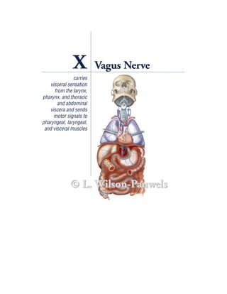
- 1. X Vagus Nerve carries visceral sensation from the larynx, pharynx, and thoracic and abdominal viscera and sends motor signals to pharyngeal, laryngeal, and visceral muscles © L. Wilson-Pauwels
- 2. X Vagus Nerve CASE HISTORY Ruth is a 46-year-old lawyer who, over the last few years, noticed a whooshing sound in her left ear when she lay on her left side at night. Ruth is a very busy woman with two teenage daughters and an active law practice, and, as the noise did not keep her from sleeping at night, she paid little attention to it. One afternoon, after playing a vigorous game of tennis with her daughter, she again became aware of the whooshing sensation in her left ear and noted that it seemed more intense with exercise. She had intended to see her family doctor but got distracted with an important defense and her daughter’s upcoming graduation. Over several months, Ruth noticed the whooshing sound was almost always present day or night, and she gradually developed problems with swallowing and a hoarse voice. Finally, Ruth made time to see her doctor. On general examination, Ruth’s doctor noted that she appeared fit and well. However, when the doctor placed a stethoscope on the base of her skull on the left side, he could hear a bruit (a whooshing sound). When he examined her cranial nerves, he found that she had an absent gag reflex on the left side and some weakness of her left sternomastoid* muscle. Ruth’s doctor immediately referred her to a neurosurgeon who was concerned that this was a glomus tumor of the jugular foramen. He sent Ruth for magnetic resonance imaging (MRI) and an angiogram. The investigations confirmed the neurosurgeon’s sus- picions and a diagnosis of a glomus jugulare tumor was made. Ruth was subsequently scheduled for surgery to have the tumor removed. ANATOMY OF THE VAGUS NERVE Vagus comes from the Latin word meaning “wandering.” The vagus nerve “wanders” from the brain stem to the splenic flexure of the colon. Not only is the vagus the parasympathetic nerve to the thoracic and abdominal viscera, it is also the largest vis- ceral sensory (afferent) nerve. Sensory fibers outnumber parasympathetic fibers four to one. In the medulla, the vagal fibers are connected to four nuclei: the spinal nucleus of the trigeminal nerve (general sensory); the nucleus of the tractus solitar- ius (visceral sensory); the nucleus ambiguus (branchial motor); and the dorsal vagal motor nucleus (parasympathetic visceral motor) (Figure X–1 and Table X–1). *Sternomastoid is a shortened form of sternocleidomastoid and will be used in this text. 182
- 3. Vagus Nerve 183 Course of the Vagus Nerve The vagus nerve emerges from the medulla of the brain stem dorsal to the olive as eight to ten rootlets caudal to those of cranial nerve IX. These rootlets converge into a flat cord that exits the skull through the jugular foramen. Two sensory ganglia, the superior (jugular) and inferior (nodosum), are located on the vagus nerve. The supe- rior ganglion is located within the jugular fossa of the petrous temporal bone, which, together with the occipital bone, forms the jugular foramen. Within the jugular foramen, the vagus nerve is in close proximity to the jugular bulb, a swelling of the proximal part of the internal jugular vein containing the jugular glomus within its adventitia (see Case History Guiding Questions, #1). The glomus jugu- lare, or tympanic body, is a collection of neuron-like cells that monitor blood oxy- gen (O2), carbon dioxide (CO2), and acidity/alkalinity (pH) levels. It is similar to the carotid body (see Chapter IX). Exiting the jugular foramen, the vagus nerve enlarges into a second swelling, the inferior (nodosum) ganglion (see Figure X–1). Nucleus of the tractus solitarius (rostral © L. Wilson-Pauwels portion) Dorsal vagal motor nucleus Inferior cerebellar peduncle Spinal nucleus of trigeminal nerve Nucleus ambiguus CUT MEDULLA CN IX CN X Olive Pyramid CN X Jugular foramen Jugular fossa Rootlet of CN XII Superior Rootlet of CN XI vagal ganglion Inferior vagal ganglion Tract of spinal trigeminal nucleus Nucleus of the tractus solitarius (caudal portion) Figure X–1 Cross-section of the medulla at the point of entry of cranial nerve X illustrating the nuclei associated with this nerve.
- 4. 184 Cranial Nerves Table X–1 Nerve Fiber Modality and Function of the Vagus Nerve Nerve Fiber Modality* Nucleus Function General sensory Of the spinal From the posterior meninges, concha, skin at the back (afferent) trigeminal tract of the ear and in the external acoustic meatus, part of the external surface of the tympanic membrane, the pharynx, and larnyx Visceral sensory Of the tractus From the larynx, trachea (caudal part), esophagus, and (afferent) solitarius thoracic and abdominal viscera, stretch receptors in the walls of the aortic arch, and chemoreceptors in the aortic bodies adjacent to the arch Branchial motor Ambiguus To the superior, middle, and inferior constrictors, levator (efferent) palati, salpingopharyngeus, palatopharyngeus, and one muscle of the tongue, the palatoglossus, via the pharyngeal plexus and to cricothyroid and intrinsic muscles of the larynx Visceral motor Dorsal vagal motor To smooth muscle and glands of the pharynx, larynx, (parasympathetic and thoracic and abdominal viscera efferent) Ambiguus To cardiac muscle *Some texts list special sense for taste as one of the components of this nerve. Because cranial nerve X carries so few taste fibers, this modality has been omitted. As the vagus nerve emerges through the jugular foramen, it lies within the same dural sheath as the accessory nerve (cranial nerve XI). For a short distance, the cau- dal branchial motor fibers of cranial nerve X travel with cranial nerve XI (some texts call these fibers the cranial root of XI). Just beyond the inferior ganglion, all the branchial motor fibers of cranial nerve X rejoin (Figure X–2). In the neck, the vagus lies posterior to and in a groove between the internal jugu- lar vein and the internal carotid artery. It descends vertically within the carotid sheath (Figure X–3), giving off branches to the pharynx, larynx, and constrictor muscles (Table X–2). The right recurrent laryngeal nerve branches from the right vagus nerve in the neck. It curves below and behind the right subclavian artery to ascend at the side of the trachea behind the right common carotid artery. The left recurrent laryngeal nerve branches from the left vagus nerve in the thorax (Figure X–4). It curves below and behind the aortic arch to ascend at the left side of the tra- chea. From the root of the neck downward, the vagus nerve takes a different path on each side of the body to reach the cardiac, pulmonary, and esophageal plexuses (consisting of both sympathetic and cranial nerve X parasympathetic axons). From the esophageal plexus, right and left gastric nerves arise to supply the abdominal vis- cera as far caudal as the splenic (left colic) flexure (see Table X–2 and Figure X–4).
- 5. Vagus Nerve 185 © L. Wilson-Pauwels Inferior cerebellar peduncle Nucleus ambiguus CUT MEDULLA CN IX Olive CN X Caudal rootlets of X CN XI Pyramid CN X Conjoint nerve CN X and CN XI Jugular foramen Rootlet of CN XII Jugular fossa Superior and inferior Rootlet of CN XI vagal ganglia CN X Accessory nucleus CN XI Figure X–2 Cross-section through the rostral (open) medulla demonstrating the branchial motor component of cranial nerve X including spinal rootlets of cranial nerve XI. GENERAL SENSORY (AFFERENT) COMPONENT The general sensory component of cranial nerve X carries sensation (pain, touch, and temperature) from the • larynx, • pharynx, • concha and skin of the external ear and external auditory canal, • external surface of the tympanic membrane, and • meninges of the posterior cranial fossa. Axons carrying general sensation from the vocal folds and the subglottis below the vocal folds accompany visceral sensory axons in the recurrent laryngeal nerve (Figures X–5 and X–6). Similarly, axons carrying general sensation from the larynx above the vocal folds accompany visceral sensory axons in the internal laryngeal nerve. The internal laryngeal nerve leaves the pharynx by piercing the thyrohyoid membrane. It ascends in the neck uniting with the external laryngeal nerve (branchial motor) to form the superior laryngeal nerve. General sensory fibers travel up the superior laryn- geal nerve to join the rest of the vagus nerve and reach the inferior vagal ganglion.
- 6. 186 Cranial Nerves Pharyngeal nerve Right vagus nerve Superior laryngeal nerve Left vagus nerve Right common carotid artery Left internal jugular vein Carotid sheath Directional and color-coded arrows indicate modalities of Right recurrent recurrent laryngeal nerves laryngeal nerve Left brachiocephalic vein Right subclavian artery Left subclavian artery Left recurrent laryngeal nerve Aortic arch Right vagus nerve Left vagus nerve to cardiac, pulmonary, to cardiac, pulmonary, and esophageal plexuses and esophageal plexuses © L. Wilson-Pauwels Figure X–3 Route of the right and left recurrent laryngeal nerves (nerve is shown in grey for clarity). General sensory fibers from the concha and the skin of the external ear, the exter- nal auditory canal, and the external surface of the tympanic membrane are carried in the auricular branch (see Figure X–6). Stimulation of the auricular nerve of cra- nial nerve X in the external auditory meatus can cause reflex coughing, vomiting, and even fainting through reflex activation of the dorsal vagal motor nucleus. Sensory branches from the meninges of the posterior cranial fossa are carried in the meningeal nerve. The peripheral processes pass into the jugular fossa and enter the superior vagal ganglion where their nerve cell bodies are located. The central processes pass upward through the jugular foramen and enter the medulla, then descend in the spinal trigeminal tract to synapse in its nucleus.
- 7. Vagus Nerve 187 Table X–2 Branches of the Vagus Nerve Location Branch Modality General Visceral Branchial Visceral Sensory Sensory Motor Motor Jugular Meningeal ✔ fossa Auricular ✔ Neck Pharyngeal ✔ ✔ ✔ ✔ Branches to carotid body ✔ Superior laryngeal ✔ ✔ ✔ ✔ Internal laryngeal ✔ ✔ ✔ External laryngeal ✔ Recurrent laryngeal (right) ✔ ✔ ✔ ✔ Cardiac ✔ ✔ Thorax Cardiac ✔ ✔ Recurrent laryngeal (left) ✔ ✔ ✔ ✔ Pulmonary ✔ ✔ Esophageal ✔ ✔ Abdomen Gastrointestinal ✔ ✔ From the nucleus of the spinal tract, second-order axons project via the ventral trigeminothalamic tract to the contralateral ventral posterior nucleus of the thala- mus. Thalamic neurons project through the internal capsule to the sensory cortex of the cerebrum (see Figure X–6, inset). VISCERAL SENSORY (AFFERENT) COMPONENT Visceral sensation is carried in the visceral sensory component of the vagus nerve. It is not appreciated at a conscious level of awareness other than as “feeling good” or “feeling bad,” unlike visceral pain that is carried in the sympathetic nervous system. Visceral sensory fibers from plexuses around the abdominal viscera converge and join with the right and left gastric nerves of the vagus (Figure X–7). These nerves pass upward through the esophageal hiatus (opening) of the diaphragm to merge with the plexus of nerves around the esophagus. Sensory fibers from plexuses around the heart and lungs also converge with the esophageal plexus and continue up through the thorax in the right and left vagus nerves.
- 8. 188 Cranial Nerves © L. Wilson-Pauwels Spinal nucleus of the trigeminal nerve Dorsal vagal motor nucleus Nucleus solitarius Nucleus ambiguus Jugular foramen Soft palate Meningeal nerve PHARYNGEAL NERVE Auricular nerve SUPERIOR LARYNGEAL NERVE Superior and inferior vagal ganglia Internal laryngeal nerve External laryngeal nerve Internal jugular vein Thyrohyoid membrane Internal carotid artery Vocal folds Carotid sheath Inferior constrictor muscle LEFT RECURRENT LARYNGEAL NERVE Esophagus CERVICAL PORTION OF LEFT VAGUS NERVE Trachea (CN X) Aortic arch Visceral motor to and visceral sensory from thorax and abdomen Figure X–4 Overview of the vagus nerve.
- 9. Vagus Nerve 189 Superior and inferior vagal ganglion Superior laryngeal nerve Internal laryngeal branches Larynx above vocal folds Vocal folds © L. Wilson-Pauwels Vagus nerve Recurrent laryngeal nerve Figure X–5 Schematic illustration depicting visceral and general sensory nerves from the larynx. The right and left vagus nerves are joined by nerves carrying visceral sensory information from the • baroreceptors (stretch receptors) in the aortic arch and chemoreceptors (measur- ing oxygen tension in the blood) in the aortic bodies; • larynx below the vocal cords in the recurrent laryngeal nerve; • larynx above the vocal folds in the internal laryngeal nerve; and • mucous membrane of the epiglottis, base of the tongue, and aryepiglottic folds in the pharyngeal plexus. The central processes of the nerve cell bodies in the inferior vagal ganglion enter the medulla and descend in the tractus solitarius to enter the caudal part of the nucleus of the tractus solitarius. From the nucleus, bilateral connections important in the reflex control of cardiovascular, respiratory, and gastrointestinal functions are made with several areas of the reticular formation and the hypothalamus.
- 10. 190 Cranial Nerves Sensory cortex © L. Wilson-Pauwels Ventral posterior nucleus of the thalamus Superior vagal gangion Spinal nucleus of the Jugular foramen trigeminal nerve Spinal nucleus of the trigeminal Soft palate nerve MENINGEAL NERVE PHARYNGEAL NERVE Superior vagal ganglion AURICULAR NERVE SUPERIOR LARYNGEAL NERVE Internal laryngeal nerve Internal jugular vein Thyrohyoid membrane Internal carotid artery Vocal folds Inferior constrictor muscle Carotid sheath LEFT RECURRENT LARYNGEAL NERVE LEFT VAGUS NERVE (CN X) Esophagus Trachea Aortic arch Figure X–6 General sensory component of the vagus nerve.
- 11. Vagus Nerve 191 to reticular formation Nucleus and ambiguus hypothalamus Hypothalamus Dorsal vagal motor nucleus Inferior vagal ganglion Nucleus of the Internal jugular tractus solitarius foramen (caudal part) Pharyngeal nerve Inferior vagal Internal laryngeal nerve off ganglion superior laryngeal nerve Tractus solitarius Right vagus Left vagus nerve nerve Common carotid artery Right recurrent Carotid sheath Inferior laryngeal nerve Left internal jugular vein vagal ganglion Superficial from baroreceptors cardiac and chemoreceptors in plexus aortic arch and bodies Right Deep cardiac plexus pulmonary nerve Left pulmonary nerve © L. Wilson-Pauwels Left cardiac nerve Right cardiac nerve Esophageal Celiac plexus plexus and hiatus Spleen Left gastric nerve Superior mesenteric Splenic flexure plexus Right gastric nerve Figure X–7 Visceral sensory component of the vagus nerve.
- 12. 192 Cranial Nerves Connections via the reticulobulbar pathway (between the reticular formation and cranial nerve nuclei in the brain stem) to the dorsal vagal motor nucleus enable the parasympathetic fibers of the vagus nerve to control these reflex responses (see Figure X–7, inset). BRANCHIAL MOTOR (EFFERENT) COMPONENT Bilateral corticobulbar fibers (fibers connecting the cortex with cranial nerve nuclei in the brain stem) are composed of axons from the premotor, motor, and other cor- tical areas. They descend through the internal capsule to synapse on motor neurons in the nucleus ambiguus, a column of cells just dorsal to the inferior olivary nucleus in the medulla. The nucleus ambiguus also receives sensory signals from other brain stem nuclei, mainly the spinal trigeminal and solitary nuclei, initiating reflex responses (eg, coughing and vomiting). Lower motor neuron axons leave the nucleus ambiguus and travel laterally to leave the medulla as eight to ten rootlets. The caudal rootlets travel briefly with cranial nerve XI, rejoining with the rostral rootlets of cranial nerve X just below the inferior vagal ganglion (see Figure X–2). The nerve leaves the skull through the jugular foramen to reach the constrictor mus- cles of the pharynx and the intrinsic muscles of the larynx (see Figures X–4 and X–8). The branchial motor fibers leave the vagus nerve as three major branches: pha- ryngeal, superior laryngeal, and recurrent laryngeal. The pharyngeal branch, the principal motor nerve of the pharynx, traverses the inferior ganglion and passes inferomedially between the internal and external carotid arteries. It enters the phar- ynx at the upper border of the middle constrictor and breaks up into the pharyn- geal plexus to supply all the muscles of the pharynx and soft palate except the stylopharyngeus (cranial nerve IX) and tensor (veli) palati (branchial motor com- ponent of V3). Therefore, it supplies the superior, middle, and inferior constrictors, levator palati, salpingopharyngeus, palatopharyngeus, and one muscle of the tongue, the palatoglossus (many are illustrated in Figure X–8). The superior laryngeal nerve branches from the main trunk of the vagus nerve at the inferior vagal ganglion distal to the pharyngeal branch. It descends adjacent to the pharynx, dividing into internal (mainly sensory) and external (motor) laryn- geal nerves. Branchial motor axons in the external laryngeal branch supply the inferior constrictor and the cricothyroid muscles. It also sends branches to the pha- ryngeal plexus. The pharyngeal plexus, supplying the palate and pharynx, is formed by branches from the external laryngeal and pharyngeal nerves, as well as branches from cranial nerve IX and the sympathetic trunk The recurrent laryngeal nerve, the third major branch, takes a different path on the right and left sides of the body (see Figure X–3). The right recurrent laryngeal nerve arises from the vagus nerve anterior to the subclavian artery, then hooks back under the artery and ascends posterior to it in the groove between the trachea and the esophagus. The left recurrent laryngeal nerve arises from the left vagus on the aortic arch. It hooks back posteriorly under the arch and ascends through the
- 13. Vagus Nerve 193 Motor cortex Corticobulbar fibers Internal capsule © L. Wilson-Pauwels Bilateral innervation to nucleus ambiguus CN IX CN X CN XI Jugular foramen (CNs IX, X, XI) Superior vagal ganglion Inferior vagal ganglion CN X PHARYNGEAL NERVE SUPERIOR LARYNGEAL NERVE Internal carotid artery External carotid artery Palatoglossus muscle External laryngeal nerve Palatopharyngeus muscle (cut) CN X Superior, middle, and inferior constrictor muscles Subclavian artery RIGHT RECURRENT LARYNGEAL NERVE Brachiocephalic artery Figure X–8 Branchial motor component of the vagus nerve.
- 14. 194 Cranial Nerves superior mediastinum to reach the groove between the trachea and the esophagus on the left side. The recurrent nerves pass deep to the inferior margin of the infe- rior constrictor muscles. The branchial motor axons supply the intrinsic muscles of the larynx (except the cricothyroid). VISCERAL MOTOR (PARASYMPATHETIC EFFERENT) COMPONENT The parasympathetic nerve cell bodies of the vagus nerve are located in the dorsal motor nucleus of the vagus (Figure X–9) and in the medial side of the nucleus ambiguus. Neurons in the dorsal vagal nucleus innervate ganglia in the gut and its derivatives (lungs, liver, pancreas), whereas neurons in the nucleus ambiguus inner- vate ganglia in the cardiac plexus. They are all influenced by input from the hypo- thalamus, the olfactory system, the reticular formation, and the nucleus of the tractus solitarius. The dorsal motor nucleus of the vagus is located in the floor of the fourth ventricle (vagal trigone) and in the central gray matter of the closed medulla. Preganglionic fibers from this nucleus traverse the spinal trigeminal tract and nucleus, emerge from the lateral surface of the medulla, and travel in the vagus nerve (see Figure X–1). Within the pharynx and larynx, the vagal preganglionic axons activate gan- glionic neurons that are secretomotor to the glands of the pharyngeal and laryngeal mucosa. Preganglionic axons are distributed to the pharyngeal plexus through the pharyngeal and internal laryngeal branches (see Figure X–4). Within the thorax, the vagi take different paths, but both break up into many branches that join plexuses around the major blood vessels to the lungs and the heart (Figure X–10). Pulmonary branches cause bronchoconstriction, and esophageal branches act to speed up peristalsis in the esophagus by activating the smooth (nonstriated) muscle of the walls of the esophagus. The axons synapse in ganglia located in the walls of the indi- vidual organs. The cell bodies of cardiac preganglionic axons are located in the medial nucleus ambiguus. Their axons terminate on small ganglia associated with the heart and act to slow down the cardiac cycle. The right and left gastric nerves emerge from the esophageal plexus. These nerves stimulate secretion by the gastric glands and are motor to the smooth mus- cle of the stomach. Intestinal branches act similarly on the small intestine, cecum, vermiform appendix, ascending colon, and most of the transverse colon. In the gut, the synapses occur in ganglia of the myenteric and submucosal plexuses of Auerbach and Meissner, respectively.
- 15. Vagus Nerve 195 © L. Wilson-Pauwels Vagal trigone Nucleus ambiguus Dorsal motor nucleus of the vagus Figure X–9 Dorsal motor nucleus of the vagus nerve (posterior brain stem). CASE HISTORY GUIDING QUESTIONS 1. What is a glomus jugulare tumor? 2. Why did Ruth hear a whooshing sound in her left ear? 3. Why did Ruth lose the gag reflex on her left side? 4. Why did Ruth develop a hoarse voice and have trouble swallowing? 5. Why was Ruth’s left sternomastoid muscle weakened? 6. What other clinical signs are seen in association with a glomus jugulare tumor? 7. Where along the course of cranial nerve X can a lesion occur? 1. What is a glomus jugulare tumor? A glomus jugulare tumor is a tumor of the glomus bodies of the jugular bulb, the proximal part of the internal jugular venous system (Figure X–11). The glomus bod- ies are paraganglia cells that are a part of the chemoreceptor system; therefore, like
- 16. 196 Cranial Nerves © L. Wilson-Pauwels Dorsal vagal motor nucleus Medulla in foramen magnum Internal jugular foramen Pharyngeal nerve Common carotid artery Carotid sheath Internal laryngeal nerve Right recurrent laryngeal nerve Internal jugular vein Aorta Superficial cardiac plexus Deep cardiac plexus Right pulmonary nerve Left pulmonary nerve Left cardiac nerve Right cardiac nerve Celiac plexus Spleen Left gastric nerve Superior mesenteric Splenic flexure plexus Right gastric nerve Figure X–10 Visceral motor component of the vagus nerve.
- 17. Vagus Nerve 197 the carotid bodies, they monitor O2, CO2, and pH. The tumor typically erodes the jugular foramen resulting in compression of cranial nerves IX, X, and XI. Women are affected more often than men, and the peak incidence is during middle adult life. The treatment involves a radical mastoidectomy with removal of the tumor, followed by radiation. 2. Why did Ruth hear a whooshing sound in her left ear? This tumor is highly vascular and, therefore, has a robust blood flow. Because it is located immediately below the floor of the middle ear, sound from the turbulent blood flow passes through the bone and stimulates the cochlea, creating a perceived whooshing noise. 3. Why did Ruth lose the gag reflex on her left side? The gag reflex involves the sensory afferents from cranial nerve IX and the motor effer- ents of cranial nerve X (Figures X–12 and X–13). If either limb of the reflex arc is damaged, the gag reflex will be lost. In Ruth’s case, the tumor compromised both the sensory (cranial nerve IX) and © L. Wilson-PauwelsPons the motor limbs (cranial nerve X) of Brain stem the gag reflex. Cochlea 4. Why did Ruth develop a hoarse voice and have trouble Eroded bone swallowing? When the muscles controlling one of Jugular canal in the vocal cords are paralysed by loss petrous temporal bone of their innervation, the cord becomes CN IX Tumor lax and cannot vibrate against the CN X other cord. As a result, the voice CN XI Internal jugular foramen (cut) becomes low pitched and hoarse. The patient, having to force increased amounts of air to set the intact cord in compressing cranial of the glomus and XI (lateraljugular bulb Figure X–11 Tumor nerves IX, X, bodies of the view showing motion, becomes short of breath when cut internal jugular foramen). speaking. Also, the impairment causes difficulty swallowing due to an inability to elevate the soft palate adequately (uni- lateral loss of levator palati muscle). This may allow food to pass up the nose. 5. Why was Ruth’s left sternomastoid muscle weakened? The sternomastoid muscle is innervated by the accessory nerve (cranial nerve XI) that exits the skull through the jugular foramen. The tumor has compressed and compromised the accessory nerve (see Figure X–11).
- 18. 198 Cranial Nerves Spinal nucleus of trigeminal nerve Nucleus ambiguus CN IX (input) General sensory from posterior third of tongue CUT MEDULLA CN X (output) Branchial motor Pyramid to striated muscles of pharynx, larynx, and tongue © L. Wilson-Pauwels Figure X–12 Gag reflex involving cranial nerve IX (input) and cranial nerve X (output). © L. Wilson-Pauwels Expelled Elevated irritant soft palate Elevated soft palate Irritant Tongue Tongue Epiglottis Constricted movement pharynx Posterior pharyngeal Closed wall glottis A B Figure X–13 The gag reflex. A , Irritant in the oropharynx stimulates the posterior tongue. As a result, cranial nerve IX's general sensory afferent nerve fibers are stimulated. B , A reflex response by cranial nerve X cell bodies in the nucleus ambiguus stimulates branchial motor efferent nerves resulting in elevation of the soft palate, closure of the glottis, and contraction of the pharyngeal wall to expel the foreign object.
- 19. Vagus Nerve 199 6. What other clinical signs are seen in association with a glomus jugulare tumor? The glomus jugulare tumor is an invasive tumor that has multiple extensions and will spread into any hole or fissure in the petrous temporal bone. Clinical signs are the result of invasion of the tumor and compression of adjacent nerves. The typical syndrome consists of a systolic bruit (abnormal sound or murmur), partial deafness, dysphagia (difficulty in swallowing), and dysphonia (difficulty in speaking) because of damage to cranial nerves VIII, IX, and X. Other neurologic findings are correlated with the extension of the tumor. If the tumor • spreads toward the foramen magnum, there may be a cranial nerve XII palsy (paresis or paralysis of tongue muscles); • invades the intrapetrous carotid canal, a Horner’s syndrome, characterized by miosis (constricted pupil), ptosis (drooping of the eyelid), enophthalmos (reces- sion of the eyeball), and redness and dry skin on the ipsilateral face, may develop due to sympathetic nerve involvement; • spreads into the inner ear, there may be vestibular (balance) and cochlear (dimin- ished hearing) involvement; or • extends directly into the external auditory canal, a vascular tumor may be visualized. 7. Where along the course of cranial nerve X can a lesion occur? A lesion of the vagus nerve can occur anywhere along its course from the cortex to the organ of innervation. An upper motor neuron lesion (UMNL) can occur any- where between the cortex and the nucleus ambiguus. Lesions that involve these fibers are typically ischemias (insufficient blood supply), infarcts, or tumors. Bilateral UMNLs involving the corticobulbar tracts affect the bulbar* musculature and are referred to as a pseudobulbar palsy. This is a misleading term as there is nothing “pseudo” about a palsy. Spastic bulbar palsy would be a better term. A lesion at the level of the nucleus ambiguus and below is a lower motor neuron lesion (LMNL). A unilateral lesion of the lower motor neuron fibers results in a bul- bar palsy (light or incomplete paralysis) of the bulbar muscles on the ipsilateral side. Lower motor neuron lesions can occur from mass lesions compressing the pons, from tumors of the jugular foramen, and from surgical mishaps following procedures in the neck area such as a carotid endarterectomy or a thyroidectomy. The LMNLs also can occur from compression of the left recurrent laryngeal nerve by lung tumors or para- tracheal lymph nodes compressing the nerve as it passes through the thorax. *The term bulbar means a swelling. In neurology, we use the term bulbar to refer to the medulla and/or brain stem. Since corticobulbar tracts are those that go from the cortex to the brain stem to synapse on nuclei there, they are named corticobulbar. Muscles supplied by these nerves are called “bulbar” muscles.
- 20. 200 Cranial Nerves CLINICAL TESTING Cranial nerve X is usually tested in conjuction with cranial nerve IX by assessment of the gag reflex. However, it is possible to test cranial nerve X in isolation. A uni- lateral lesion of the nerve results in lowering and flattening of the palatal arch on the affected side. Therefore, when examining the tenth nerve, one should observe the posterior pharynx at rest and then on phonation. On phonation, there is con- traction of the superior pharyngeal muscle. Unilateral paresis of the superior pha- ryngeal constrictor results in deviation of the uvula to the normal side and also a pull of the posterior pharyngeal wall toward the intact side (Figure X–14). Because the damaged side cannot counter the pull of the intact side, the motion resembles that of swagging or drawing a curtain (of pharyngeal muscles) to the unaffected side, as you would swag a drape to the side. (See also Cranial Nerves Examination on CD-ROM.) © L. Wilson-Pauwels Arch of soft palate (dropped) Uvula deviated to normal intact side Figure X–14 Lower motor neuron lesion (LMNL) on the left side.
- 21. Vagus Nerve 201 ADDITIONAL RESOURCES Bradley WG, Daroff RB, Fenichel GM, Marsden CD. Neurology in clinical practice. 2nd ed. Toronto: Butterworth-Heinemann; 1996. p. 251–63. Brodal A. Neurological anatomy in relation to clinical medicine. 3rd ed. New York, Oxford: Oxford University Press; 1981. p. 460, 464, 712. Fitzerald MTJ. Neuroanatomy basic and clinical. 3rd ed. Toronto: W.B. Saunders Company Ltd; 1996. p. 148–50. George B. Jugulare paragangliomas. Acta Neurochir (Wien) 1992;118:20–6. Kandel ER, Schwartz JH, Jessell TM. Principles of neuroscience. 3rd ed. New York: Elsevier; 1991. p. 772. Kiernan JA. Barr’s the human nervous system. 7th ed. Phildelphia, New York: Lippincott- Raven; 1998. p. 169–74. Kramer W. Glomus jugulare tumors. Handbook of clinical neurology. Vol 18. Amsterdam, Holland: Baillière and Tindall; 1975. p. 435–55. Lindsay WK, Bone I, Callander R. Neurology and neurosurgery illustrated. 3rd ed. New York: Churchill Livingstone; 1997. p. 172–3, 343. Nolte J. The human brain. 4th ed. Toronto: Mosby; 1999. p. 291, 306. Sillars HA, Fagan PA. The management of multiple paraganglioma of the head and neck. Laryngol Otol 1993;107:538–42.