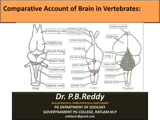
Comparision of brain in vertebrates

The vertebrate brain The vertebrate brain is the main part of the central nervous system. The brain and the spinal cord make up the central nervous system, In most of the vertebrates the brain is at the front, in the head. It is protected by the skull and close to the main sense organs. Brains are extremely complex and the part of human and animal body. The brain controls the other organs of the body, either by activating muscles or by causing secretion of chemicals such as hormones and neurotransmitters. Muscular action allows rapid and coordinated responses to changes in the environment. The brain of an adult human weights about 1300–1400 grams . In vertebrates, the spinal cord by itself can cause reflex responses as well as simple movement such as swimming or walking. However, sophisticated control of behaviour requires a centralized brain. The structure of all vertebrate brains is basically the same. At the same time, during the course of evolution, the vertebrate brain has undergone changes, and become more effective. In so-called 'lower' animals, most or all of the brain structure is inherited, and therefore their behaviour is mostly instinctive. In mammals, and especially in man, the brain is developed further during life by learning. This has the benefit of helping them fit better into their environment. The capacity to learn is seen best in the cerebral cortex. Three principles The brain and nervous system is essentially a system which makes connections. It has input from sense organs and output to muscles. It is connected in several ways with the endocrine system, which makes hormones, and the digestive system and sex system. Hormones work slowly, so those changes are gradual. The brain is a kind of department store. It has, all inter-connected, departments which do different things. They all help each other gather senses. Much of what the body does is not conscious. Basically, much of the body runs on automatic (breathing, heart beat, hungry, hair growth) adjusted by the autonomic nervous system. The brain, too, does much of its work without a person noticing it. The unconscious mind refers to the brain activities which are hardly ever noticed.

Empfohlen
Weitere ähnliche Inhalte
Was ist angesagt?
Was ist angesagt? (20)
Ähnlich wie Comparision of brain in vertebrates
Ähnlich wie Comparision of brain in vertebrates (20)
Mehr von Govt.college,Nagda, ujjain.M.P
Mehr von Govt.college,Nagda, ujjain.M.P (20)
Kürzlich hochgeladen
Kürzlich hochgeladen (20)
Comparision of brain in vertebrates
- 1. Comparative Account of Brain in Vertebrates: Dr. P.B.Reddy M.Sc,M.Phil,Ph.D, FIMRF,FICER,FSLSc,FISZS,FISQEM PG DEPARTMENT OF ZOOLOGY GOVERTNAMENT PG COLLEGE, RATLAM.M.P reddysirr@gmail.com
- 5. The vertebrate brain The vertebrate brain is the main part of the central nervous system. The brain and the spinal cord make up the central nervous system, In most of the vertebrates the brain is at the front, in the head. It is protected by the skull and close to the main sense organs. Brains are extremely complex and the part of human and animal body. The brain controls the other organs of the body, either by activating muscles or by causing secretion of chemicals such as hormones and neurotransmitters. Muscular action allows rapid and coordinated responses to changes in the environment. The brain of an adult human weights about 1300–1400 grams . In vertebrates, the spinal cord by itself can cause reflex responses as well as simple movement such as swimming or walking.However, sophisticated control of behaviour requires a centralized brain. The structure of all vertebrate brains is basically the same. At the same time, during the course of evolution, the vertebrate brain has undergone changes, and become more effective. In so-called 'lower' animals, most or all of the brain structure is inherited, and therefore their behaviour is mostly instinctive. In mammals, and especially in man, the brain is developed further during life by learning. This has the benefit of helping them fit better into their environment. The capacity to learn is seen best in the cerebral cortex.
- 6. The nerve fibers Agatha are not covered by the myelin sheath (a fatty insulating layer) found in all higher vertebrates. This means that nervous conduction is slow and the complex nervous connections found in higher forms are impossible in these early vertebrates.
- 8. Three principles 1. The brain and nervous system is essentially a system which makes connections. It has input from sense organs and output to muscles. It is connected in several ways with the endocrine system, which makes hormones, and the digestive system and sex system. Hormones work slowly, so those changes are gradual. 2. The brain is a kind of department store. It has, all inter- connected, departments which do different things. They all help each other gather senses. 3. Much of what the body does is not conscious. Basically, much of the body runs on automatic (breathing, heart beat, hungry, hair growth) adjusted by the autonomic nervous system. The brain, too, does much of its work without a person noticing it. The unconscious mind refers to the brain activities which are hardly ever noticed.
- 9. Vertebrate brain regions Several brain areas have kept their identities across the whole range of vertebrates. Starting from the back (or, in humans, the underneath part) the regions are: • The medulla and the spinal cord deal with various autonomic functions. These include the heart beat and blood pressure, breathing, and vomiting. •The pons is a relay station, carrying messages between the cerebrum and the medulla and cerebellum. •The hypothalamus is a small region at the base of the forebrain. It is the central control station for sleep/wake cycles, control of eating and drinking, control of hormone release, and many other functions. It sits immediately above the pituitary gland, and secretes hormones into the gland. These hormones inhibit or stimulate the pituitary gland. The gland, in turn, makes hormones which affect the rest of the body. •The thalamus sits above the hypothalamus, and below the cerebral cortex. • It is a collection of nuclei with various functions. It acts as a relay station, gathering sense information of all kinds (except olfactory) and passes it on to the cerebral cortex. Also, it has a role in consciousness and sleep. There are action systems for several types of behaviour, including eating, drinking, defecation, and copulation.
- 10. •The cerebellum adjusts the output of other brain systems to make them more precise. Removal of the cerebellum does not prevent an animal from doing anything in particular, but it makes actions hesitant and clumsy. This precision is not built-in, but learned by trial and error. •The tectum, often called 'optic tectum', directs actions to points in space. It gets strong visual inputs, and inputs from other senses which are useful in directing actions, such as auditory input in owls, input from the thermo sensitive pit organs in snakes, etc. In some fish, such as lampreys, it is the largest part of the brain. •The hippocampus is, strictly speaking, only in mammals, but it has counterparts in all vertebrates. It is involved in spatial memory and navigation in fishes, birds, reptiles, and mammals. •The basal ganglia are a group of structures in the forebrain, which are strongly connected to the cerebral cortex and the thalamus. The main function of the basal ganglia seems to be selecting actions. They send inhibitory signals to all parts of the brain that can generate actions. In the right circumstances they release the inhibition, so that the action-generating systems can execute their actions. Rewards and punishments exert most important neural effects on the basal ganglia. •The cerebral cortex is a layer of grey cells that is on the surface of the forebrain. It is involved in multiple functions, including olfaction and spatial memory. In mammals, where it dominates the brain, it controls functions from many subcortical areas. •The olfactory bulb is a special structure that processes olfactory sensory signals (smells), and sends its output to the olfactory part of the cerebral cortex. It is a major brain component in many vertebrates, but much reduced in primates.
- 11. Brain is called as cephalon. It is divided into three parts, namely, prosencephalon, mesencephalon and rhomb encephalon. As the brain develops further by increasing the number of neurons, it further divides into different parts, each one having assigned its own specific function. Prosencephalon divides into Telencephalon and Diencephalon, the former includes olfactory lobes (Rhinencephalon) and Cerebral hemispheres that coordinate the activities of the entire brain. The roof of cerebrum is called Pallium and the floor that generally contains nerve fibres is known as corpus striatum. Diencephalon is a small part of brain, generally covered by enormously enlarged cerebral hemispheres. This is an extremely important part of brain which functions as switch board to cerebrum. Dorsal part of diencephalon is called epithalamus and the ventral part hypothalamus while the lateral parts are called thalami that contain relay centres to connect dorsal and ventral parts of thalamus. Anterior part of epithalamus contains a glandular area called anterior choroid plexus (Tela Choroidea) which secretes cerebro-spinal fluid. Two dorsal processes of epithalamus, the anterior paraphysis supports parietal body and the posterior epiphysis bears pineal body. These two bodies function as photoreceptors in lower vertebrates and gradually transform into endocrine organs and biological clock in higher vertebrates. The ventral hypothalamus has the optic chiasma (crossing of optic nerves) on the anterior side and a ventral median evagination called infundibulum which supports pituitary gland or hypophysis. There is an olfactory area, mammillary body on the posterior side of hypothalamus.
- 12. Mesencephalon is concerned with sight and hearing. Its dorsal side is called Tectum and the ventral fibre bundles are called Cruracerebri or cerebral peduncle. The tectum has a pair of bulging optic lobes on the anterior side and a pair of auditory lobes on the posterior side. In lower animals auditory lobes are insignificant and optic lobes are prominent. This is called Corpora bigemina. Higher vertebrates such as mammals and snakes have corpora quadrigemina, which means they have optic and auditory lobes of equal size. Metencephalon is called cerebellum which is quite enlarged in active animals. In mammals cerebellum contains bundles of branching fibres of white matter called ArborVitae. The bulging ventral side of cerebellum is called pons varolli and it contains criss-crossing fibres of neurons Myelencephalon or medulla oblongata is the posterior part of the brain which does not undergo much modification in vertebrates since it controls the autonomic functions of body. The ventral side contains RAS (Reticular Activating and Inhibiting System) which keep the brain attentive and awake. Dorsal side exhibits the posterior choroid plexus, which secretes cerebrospinal fluid that flows into the brain ventricles and to meninges through a median Foramen of Megendie and the paired Foramina of Luschka. Medulla is attached with cranial nerves which bring sensory impulses from the body.
- 13. Brain is hollow inside. The cavities are called ventricles which are lined by ciliated epithelium, ependyma. These cavities help to communicate each lobe within the brain. These structures are responsible for the production, transport and removal of cerebrospinal fluid, which bathes the central nervous system. Ventricles of the two cerebral hemispheres are called lateral ventricles, or Telocoel or I and II ventricles which are connected together with a foramen of Monro. The third ventricle extends from diencephalon to mesencephalon and the IV ventricle is larger inside metencephalon and myelencephalon. The third and fourth ventricles are connected together by a tube-like connection called Iter or aqueduct of Sylvius. The ventricular system
- 14. MENINGES Meninges are protective layers around the brain. The outermost layer is fibrous duramater (meaning tough mother) which, though tightly attached to the periostial layer of skull. The second layer under duramater is Arachnoid, so named because of spider web like appearance due to presence of villi for the absorption of cerebrospinal fluid. Between the duramater and arachnoid exists the subdural space and between arachnoid and the lower piamater is the subarachnoid space. The innermost layer of meninges is the delicate piamater which is intimately attached with the brain tissue and extends deep into the sulci and fissures. It carries blood vessels and nerves. The three separate meningal layers are found in mammals only, while in amphibia, reptiles and birds, arachnoid and piamater fuse to form a single pia- arachnoid layer below the subdural space. Fishes have a single meninx primitiva that is separated from the skull bone by perimeningeal
- 15. CYCLOSTOME BRAIN Cyclostome brain is very primitive owing to their parasitic and detritus feeding habits. Cerebral hemispheres are small and smooth. Olfactory lobes are well developed as these animals detect suitability of their hosts by acute sense of smell. Thalamus is enlarged with a prominent median olfactory area called habenula. Optic lobes are small because of primitive or rudimentary eyes. Cerebellum which is related with balance and posture is reduced. Medulla oblongata is quite well developed and receives six pairs of cranial nerves. pons varolli on the ventral side is absent. Pineal and parietal bodies are present in lampreys but absent in hag fishes. FISH BRAIN Active bony fishes and sharks have well developed brain but bottom dwelling fishes have reduced brain organs. Olfactory lobes are large in sharks and they can detect their injured prey by the smell of blood from a distance of about a kilometer. But in majority of bony fishes optic lobes are reduced. Cerebral hemispheres are quite large but smooth and white. Pineal and parietal bodies are generally reduced in fishes. On the ventral side of diencephalon, there is saccus vaculosus posterior to the pituitary that serves as sense organ. Optic lobes are well developed but in deep sea fishes they are reduced. Cerebellum is highly enlarged in sharks as well as in active bony fishes and also has lateral extensions called restiform bodies or auricular lobes which connect medulla with cerebellum. Cerebellum is smaller in rays, lung fishes, ganoid fishes and deep sea fishes. Medulla oblongata has no particular variation except in deep sea fishes in which there are large vagal lobes on the lateral side which receive impulses from taste buds that are scattered all over the body as pit organs.
- 16. URODELE BRAIN Urodele brain is primitive and reflects their sluggish nature and under-developed sense organs. Olfactory lobes, optic lobes and cerebellum are reduced and cerebral hemispheres are also small and smooth. Pineal and parietal bodies are present but reduced. There is no saccus vaculosus and corpus striatum is weak. ANURAN BRAIN Frogs and toads possess a better developed brain as compared with urodeles. Olfactory lobes are large and fused at base that gives better sense of smell. Cerebral hemispheres are larger with a centralized gray area called Archipallium and white area are called paleo pallium. Parietal body is reduced but pineal is well developed. Ancient amphibians possessed a third eye over the pineal-parietal complex. Optic lobes are well developed but there are no auditory lobes. Optic lobes form corpora bigemina. Cerebellum is reduced but medulla is enlarged. REPTILIAN BRAIN Brain becomes large by the enlargement of corpus striatum. Cerebral hemispheres are large and oval but the surface is white and smooth. crocodiles develop gray matter called neopallium similar to mammals. Olfactory lobes are well developed in snakes and lizards which have olfactory sense organs on the tongue, but reduced in turtles and crocodiles. Parietal body is well developed in lizards and in Sphenodon it lies under a lens-like transparent area called the third eye. Pineal body is absent in crocodiles. Optic lobes are well developed in all reptiles and corpora quadrigemina is found only in snakes. Cerebellum is reduced in all reptiles owing to their creeping habit.
- 17. BIRD BRAIN Bird brain is characterized by enormous enlargement of cerebral hemispheres, optic lobes and cerebellum. Cerebral hemispheres become enlarged owing to enlargement of corpus striatum which is called hyper striatum but pallium is thin and surface has only white matter. Olfactory lobes are highly reduced. Optic lobes are enormous for the best eye sight in animal kingdom. Parietal body is absent and pineal small in most of the birds. Cerebellum is trilobed and highly enlarged. The middle lobe of cerebellum is called vermis as it has transverse folds and the lateral lobes are called flocculi. MAMMALIAN BRAIN Mammalian brain is highly developed and keeps complete control over the body functions. Cerebral hemispheres are enormously enlarged and the surface is folded into depressions (sulci) and raised portions (gyri) so that large surface area can be accommodated in the small space of the skull. Gray matter has spread to the surface which is called neopallium. Two cerebral hemispheres are connected by thick bundles of nerve fibres called corpus callosum, which is not found in monotremes and marsupials. Olfactory lobes are highly enlarged, so that mammalian brain is sometimes called nose-brain. Parietal body is absent and pineal is usually present except in animals like Armadillo, Sirenia and Edentates. Mammals being active animals, cerebellum is highly enlarged and trilobed and all lobes possess gyri and sulci. Nerve cells form bundles of branching fibres called ArborVitae. Medulla is short as compared to the large brain but ponsvarolli is enlarged.
- 18. spinal cord and spinal nerves The spinal cord is a long, fragile tube like structure that begins at the end of the brain stem and continues down almost to the bottom of the spine. The spinal cord consists of nerves that carry incoming and outgoing messages between the brain and the rest of the body. It is also the center for reflexes, such as the knee jerk reflex The spinal cord is uniformly gray in color with the nerve cell bodies lying close to the central canal. Connections with other cells are made around the outside of this gray matter in what corresponds to the white matter of higher vertebrates. The roots of the spinal nerves do not join just outside the spinal cord as they do in other vertebrates.
- 19. Cranial capacity is a measure of the volume of the interior of the skull of those vertebrates. Brain-to-body mass ratio, also known as the brain-to-body weight ratio. It is the ratio of brain mass to body mass, which is hypothesized to be a rough estimate of the intelligence of an animal. A more complex measurement, encephalization quotient, takes into account allometric effects of widely divergent body sizes across several taxa. The raw brain-to-body mass ratio is however simpler to come by, and is still a useful tool for comparing encephalization within species or between fairly closely related species. Brain-to-body mass ratio