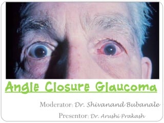
Angle closure glaucoma
•Als PPT, PDF herunterladen•
94 gefällt mir•13,933 views

Angle closure glaucoma

Empfohlen
Weitere ähnliche Inhalte
Was ist angesagt?
Was ist angesagt? (20)
Andere mochten auch
Andere mochten auch (6)
Ähnlich wie Angle closure glaucoma
Ähnlich wie Angle closure glaucoma (20)
Mehr von Arushi Prakash
Kürzlich hochgeladen
Kürzlich hochgeladen (20)
Angle closure glaucoma
- 1. Angle Closure Glaucoma Moderator: Dr. Shivanand Bubanale Presentor: Dr. Arushi Prakash
- 2. Contents Glaucoma- definition - history Angleclosureglaucoma–definition classification Grading of anglewidth Mechanismsof angleclosureglaucoma Epidemiology Provocativetests Acuteprimary angleclosureglaucoma clinical presentation treatment sequalae Plateau irisconfiguration Pseudoplateau irissyndrome
- 3. Contents Phacomorphic Glaucoma Secondary angle closure glaucomas Neovascular glaucoma Iridocorneal endothelial syndrome Posterior polymorphousdystrophy Epithelial downgrowth Fibrovascular ingrowth Flat anterior chamber Iridoschisis Inflammatory glaucomas Ciliary body glaucoma Intraocular tumors Nanophthalmos
- 4. Contents Posterior Scleritis Central retinal vein occlusion Post ocular surgery glaucomas Retinopathy of prematurity Microspherophakia
- 5. Glaucoma Glaucoma is a group of disorders characterized by a progressive optic neuropathy resulting in a characteristic appearance of the optic disc and specific pattern of irreversible visual field defects that are associated frequently but not invariably with raised intraocular pressure.
- 6. History of GlaucomasHistory of Glaucomas 08/22/15
- 7. History of Glaucomas Term acute glaucoma first used by Lawrence to describe severe ocular inflammation MacKenzie emphasized firmness to touch Von Graefe observed progressive optic disc cupping and stressed the importance of elevated intraocular pressure and the beneficial effect of iridectomy Curran and Banzinger introduced the concept of physiologic obstruction of aqueous humor flow through the pupil from the posterior chamber to the anterior chamber (pupillary block)
- 8. Curran reported success with surgical peripheral iridectomy in eyes with shallow anterior chambers but not in eyes with chambers of normal dephth The introduction of gonioscopy and the recognition of synechial angle closure provided the basis for the differentiation of open angle and angle closure glaucomas
- 9. Rosengren demonstrated that anterior chamber in eyes with “acute congestive glaucoma” was significantly shallower than in normal eyes Lowe investigated biometry of eyes with angle closure glaucoma And introduced the concept of polygenic inheritance Becker introduced acetazolamide
- 10. Before that, eyes with acute angkle closure glaucoma were prepared for surgery by intensive miotic therapy, purges and the application of leeches Use of hyperosmotic agents to withdraw fluid from the eye began with intravenous urea then, intravenous mannitol and finally, oral agents
- 11. Surgical treatment of angle closure glaucoma has been revolutionized with the development of laser iridotomy and iridoplasty.
- 12. Angle closure Glaucoma the term “angle closure” refers to occlusion of the trabecular meshwork by the peripheral iris, obstructing aqueous outflow. It may be- Primary- in an anatomically predisposed eye Secondary- to another ocular condition
- 13. Nomenclature and Classification Historically the angle-closure glaucoma nomenclature has been confusing: conditions were sometimes classified by the time course of the disease, sometimes by the effects of the angle closure, and sometimes by the presumptive pathophysiology of the angle closure
- 14. Nomenclature and Classification Angle-closure glaucoma has been described by the adjective pairs congestive/non-congestive and compensated/uncompensated. These terms have been abandoned because they lack specificity. The congestion and corneal decompensation are usually a function of the rapidity with which the pressure rises, or reflect underlying causal phenomena such as uveitis. Similarly the terms acute, subacute, and chronic have often been used to reflect the time course and/or presence of symptoms. Abrupt and total angle closure is acute; recurrent and self- limiting episodes of closure with elevated intraocular pressure (IOP) are subacute; and asymptomatic elevated
- 15. Twenty-first century consensus classification The new classification of primary angle-closure disease relies on three simple categories: IOP measurement, Gonioscopy, and disc and visual field evaluation. In other words, the presenting patient’s clinical examination alone determines the staging of the disease, regardless of the presence, absence, or reliability of symptom history, alleged duration, intermittency of problems, etc.
- 16. Twenty-first century consensus classification 1. Primary angle closure SUSPECT (PAC suspect): greater than 270° of irido-trabecular contact absence of peripheral anterior synechiae (PAS) normal IOP,disc,and visual field. In other words,the suspect eye has normal IOPs, optic nerves and visual fields, i.e., no signs of clinical glaucoma, but whose angle before indentation gonioscopy is graded as a Shaffer grade 2 or less, without PAS on compression. The angle is at risk.
- 17. Twenty-first century consensus classification 2. Primary angle CLOSURE (PAC): greater than 270° of irido-trabecular contact with either elevated IOP and/or PAS normal disc and visual field examinations. In other words, angle closure demonstrates irido-trabecular contact in 75% of the angle, with either PAS or elevated IOPs, but without disc and visual field changes. The angle is abnormal in structure (PAS) or function (elevated IOP).
- 18. Twenty-first century consensus classification 3. Primary angle-closure GLAUCOMA (PACG): Greater than 270° of irido-trabecular contact elevated IOP optic nerve and visual field damage. In other words, angle closure glaucoma manifests the criteria of closure above,plus demonstrable disc and/or visual field changes. The angle is abnormal in structure and function,with optic neuropathy.
- 19. 08/22/15 To apply this classification mastery of gonioscopy is required Irido-trabecular contact needs to be identified as present or absent, and then discriminated as either appositional (by indenting and revealing angle structures) or synechial, while documenting the latter’s extent (in terms of degrees or total clock hours) A goniolens larger than the corneal diameter may allow better resolution of angle structures, but successful indentation to view deeper into the angle is usually not possible.
- 20. Newer Imaging Technologies 08/22/15 1. Ultrasonic biomicroscopy (UBM) With tissue penetration of 4 mm, a UBM’s resolution typically includes angle structures as well as imaging of the anterior lens and anterior ciliary processes; images appear as radial slices of one portion of the angle Ultrasonic biomicroscopy of angle closed (star) in darkness-induced dilation Ultrasonic biomicroscopy of angle opened (star) in light-induced miosis.
- 21. Newer Imaging Technologies 08/22/15 2.Anterior segment ocular coherent tomography (AS-OCT) uses infrared light while examining a sitting patient without direct ocular contact, Images comprise a 180°-diameter slice of the anterior segment, currently limited to but a few clock hours (e.g., 3–9 o’clock scan), but dramatically capture the pupil and irido trabecular configuration in high definition.Anterior segment ocular coherent tomography 180°image of narrow angles pre iridotomy
- 22. Classification by mechanisms Angle closure can be caused by one or a combination of the following: (1) Abnormalities in the relative sizes or positions of the anterior segment structures (2) Abnormalities in the absolute sizes or positions of the anterior segment structures (3) Abnormal forces in the posterior segment that alter the anatomy of the anterior segment
- 23. Mechanisms of Angle Closure Glaucoma I. Pupillary Block A. Relative pupillary block (primary angle closure) B. Miotic- induced angle closure C. Posterior synechiae 1. Crystalline lens 2. Intraocular lens 3.Anterior hyaloid face
- 24. Mechanisms of Angle Closure Glaucoma II.Plateau Iris A.True plateau iris B. Pseudoplateau iris- iris and ciliary body cysts
- 25. 08/22/15 III. Lens Induced angle closure A. Intumescent lens (phakomorphic) B.Anterior lens subluxation 1.Trauma 2. Exfoliation syndrome 3. Hereditary disorders C. Drug sensitivity e.g. Sulfonamides
- 26. Mechanisms of Angle Closure Glaucoma IV. Malignant (ciliary block) glaucoma A. Phakic B. Pseudophakic C.Aphakic
- 27. Mechanisms of Angle Closure Glaucoma D.Assosiated with other conditions 1.After panretinal photocoagulation 2.After scleral buckling procedures 3.After central retinal vein occlusion 4. Intraocular tumors 5. Posterior scleritis 6. Retrolenticular tissue contracture a. Retinopathy of prematurity b. Persistent hyperplastic primary vitreous 7. Uveal effusion from adjacent inflammation a. Posterior scleritis b.Aquired immunodeficiency syndrome
- 28. Pupillary block Glaucoma 08/22/15 Pupillary block is the fundamental mechanism underlying the spectrum of PrimaryAngle Closure disease It may be absolute, as when then iris is completely bound down to the lens by posterior synechiae But, most often is a functional block, termed relative pupillary block.
- 29. Pupillary block Glaucoma 08/22/15 Its pathophysiology involves: (1) lens– iris apposition at the pupil, with resultant bowing forward of the peripheral iris as aqueous pressure builds up in the posterior chamber; and (2) an anatomically predisposed eye that allows the anterior displaced peripheral iris to occlude the trabecular meshwork.
- 30. 08/22/15
- 31. 08/22/15
- 32. Other Demographic risk Factors 08/22/15 • AGE • Commonly seen with the most frequency in the 6th and 7th decades of life • Several age-associated changes can include progressive relative pupillary block from a combination of 1. increasing lens thickness, 2. more anterior positioning of the lens, 3. and pupillary miosis
- 33. 08/22/15 These age-related changes increase the contact between the iris and lens, potentiate pupillary block and reduce anterior chamber depth and volume. Estimated central anterior chamber depth decreases 0.01mm/year
- 34. 08/22/15 however, PACG with pupillary block can occur in patients of any age, and rarely even in children – though the etiologies among the young are almost always developmental or secondary
- 35. 08/22/15 GENDER 2 to 3 times more commonly seen in women than men Because of shallow anterior chambers
- 36. HEREDITY Most cases of PACG with pupillary block are sporadic in nature Shallow anterior chambers and narrow angles have been reported as more common in relatives of patients with PACG than in individuals whose relatives do not have the disorder
- 37. Refractive error 08/22/15 prevalence much higher in individuals with hyperopic eyes, which typically have shallow anterior chambers and short axial lengths Although rare, angleclosure glaucoma can occur in myopic eyes
- 38. Other Risk factors 08/22/15 Occurs more commonly in the winter months. attributed to low levels of illumination, increased cloudiness, changeable weather, Central corneal thickness – a recently recognized risk factor for POAG – does not seem to have an association with PACG
- 39. Ocular risk factors and mechanisms 08/22/15 Ocular risk factors cluster around a variety of findings, each of which reflects smaller ocular dimensions: 1. Shallow anterior chamber both centrally and peripherally. 2. Decreased anterior chamber volume. 3. Short axial length of the globe. 4. Small corneal diameter. 5. Increased posterior corneal curvature (i.e., decreased radius of posterior corneal curvature). 6. Decreased corneal height.
- 40. 08/22/15 7.Anterior position of the lens with respect to the ciliary body. 8. Increased curvature of the anterior lens surface. 9. Increased thickness of the lens. 10. More anterior insertion of the iris into the ciliary body, giving a narrower approach to the angle recess, and possible anomalies of iris histology. 11.Thinning of the ciliary body is reportedly associated with anterior movement of the lens, increased lens thickness and decreased anterior chamber depth.
- 41. 08/22/15 • Three measures in particular show particularly high correlations with angle-closure disease: • (1) reduced axial anterior chamber depth and volume • (2) thicker lens; and • (3) steeper radii of corneal curvature
- 42. • The biometric peculiarities of eyes predisposed to angle- closure glaucoma are accentuated by three trends associated with aging. • First, the lens grows in thickness throughout life. • Second, the lens assumes a more anterior position with age. • Third, the pupil becomes increasingly miotic with age. • All of these • age-associated changes increase the contact between the iris and lens, potentiate pupillary block, and reduce anterior chamber depth and volume. It is estimated that central anterior chamber depth decreases 0.01 mm/year. 08/22/15
- 43. 08/22/15 Somehow the junction of the lens and iris at the pupillary plane modulates the flow of aqueous from the posterior to the anterior chamber, but it apparently is not a simple matter of direct contact between lens and iris it includes- iris–lens channel: It is an extremely thin (<5 microns), fluid-filled, flat, doughnut shaped passage between the posterior iris surface and the anterior lens, circumferentially extending beyond the edges of the pupil.
- 44. 08/22/15 This dynamic and pulsatile fluid‘structure’ provides normal resistance to aqueous flow from the posterior to anterior chambers. This thus functions as a relative one-way valve to sustain a minimally higher pressure in the posterior chamber than in the anterior chamber, hence directing anterior flow forward The resistance to flow has classically been referred to as relative pupillary block
- 45. if the peripheral iris bows forward enough to cover the trabecular meshwork, the normal outflow of aqueous humor from the anterior chamber would be blocked and the IOP could increase. Angle closure disease typically occurs in eyes with small anterior segments in which even a relatively small forward bow of the peripheral iris may contact the trabecular meshwork. 08/22/15
- 46. 08/22/15
- 47. 08/22/15 Moderate pupillary dilation is historically the most recognizable cause of increased pupillary block, frequently due to pharmacologic dilation. the posterior vector of force of the iris sphincter muscle reaches its maximum when the pupil is moderately dilated to a diameter of 3.0–4.5 mm. Also, in a midilated pupil, the peripheral iris is under less tension and is more easily pushed forward into contact with the trabecular meshwork. Lastly, dilation may also thicken and bunch the peripheral iris in the angle.
- 48. In contrast, when the pupil is widely dilated,there is little or no contact between the lens and the iris and minimum pupillary block. Therefore, acute angle-closure glaucoma rarely occurs while the pupil is in the actual process of dilating due to mydriatic eye drops: the dilation occurs rapidly enough that pupillary block does not have time to develop. Rather, pupillary block‘classically’ occurs as the pupil constricts over hours following dilation, presumably because the mid-dilation is prolonged as the mydriatic effect slowly reverses. 08/22/15
- 49. 08/22/15 Pupillary block can also be increased by marked pupillary miosis There are several everyday life‘triggers’ for precipitating attacks of acute PACG emotional upset (e.g., bad news, pain, fear, illness, an accident) or dim illumination (e.g., in a restaurant or theater). Emotional upset is thought to dilate the pupil through increased sympathetic tone to the iris dilator muscle, whereas dim illumination dilates the pupil through decreased cholinergic tone to the iris sphincter muscle. Similarly, the forward movement of the lens, which occurs in a variety of situations such as reading, changes in body position, and miotic therapy, has been implicated as a trigger. Diurnal variations in the anterior chamber depth with parasympathetic fluctuations and pupillary diameter, and diurnal variations in aqueous secretion have also been suggested as contributory factors
- 50. Provocative tests 08/22/15 These were designed to elevate IOP in conjunction with occlusion of the angle, so as to indicate which eyes‘at risk’ merit surgical intervention. Most provocative tests were designed to resemble ‘physiologic’ situations, in the hope that the test would mimic the natural history of the condition.
- 51. Provocative tests 1) Mydriatic stimulation- A weak, short-acting parasympatholytic agent such as tropicamide 0.5% or a weak sympathomimetic such as hydroxyamfetamine, to mildly dilate the pupil (raise the IOP >8mmhg is consider positive)
- 52. (2) Dark room testing to induce physiologic miosis (3) A prone test with the head resting on one’s arms on a table shifting the lens anteriorly without dilation; (4) Complex pharmacologic provocations, mixture of cycloplegics or mydriatics with pilocarpine.
- 53. Subacute or intermittent PACG presents with -unilateral headache -blurring of vision -unbroken coloured haloes With spontaneous resolution of symptoms
- 54. Acute PACG 1)Unilateral sudden onset of pain or aching on the side of the affected eye. 2)This pain is accompanied by blurred vision or colored haloes around lights 3)Ocular congestion and corneal edema 4) and sometimes nausea, vomiting, and sweating. The pain usually occurs in the trigeminal distribution, and is locally experienced by the patient as in the eye; or it can manifest as referred pain in the orbit, head, ear, sinuses, or teeth.
- 55. Physical findings in acute angle-closure glaucoma with pupillary block 08/22/15 • Findings during an acute attack of angle-closure glaucoma • Two of the following symptom sets: Periorbital or ocular pain Diminished vision Specific history of rainbow haloes with blurred vision IOP higher than21 mmHg • plus three of the following findings: Ciliary flush Corneal edema Shallow anterior chamber Anterior chamber cell and flare Mid-dilated and sluggishly reactive pupil Closed angle on gonioscopy Diminished outflow facility Hyperemic and swollen optic disc Constricted visual field
- 56. Acute congestive angle-closure glaucoma • Severe corneal oedema • Complete angle closure (Shaffer grade 0) • Dilated, unreactive, vertically oval pupil • Shallow anterior chamber • Ciliary injection Signs
- 57. 08/22/15 • Findings suggesting previous episodes of acute angle- closure glaucoma Peripheral anterior synechiae Posterior synechiae to lens Glaukomflecken Sector or generalized iris atrophy Optic nerve cupping and/ or pallor Visual field loss Diminished outflow facility
- 58. Differential diagnosis of acute angle- closure glaucoma 08/22/15 • Evidence of compromised angle on gonioscopy or shallow anterior chamber Ciliary block glaucoma (aqueous misdirection or malignant glaucoma) Neovascular glaucoma Iridocorneal endothelial syndrome Plateau iris syndrome with angle closure Secondary angle closure with pupillary block (e.g., posterior scleritis) Cilio-choroidal detachments (bilateral)
- 59. 08/22/15 • High-pressure open-angle glaucomas masquerading as acute angle closure Glaucomatocyclitic crisis Herpes simplex keratouveitis Herpes zoster uveitis Sarcoid uveitis Pigmentary glaucoma Exfoliative glaucoma (may have associated angle closure) Post-traumatic glaucoma Phacolytic glaucoma Steroid-induced glaucoma
- 61. Primary angle-closure suspect 1)Prophylactic laser iridotomy is recommended iridotomy widens the angle by about two grades 2)If significant ITC persists after iridotomy -laser iridoplasty -lens extraction
- 62. Treatment of acute PACG IMMEDIATE MEDICALTHERAPY PROTECTION OF FELLOW EYE LASER IRIDOTOMY IN BOTH INVOVED AND FELLOW EYE LONGTERM GLAUCOMA SURVEILLANCE AND IOP MANAGEMENT OF BOTH EYES
- 63. Hyperosmotic agents Raise serum osmotic pressure removing fluid from eye especially from vitreous In addition, vitreous dehydration allows lens to move posteriorly deepeningAC and facilitating opening of the angle Pressure decreases within 30-60 minutes after administration and effect lasts 5-6 hrs Oral glycerol 50% 1-1.5g/kg Isosorbide 1.5-2g/kg in diabetics (not metabolized) Mannitol 20% 1-2g/kg given intravenously over 45 minutes has a greater hypotensive effect. Adverse effects- thirst, headache. Hyperosmolar coma can be a serious complication due to severe CNS dehydration. Patients with renal or cardiovascular disease or those already dehydrated by vomiting are at risk. 08/22/15
- 64. Inhibitors of Aqueous Humor Secretion Carbonic anhydrase inhibitors Acetazolamide Highly effective in angle closure glaucomas May open some closed angles even in prescence of ischemic iris atrophy and paralysis of pupil Aim is to give large dose quickly- 500mg intravenously, advantageous when vomiting is present, onset is rapid May be given orally but onset of action is not rapid After oral therapy plasma levels last for 4 to 6 hours Adverse reactions are uncommon but there is a remote chance of sensitivity since it is a sulphonamide 08/22/15
- 65. 08/22/15
- 66. β blockers - Beta adrenergic antagonists are additive with acetazolamide but have a more prolonged onset of action than intravenous acetazolamide in acute angle closure glaucoma. They are more useful in later stages of treatment and in maintaining reduced intraocular pressure before laser iridotomy α adrenergic agonists – by means of vasoconstriction they reduce aqueous production and increase uveoscleral flow. 08/22/15
- 67. DRUGS THAT INCREASE AQUEOUS OUTFLOW Prostaglandin Analogs- Low concentrations PGF2α lower intraocular pressure without inducing ocular inflammation by increasing uveoscleral outflow, by increasing permeability of tissues in cilliary muscle or by an action on episcleral vessels. Side effects include increased iris pigmentation, thickening and darkening of eyelashes. No systemic side effects 08/22/15
- 68. Topical steroids -To reduce anterior chamber inflammation and the chance of both anterior and posterior synechiae formation, may be administered after the acute attack is broken
- 69. ROLE OF PILOCARPINE in pacg ?? WHAT MAKESTHE USE OF PILOCARPINE APPEALING? - It constricts pupil, which‘retracts’ the peripheral iris from its contact with the angle - So Miotic agents’ were used in managing acute disease, and as preventive therapy for the fellow eye or until an iridotomy can be arranged
- 70. But prophylactic’ pilocarpine in these fellow eyes may have been the precipitating cause of PACG Two major intraocular mechanic situations that miotics exacerbate: (1) Pilocarpine’s effects on ciliary body and zonular responses allow the lens shape to become more convex, while facilitating its anterior movement, thus shallowing the anterior chamber, with peripheral compromise of the irido- trabecular angle
- 71. (2) Miotics induce increased convexity of the iris as the lens advances which, with the simultaneous miotic-induced miosis, predisposes the lens–iris channel to greater resistance to aqueous flow. The result is frequently an inadvertent worsening of pupillary block.
- 72. SOWHERE PILOCARPINE CAN BE USED? Before immediate laser iridotomy or iridoplasty -To induce miosis to maximally stretch the peripheral iris In plateau iris
- 73. IT IS NOT EFFECTIVE IN ACUTE ATTACKS - The IOP is so high that the pupillary sphincter muscle is ischemic and unresponsive to topical miotic agents.
- 74. WHENTO CONSIDERTHE ACUTE ATTACK BEEN TERMINATED? - Until the IOP is reduced and sustained at the lowest possible levels. Because IOP may fall temporarily with medical treatment as profound hyposecretion and hypotony can follow an acute attack of PACG - The angle is as open as physically possible on gonioscopy.
- 75. Slit-lamp maneuvers in management of acute PACG To often accelerate corneal clarity Such Maneuvers include: 1) Axially depressing the central cornea with a small gonioprism (e.g.,the Zeiss four-mirror lens) or with a blunt instrument(e.g., a muscle hook or moist cotton swab),
- 76. 2.Applying digital massage to the globe after retrobulbar anesthesia. This technique is used to dehydrate the vitreous cavity and lower IOP 3. Performing an anterior chamber paracentesis,under topical anesthesia,with a small-gauge disposable sterile needle
- 77. Laser interventions for acute PACG 1.Peripheral laser iridotomy, 2.A peripheral iridoplasty (gonioplasty) 3.A pupilloplasty with Nd:YAG or argon instruments, is the definitive treatment for acute angle- closure
- 78. WHENTO DO IRIDOTOMY? DIRECTLY- if an acute attack terminated by medical means OR WAIT 1-2 DAYS- For the cornea to clear and intraocular inflammation to subside
- 79. 08/22/15
- 80. ROLE OF IRIDECTOMY Surgical iridectomy - Is a relatively safe and simple procedure BUT invasive - complications such as cataract, bleeding, and endophthalmitis can accur
- 81. Surgical iridectomy reserved for situations such as: 1)The laser fails to produce a patent iridotomy; 2) Laser iridotomies repeatedly close; 3) A laser is neither available nor functioning properly; 4) Opacities of the cornea interfere with laser treatment; 5)The patient is uncooperative or unable to sit at the slit lamp
- 82. 2) Peripheral laser iridoplasty (gonioplasty) can be use in a)Acute PACG from pupillary block -unresponsive to medical treatment where a perforating laser iridotomy is precluded by excessive shallowing of the anterior chamber, inflammation, or corneal edema b) plateau iris syndrome; and c) acute phacomorphic angle closure.
- 83. 3) Laser pupilloplasty PRINCIPAL It interrupt pupillary block by distorting the pupil into a tear-shaped configuration, focally interrupting decreased flow through the iris– lenticular junction, thus facilitating aqueous to flow into the anterior chamber
- 84. SURGICAL MANAGEMENT When to go for surgical option? If IOP elevation persists despite a patent laser iridotomy and subsequent medical therapy 1)filtering surgery alone; 2)lens extraction alone, with or without goniosynechialysis; 3) combined lens extraction with trabeculectomy; 4)or goniosynechialysis alone
- 85. MANAGEMENT OF FELLOW EYE -Is initiated with medical treatment to reduce the IOP, until the acute attack is resolved and -A prophylactic iridotomy can be performed --vigorous surveillance with periodic gonioscopic, disc, and field assessments
- 86. Instructions to patient Need for lifelong care even when iridotomy has apparently‘cured’ their acute glaucoma. Once optic nerve damage is demonstrated after an acute attack – i.e.PACG is manifest – iridotomy won’t sufficiently control pressure, and supplemental medical therapy or surgery is required. More frequent gonioscopy examination is required
- 88. Sequelae of acute PACG Elevated IOP- -may occur as a result of release of pigment and other debris, -incomplete or sealed iridotomy, -unrecognized plateau iris syndrome, - inflammation, -extensive PAS, - corticosteroid administration
- 89. Visual field loss -Typical glaucomatous visual field loss following an acute attack of PACG occurs in the minority of eyes (about 40%) -Nerve fiber layer loss can be demonstrated in most patients whose attack’s duration was longer than 48 hours.
- 90. --Visually significant cataract --Corneal damage --Loss of endothelial cells -- superficial corneal surface burns - Corneal decompensation
- 91. Plateau iris --Plateau iris refers On gonioscopy, that the iris assumes a steep approach at its insertion before flattening centrally plateau iris is divided into two entities 1)‘plateau iris configuration’ 2)‘plateau iris syndrome
- 93. On Gonioscopy, The iris appears flat from the pupillary margin to the periphery and creates a narrow angle recess and the potential for angle closure Koeppe gonioscopy- double hump or S-sign of a peripherally elevated roll of iris is seen ,where the forwardly positioned ciliary processes prevent the peripheral iris from falling backward in the supine position
- 94. many cases, plateau iris configuration is accompanied by some degree of pupillary block, which exacerbates the problem. Therefore, it is often not possible on clinical grounds to differentiate between a pupillary block PAC and a plateau iris configuration embarrassing the angle until after an iridotomy has been performed. In conditions precipitated by pupillary block, iridotomy will cause the iris to fall back and the peripheral chamber to deepen; whereas in plateau iris, the peripheral angle remains unchanged. 08/22/15
- 95. Patients with plateau iris configuration (and plateau iris syndrome) should be warned to avoid agents which have a potential for dilating the pupil These include any medication with anticholinergic activity such as antihistamines, phenothiazine antianxiety agents, antidepressants, and drugs used for incontinence and for diarrhea 08/22/15
- 96. -Plateau iris syndrome refers to the development of angle closure either spontaneously or after pharmacologic dilation ,in an eye with a patent iridotomy angle closure occurs because the ciliary processes are rotated forward.
- 97. presentation - -Seen in young patients - - Aged 30–40 years old - Occurs equally among men and women presents with or without symptoms occurring days, weeks, months, or years after iridotomy or other intraocular surgery If not diagnosed and treated properly, repeated episodes of angle closure can progress to PAC or PACG: increasing numbers of PAS, persistent elevation of IOP, and ultimate glaucomatous damage
- 98. Differential diagnosis of plateau iris syndrome 1)Extensive PAS due to any cause; 2)Imperforate or occluded iridotomy with persistent pupillary block 3) Multiple cysts of the iris or ciliary body 4) Ciliary block glaucoma 5) Open-angle glaucoma with anatomically narrow angles(as with increasing cataract formation); and the effect of chronic topical corticosteroids or cycloplegic agents
- 99. treatment Iridotomy. Usually the first treatment for plateau iris syndrome Peripheral iridoplasty – If UBM imaging is available, it may be the initial Treatment since iridotomy often does not change the anterior Segment anatomy Miotic agents can be tried to minimize pupillary dilation (e.g., pilcocarpine 1–2% 2–4 times a day)
- 100. Argon laser peripheral iridoplasty (gonioplasty) - if the response to miotics is inadequate, -or if the patient is unable or unwilling to use them longterm Filtration surgery,or glaucoma drainage devices - If iridoplasty plus medical therapy cannot control the IOP adequately
- 101. Cataract extraction with IOL implant -Effective in allowing the ciliary processes to move posteriorly and improve pressure control; but apposition has been documented to persist even after lens removal
- 102. Care to be taken after an iridotomy done in plateau iris Eyes should be examined periodically for signs of elevated IOP, peripheral synechia and glaucomatous damage. Pupillary dilation should be undertaken with care, even after iridotomy
- 103. •Primary cysts of the iris and ciliary body usually arise from the epithelial layers. •The cysts can be single or multiple and involve oneor both eyes •In most cases the cysts remain stationary and cause no harm. •In rare cases the cysts are sufficient in size and number to lift the iris forward and cause angle-closure glaucoma without pupillary block.
- 104. MANAGEMENT If the cysts causing angle closure are visible -Punctured with an argon or Nd:YAG laser If the cysts are not visible -If UBM has identified their location it is possible to puncture them by first doing a laser iridotomy over the involved area
- 105. Medical therapy - May be required after cyst puncture if extensive PAS are present. Filtering surgery -In a few cases where the cysts cannot be treated with a laser
- 106. PHACOMORPHIC GLAUCOMA An abnormal lens either compromises 1)The lens–iris channel (pupillary block) - can develop slowly with an age-related cataract or -rapidly with a traumatic, swollen cataract. 2)Mechanically pushes the peripheral iris forward into the angle structures
- 107. Usually unilateral and resembles PACG, except for the presence of Intumescent lens and A normal anterior chamber depth in the fellow eye.
- 108. TREATMENT Medical treatment is identical to that of PACG The definitive treatment -cataract extraction. Role of iridotomy If cataract extraction is not possible -because of extenuating circumstances (e.g.,gravely ill patient )or must be delayed
- 110. Secondary angle-closure glaucoma When a related or identifiable ophthalmic condition is known to be present with the onset of angle closure, it is referred to as secondary. (replaces the category of‘combined (mixed) mechanism glaucoma’) Two different fundamental mechanisms: 1)Anterior pulling mechanism 2)Posterior pushing mechanism
- 111. 1)Anterior pulling mechanism With the anterior pulling mechanism, the peripheral iris is pulled forward onto the trabecular meshwork by the contraction of a membrane, inflammatory exudate, or fibrous band
- 112. As the membrane, band, or inflammatory material contracts, it acts like a zipper to form permanent PAS, which can be spotty and irregular or diffuse and quite regular. Pupillary block plays little or no role in this mechanism. 08/22/15
- 113. Neovascular Glaucoma caused by a fibrovascular membrane that develops on the surface of the iris and the angle. At first the membrane merely covers the angle structures, but then it contracts to form PAS Almost always associated with some form of ocular ischemia Has also been refered to as – thrombotic glaucoma, hemorrhagic glaucoma, diabetic hemorrhagic glaucoma, congestive glaucoma, and rubeotic glaucoma 08/22/15
- 114. important to distinguish the terms neovascular glaucoma and rubeosis iridis pathogenesis of neovascular glaucoma is that retinal ischemia liberates angiogenic factors that diffuse forward and induce new vessel formation on the iris and in the angle. Capillary occlusion or ischemia appears to be the initiating event in this process, similar to the production of an angiogenic factors by solid tumors 08/22/15
- 115. Ocular vascular disease Central retinal vein occlusion Central retinal artery occlusion Branch retinal vein occlusion Branch retinal artery occlusion Sturge-Weber syndrome with choroidal hemangioma Leber’s miliary aneurysms Sickle cell retinopathy Diabetes mellitus Extraocular disease Carotid artery disease/ligation Ocular ischemia Aortic arch syndrome Carotid-cavernous fistula Giant cell arteritis Pulseless disease 08/22/15
- 116. Assorted ocular diseases Retinal detachment Eales’ disease Coats’ disease Retinopathy of prematurity Persistence and hyperplasia of the primary vitreous Retinoschisis Glaucoma Open-angle Angle-closure Secondary Norrie’s disease Stickler’s syndrome 08/22/15
- 117. Trauma Essential iris atrophy Neurofibromatosis Lupus erythematosus Marfan’s syndrome Recurrent hemorrhages Vitreous wick syndrome Ocular neoplasms Malignant melanoma Retinoblastoma Optic nerve glioma associated with venous stasis Metastatic carcinoma Reticulum cell sarcoma Medulloepithelioma Squamous cell carcinoma conjunctiva Angiomatosis retinae 08/22/15
- 118. Ocular inflammatory disease Chronic uveitis Endophthalmitis Sympathetic ophthalmia Syphilitic retinitis Vogt-Koyanagi-Harada syndrome Ocular therapy Cataract excision (especially in diabetics) Vitrectomy (especially in diabetics) Retinal detachment surgery Radiation Laser coreoplasty 08/22/15
- 119. Diabetes mellitus is associated with about one-third of the cases of neovascular glaucoma; Central retinal vein occlusion with another third; and a variety of conditions with the last third – with carotid occlusive disease being the most common in the last group 08/22/15
- 120. Clinical Presentation acute onset of pain, tearing, redness, and blurred vision some cases (weeks to months) before the onset of the pain and redness ciliary injection, a hazy cornea from epithelial edema, a deep anterior chamber with moderate flare, a hyphema, a small pupil, and new vessels on the iris and in the angle. Clinically the new vessels are first detected as small tufts at the pupillary margin. Occasionally new vessels are seen first in the angle if the tufts near the pupil are obscured by dark iris pigment
- 121. The neovascularization progresses over the iris surface and into the angle.The new vessels extend from the iris root across the ciliary body and scleral spur, where they arborize over the trabecular meshwork. may be difficult to distinguish new vessels from normal iris vessels 08/22/15
- 122. Normal iris vessels Neovascularization Uniform size irregular size Radial course Irregular course Do not branch within the iris Branch frequently Lie in the stroma Lie on the iris surface 08/22/15
- 123. 08/22/15 Stages of neovascular glaucoma. (A) Pre-glaucoma stage with new vessels appearing at pupillary margin and in angle. (B) Open-angle glaucoma stage with new vessels spreading and fibrovascular tissue covering angle. (C) Heavy neovascularization and extensive peripheral anterior synechiae. (D) Regression stage with angle sealed and vessels less visible.
- 124. AN ACUTE EPISODE IN NVG Treated by -Maximal IOP-reduction medical therapy, -Atropine for relief and to maximally dilate the pupil before iris mobility is lost, -Corticosteroid. -Panretinal photocoagulation or retinal cryoablation to prevent total angle closure.
- 125. If the eye has good visual potential, and if the neovascular membrane has regressed -Filtering surgery can be successful, especially when augmented with antimetabolites. -Glaucoma drainage implants appear to be Successful
- 126. Eyes with limited or no vision can often be made comfortable using -Cycloplegic agents -Topical corticosteroids regardless of the IOP.
- 127. Cyclodestructive procedures May be appropriate if the patient infirm for surgery, has too little visual potential to procede with filtration or tube surgery, or requires immediate pain relief.
- 128. Cyclocryotherapy is usually applied at -60°C to -80°C, using a large-tip probe with its anterior edge 2.5 mm posterior to the limbus. Six to eight 60-second freezes are placed over half of the Circumference of the ciliary body. Complications iridocyclitis, hypotony, pain, cataract, and phthisis bulbi.
- 129. Diode laser cyclodestruction for patients with poor vision or poor visual prognosis and for those for whom a glaucoma drainage device operation may be inadvisable Retrobulbar alcohol injections and enucleation are appropriate treatments for eyes either with no useful vision or with intractable pain that does not respond to medical therapy and ciliodestructive procedures
- 130. Goniophotocoagulation direct laser treatment to new vessels in the angle it may be a useful adjunct to panretinal photocoagulation in certain situations.ountered when full panretinal photocoagulation is not totally successful in reducing the angiogenic stimulus. Intravitreal bevacizumab-through pars plana
- 131. Iridocorneal endothelial syndrome Fundamental defect is in the corneal endothelium, that forms a membrane over the anterior surface of the iris and the angle structures. this membrane on contracting , distorts the iris and closes the angle Clinical presentation Clinical entities – 1) Progressive iris atrophy, 2)Chandler’s syndrome, 3)Cogan-Reese syndrome
- 133. early to mid adult life, White> Blacks Women> Men The patients usually notice change in iris or pupil, disturbance in their vision, or mild ocular discomfort. Few familial cases, mostly medical and family hostories unrevealing 08/22/15
- 134. Progressive (essential) iris atrophy - corectopia and ectopion uvea(The synechiae lift the iris off the surface of the lens The iris dissolution begins as a patchy disappearance of the stroma and progresses to full-thickness holes ‘stretch holes’- in quadrants away from pupillary displacement due to traction ‘melt holes’- without correctopia and ischaemic in nature
- 135. Early essential iris atrophy
- 136. Advanced essential iris atrophy with polycoria.
- 137. Chandler’s syndrome Chandler’s syndrome is the most common variant of the ICE - Marked corneal changes - Corectopia is minimal or absent. The endothelium has a hammered silver appearance. In contrast to the marked corneal changes, the iris involvement is generally mild and limited to superficial stromal dissolution - Peripheral anterior synechiae form, but they are not as diffuse and do not extend as far anteriorly as in progressive iris atrophy. For this reason glaucoma is often mild
- 139. The Cogan-Reese, or iris nevus, syndrome
- 140. Occurrence of pigmented lesions of the iris Pedunculated iris nodules diffuse pigmented lesions Some eyes have both 08/22/15
- 141. treatment Hypertonic solutions or soft contact lenses - If corneal edema produces pain or reduced vision if IOP is reduced eventually require - penetrating keratoplasty Goniotomy Filtering surgery or glaucoma drainage devices are often required to control glaucoma
- 142. Posterior polymorphous dystrophy Glaucoma occurs in 10–15% of patients -presents with corneal edema, iris atrophy, mild corectopia, and iridocorneal adhesions -maintain good vision throughout lives -cluster or linear arrangement of vesicles in the posterior cornea surrounded by a gray haze
- 143. treatment Most cases of posterior polymorphous dystrophy require no treatment. Eyes with glaucoma are treated with medication and then filtering surgery as necessary
- 144. Epithelial downgrowth Epithelial downgrowth (also called epithelial ingrowth) occurs when an epithelial membrane enters an eye through a wound and then proliferates over the corneal endothelium, trabecular meshwork,anterior iris surface, and vitreous face
- 145. Cataract surgery is the most common cause of epithelial downgrowth. also been reported after penetrating keratoplasty, glaucoma surgery, penetrating trauma, and unsuccessful removal of epithelial cysts of the anterior segment
- 146. presentation . evidence of current or past wound leak and are hypotonic if the fistula is still functional. - a grayish white membrane with a scalloped, thickened leading edge on the posterosuperior corneal surface.The cornea overlying the membrane may be edematous, and the iris may be drawn up to the old wound or incision.
- 147. diagnosis Clinically on examination Involvement of the iris is dramatically demonstrated when it is treated with large, low-energy argon laser burns (200–500 m, 100–400 mW, 0.1 second), which turn the epithelial membrane white. (Normal iris does not respond this way.) Aqueous humor can be aspirated, passed through a Millipore filter, and then examined to identify epithelial cells
- 148. treatment The treatment of epithelial downgrowth is often difficult and unrewarding. The corneal portion of the membrane can then be destroyed with cryotherapy or chemical cauterization. The affected iris can be excised, and cryotherapy can be applied to any remaining membrane on the ciliary body and retina
- 149. alternative approach is to do an en-bloc excision of all involved tissues.
- 150. Fibrovascular ingrowth Fibrovascular tissue can grow into an eye if there is an open wound after penetrating trauma or surgery occurs more frequently if the trauma or surgery is associated with hemorrhage, inflammation, or incarcerated tissue Fibrovascular ingrowth usually causes glaucoma when the membrane covers the angle and then contracts to form peripheral anterior synechiae Other factors contributing to the glaucoma include uveitis or pupillary block.
- 151. treatment Medical therapy. In some cases posterior glaucoma drainage device implantation or cyclophotocoagulation
- 152. Flat anterior chamber A flat anterior chamber after penetrating trauma or surgery can lead to the formation of PAS and secondary angle-closure glaucoma without pupillary block In most of the studies secondary angle-closure glaucoma was common if the anterior chamber was flat for 5 days or longer
- 153. TREATMENT Following re-formation of a flat anterior chamber, the residual secondary angle-closure glaucoma is treated with standard medical therapy.
- 154. 08/22/15 Malignan t Glaucoma Choroidal Detachme nt Pupillary Block Suprachor oidal Hemorrha ge Wound Leak Central anterior chamber Flat or shallow Shallow May be normal Flat or shallow Flat or shallow Intraocular pressure Normal or elevated Low Normal or elevated Normal or elevated Low Fundus appearance Usually normal Large, smooth, brown mass Usually normal Dark brown or dark red elevation Choroidals may be present Suprachoro idal fluid Absent Present Absent Present Absent Relief by drainageof suprachoroi No Yes No Yes No
- 155. 08/22/15 Malignan t Glaucoma Choroidal Detachme nt Pupillary Block Suprachor oidal Hemorrha ge Wound Leak Relief by iridectomy No Yes No Yes No Patent iridectomy Yes Yes No Yes Yes Onset Usually within days, but may be months Usually within first week Anytime Immediatel y or within first few days Usually within first few days but may be late with adjunctive antimetabo lites
- 156. Iridoschisis usually bilateral and tends to involve the lower iris quadrants Glaucoma occurs in about 50% of the patients and is usually related to the development of PAS in the region of the iris strands. Elevated IOP and iridoschisis are usually managed by medical therapy. A laser iridotomy should be performed( If pupillary block)
- 157. INFLAMMATION Inflammation can produce glaucoma through a variety of mechanisms, Including increased viscosity of the aqueous humor, Obstruction of the trabecular meshwork by inflammatory cells and debris, Scarring of the outflow channels , Elevated episcleral venous pressure, Forward displacement of the lens–iris diaphragm, Pupillary block from posterior synechiae
- 158. PATHOGENESIS 1)Peripheral anterior synechiae 2)Posterior synechiae 3)Forward rotation of ciliary body
- 159. Management Pupillary dilatation- (i) gives comfort and rest to the eye by relieving spasm of iris sphincter and ciliary muscle, (ii) prevents the formation of synechia and may break the already formed synechia, (iii) reduces exudation by decreasing hyperaemia and vascular permeability and (iv) increases the blood supply to anterior uvea by relieving pressure on the anterior ciliary arteries
- 160. Corticosteroids Immunosuppresive theraphy- Antiglaucoma medical theraphy Surgical theraphy 1)Iridotomy 2)Argon laser trabeculoplasty 3)Filtration surgery 4)Antifibrotic filtration surgery 5)Cyclodestructive surgery
- 161. Posterior pushing (or rotational) mechanism The peripheral iris is displaced forward by the lens, vitreous, or ciliary body The posterior pushing mechanism is often accompanied by swelling and anterior rotation of the ciliary body, which further acts to close the angle.
- 162. the role of the ciliary body in ‘pushing’ forms of secondary angle-closure glaucoma 1) When the anterior uveal tract swells from inflammation or vascular congestion, the ciliary ring is narrowed, which reduces tension on the zonules, permits the lens to come forward, and displaces the peripheral iris.
- 163. 2)When the ciliary body swells, it also rotates forward about its attachment to the scleral spur, which again loosens the Zonules and displaces the root of the iris 3) Ciliary body swelling is often accompanied by the acumulation of suprachoroidal and supraciliary fluid, which further rotates the ciliary body and iris root into the angle
- 164. Ciliary block glaucoma (aqueous misdirection,hyaloid block glaucoma and posterior aqueous entrapment) -commonly complication of a filtering procedure in eyes with pre-existing angle-closure glaucoma or shallow anterior chambers(2–4% of patients ) - ‘Triggers’ laser iridotomy, miotic usage, infectious endophthalmitis, retinal conditions, hyperplastic ciliary processes
- 165. OCULAR MANIFESTATIONS - Red, painful eye is most commonly seen after surgery for acute angle-closure glaucoma. -The condition usually occurs immediately after surgery but may occur during surgery or months to years later; -Its development often corresponds to the cessation of cycloplegic therapy or the institution of miotic drops
- 166. -The key is that the IOP is elevated and the anterior chamber is axially shallow without iris Bombe. -A high index of suspicion is necessary to make the appropriate diagnosis, since initially the IOP may not be elevated much.
- 167. DIAGNOSIS Patent iridectomy must be established (to rule out pupillary block) High-resolution ultrasound biomicroscopy It reveals anterior rotation of the ciliary body against the peripheral iris and forward displacement of the posterior chamber intraocular lens, as well as a shallow central anterior chamber, all of which are reversible
- 168. DIFFERENTIAL DIAGNOSIS 1) Pupillary block-patent iridectomy will rule out 2) Suprachoroidal hemorrage - USG or Ophthalmoscopy reveal the presence of single or multiple elevations of choroid 3)Serous choroidal effusion
- 169. treatment To move the lens-iris diaphragm back and relax the ciliary muscle -Mydriatics and Cycloplegics To decrease aqueous production -Topical -blockers, oral or topical carbonic anhydraseβ inhibitors, and -agonistsα
- 170. FELLOW EYE If a narrow angle is present in the fellow eye, a laser peripheral iridectomy is performed before any other surgical procedures
- 171. Nd:YAG) laser may be used in aphakic and pseudophakic patients Pars plana vitrectomy When medical or laser therapy fails, or in phakic eyes for which laser treatment is not a good option,
- 172. Intraocular tumors Ocular malignant melanoma is frequently associated with glaucoma Mechanisms, 1) Direct extension into the trabecular meshwork 2) Seeding of tumor cells into the outflow channels 3)Obstruction of the meshwork by pigment or pigment-laden macrophages, 4)Neovascularization, 5)PAS,iridocyclitis, and hyphema
- 173. IRIS MELANOMA INVADING ANGLE
- 174. LEIOMYOMA
- 175. Nanophthalmos Refers to an eye that is normal in shape but small in size. Ratio of the volume of the lens to the volume of the eye is 10–25% instead of the normal 3–4%.
- 176. FACTORS IN DEVLOPMENT OF GLAUCOMA IN NANOPHTHALMOS 1) Little anatomic reserve 2) Development of a choroidal effusion.(as thick sclera obstructing flow through the vortex veins) Frequently develop angle-closure glaucoma in the fourth to sixth decades of life.
- 177. management Laser iridotomy-if narrowing of angle present Laser gonioplasty-if angle not does not deepen on laser iridotomy Antiglaucoma theraphy Unroofing of at least two vortex veins to relieve venous obstruction
- 178. POSTERIOR SCLERITIS Posterior scleritis causes Choroidal effusion, Swelling, and anterior rotation of the ciliary body, as well as secondary angle- closure glaucoma without pupillary block. Management- Antiglaucoma theraphy to control IOP Systemic NSAIDS Topical and systemic corticosteroids
- 179. Central retinal vein occlusion It interferes with the venous drainage of the uveal tract, causing swelling and anterior rotation of the ciliary body. It also causes transudation of fluid into the choroid, retina, and vitreous.The fluid acts like an acutely developing posterior mass that Displaces the lens–iris diaphragm causing glaucoma
- 180. Management Antiglaucoma theraphy Laser iridotomy if pupillary block present Laser gonioplasty If ischemia is present –retinal ablation should be performed
- 181. Scleral buckling procedure After a scleral buckling procedure, it is common for the anterior chamber to be shallow for a few days. In 1–2% of eyes, this condition progresses to angle-closure glaucoma without pupillary block. Most cases of postscleral buckling angle closure glaucoma can be treated medically until the anterior chamber deepens spontaneously
- 182. Panretinal photocoagulation Photocoagulation may break the blood–ocular barriers and cause a transudation of fluid into the retina, choroid, and vitreous Most individuals with angle closure after PRP are asymptomatic,although an occasional patient complains of ocular discomfort or headache Most cases managed medically
- 183. Retinopathy of prematurity Mechanisms include Neovascularization, Pupillary block, and Pushing forward of the lens–iris diaphragm
- 184. POSTERIOR PUSHING MECHANISM WITH PUPILLARY BLOCK-dislocated lens
- 185. Microspherophakia -Pupillary block and glaucoma occur through one of two mechanisms: either the lens dislocates into the pupil and anterior chamber or long zonules allow the lens to come into pupil.
- 186. y 08/22/15 # Becker-Shaffer's Diagnosis and Therapy of the Glaucomas; 8th Ed. # Shields Textbook of Glaucoma; 2nd Ed. # American Academy of Ophthalmology BCSC. Section 10; 2011-2012 # Parson's Diseases of the Eye; 21st Ed. # Comprehensive Ophthalmology by A K Khurana; 4th Ed.
- 187. 08/22/15
Hinweis der Redaktion
- ----------First tell ---------In prence of these risk factors goal become to predict which eye will proceed towards disease…provocative test were designed
- But, each of these ‘provocative’ tests produc enough false positives and false negatives
- ……(coloured halos are due to refractive and diffractive changes of corneal edema with blue-green colors are central and the yellow-red colors are peripheral
- In plateau iris - to effect mild miosis thus stretching the peripheral iris and helping to open the angle
- 1)LASER IRIDOTOMY PRINCIPAL Once free communication exists between the posterior and anterior chambers there is an Insufficient pressure differential to push the peripheral iris forward against the trabecular Meshwork Rarely, acute attacks persist despite a patent iridotomy, nearly always necessitating surgery for cataract extraction and/or filtration.
- Cause contraction of peripheral iris and widens angle
- After the treatment is over…
- -Visually significant cataract (nearly all of whom were treated with iridotomies following acute attack) -Corneal damage can occur both from the acute attack itself and to a limited extent, from the laser iridotomy treatment. -Loss of endothelial cells also correlates with other indicators of ocular damage, including visual field loss and optic disc cupping -superficial corneal surface burns, especially if a contact lens is not used during argon laser iridotomy; however, usually disappear within a few days. -corneal endothelial cells loss The Nd:YAG laser can cause, especially if a contact lens is not used or if the treated iris is very close to the corneal endothelium -Corneal decompensation In patients with pre-existing endothelial Dystrophy are more susceptible to effect of laser iridotomy
- 20% of eyes with ‘noncongestive’ forms of angle-closure glaucoma are atypical in that they had normal central anterior chamber depths, little bombé, and minimal pupillary block seen in plateau iris
- Plateau iris configuration with relatively deep central anterior chamber and shallow peripheral anterior chamber. The plane of the iris is flat until near its insertion, where it takes a sharp, angled turn.
- Ciliary body cysts. Ultrasound biomicroscopy image
- rare cases the cysts are sufficient in size and number to lift the iris forward and cause angle-closure glaucoma without pupillary block.
- Why its important to identify this condition? -Not only because they are common causes of glaucoma, but rather because they are capable of producing severe elevations of intraocular pressure (IOP) and marked loss of vision.
- - Neovascular glaucoma can be prevented or treated satisfactorily in terms of patient comfort, although restoration of visual function is uncommon
- If cyclocryotherapy fails to reduce IOP, the treatment can be repeated over the same quadrants of the ciliary body and extended slightly. At least one-quarter of the ciliary body should remain untouched to reduce the incidence of phthisis bulbi. Cyclocryotherapy itself has been replaced by either trans-scleral laser cyclophotocoagulation or endocyclophotocoagulation
- total closure of angle with thinning of iris, pupillary displacement, and hole formation
- Ischemia may be a secondary phenomenon producing ‘melt holes
- HAMMERED SILVER ENDOTHELIAL CHANGES IN CHANDLERS SYNDROME
- DIFFUSE IRIS NAEVUE AND MULTIPLE IRIS NODULES -----COGAN REES SYNDRM differentiated from progressive iris atrophy and Chandler’s syndrome by the occurrence of pigmented lesions of the iris.
- ……….FIRST TELL……..Epithelial downgrowth usually is seen as a low-grade persistent postoperative inflammation, including conjunctival injection, photophobia, discomfort, and aqueous humor cells
- The invading fibrovascular tissue grows over the corneal endothelium, anterior iris surface, vitreous face, and angle, where it contracts to form PAS.
- …………FIRST TELL………Iridoschisis is a patchy dissolution of the iris in which the anterior stroma separates from the posterior stroma and muscle layer. The anterior stroma then splits into strands that project into the anterior chamber and sometimes touch the cornea
- IN ND YAG ----to create a large peripheral iridectomy and anterior hyaloid rupture to release the trapped aqueous from the vitreous and re-establish normal aqueous flow. [6] [9] Several openings are made peripherally - that is, not directly behind the optic. [ since the underlying etiology is an abnormal relationship between the hyaloid and the ciliary processes. IN PARS PLANA------debulk the vitreous and possibly also to disrupt the anterior hyaloid face
- LEIOMYOMA PUSHING THE PERIPHERAL IRIS FORWARD AND CLOSING OFF THE CHAMBER ANGLE
- Nanophthalmic eyes have a short anteroposterior length (,20 mm) and a small corneal diameter (Table 16–2). In contrast, the lens is normal, or even somewhat large, in size
- -buckleitself displaces the lens, iris, and vitreous -encircling band causes a temporary interference with the venous drainage of the uveal tract, which leads to swelling and anterior rotation of the ciliary body and accumulation of supraciliary and suprachoroidal fluid.
- This fluid may act as an acutely developing mass and displace the lens–iris diaphragm forward. Pupillary block may contribute to the development of glaucoma
- Dislocated or subluxated lens inducing pupillary block
- -