Radial artery pseudoaneurysm
•
0 gefällt mir•234 views

1) A 74-year-old male developed a large radial artery pseudoaneurysm following transradial coronary angiography. Ultrasound-guided compression was unsuccessful in treating the pseudoaneurysm. 2) Due to the large size of the pseudoaneurysm and failure of conservative treatment, the patient underwent surgical repair. 3) At one month follow up after surgical repair, the radial artery patency was restored with complete healing of the access site and no recurrence of the pseudoaneurysm.

Empfohlen
Weitere ähnliche Inhalte
Was ist angesagt?
Was ist angesagt? (20)
Ähnlich wie Radial artery pseudoaneurysm
Ähnlich wie Radial artery pseudoaneurysm (20)
Mehr von Ramachandra Barik
Mehr von Ramachandra Barik (20)
Kürzlich hochgeladen
Kürzlich hochgeladen (20)
Radial artery pseudoaneurysm
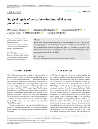
- 1. Clin Case Rep. 2020;00:1–4. | 1wileyonlinelibrary.com/journal/ccr3 1 | INTRODUCTION The TRA is getting popular because of a fewer number of complications at the site in comparison with femoral access. RAP is a rare complication of TRA.1 Lack of close follow-up and inadequate compression immediately after sheath re- moval can lead to RAP. The various approaches to treat RAPs are ultrasound-guided compression bandage,2 thrombin injec- tion into the sac of the RAP,3 surgical repair,4 and implanta- tion of covered stent.5 Recently, in a novel case report, a long dwelling catheter-based approach has been reported to suc- cessfully repair RAP.6 However, all these above approaches are in the learning curve because of rare incidence of RAPs. To reduce access site complications and preservation of pa- tency of radial artery, some trans-radialists are using distal TRA (dTRA) access.7 We report a case of a 74-year-old male who had a large ra- dial artery pseudoaneurysm which was managed by surgical repair because the ultrasound-guided compression was not successful. 2 | CASE REPORT A 74-year-old male was referred to evaluate cardiac sta- tus regarding surgical fitness to undergo radical excision of malignant melanoma over face under general anesthe- sia as the patient had shortness of breath on exertion and resting T-wave inversion in precordial leads in 12 lead ECG (Figure 1). He was not a diabetic patient. Coronary angiogram was done through the right radial artery. Radial artery puncture was done by using 20-gauge Jelco nee- dle with a single attempt of puncture using transfixation technique.A 5Fr sheath was secured in place after giving radial artery cocktail containing 5000 IU of unfraction- ated heparin(UFH), 200 µg of Nitroglycerin, and 4 mL 2% Lidocaine without Adrenaline. The coronary angiogram showed mild coronary artery disease. Hemostasis at the transradial access site was tried to be achieved using ad- equate indigenous compression bandage made using cot- ton and gauge, which is a routine practice in our CATH Lab, which provides almost hundred percent hemostasis. Received: 8 October 2019 | Revised: 28 November 2019 | Accepted: 8 December 2019 DOI: 10.1002/ccr3.2643 C A S E R E P O R T Surgical repair of postcatheterization radial artery pseudoaneurysm Dibyasundar Mahanta1 | Rudraprasad Mahapatra2 | Ramachandra Barik1 | Jogendra Singh1 | Siddhartha Sathia2 | Satyapriya Mohanty2 This is an open access article under the terms of the Creative Commons Attribution-NonCommercial-NoDerivs License, which permits use and distribution in any medium, provided the original work is properly cited, the use is non-commercial and no modifications or adaptations are made. © 2020 The Authors. Clinical Case Reports published by John Wiley & Sons Ltd. 1 Department of Cardiology, All India Institute of Medical Sciences (AIIMS), Bhubaneswar, India 2 Department of Cardiothoracic Surgery, All India Institute of Medical Sciences (AIIMS), Bhubaneswar, India Correspondence Dibyasundar Mahanta, Department of Cardiology, All India Institute of Medical Sciences (AIIMS), Bhubaneswar, India. Email: drdibyasundar@gmail.com Abstract Radial artery pseudoaneurysm (RAP) at the site of transradial access (TRA) for coro- nary angiography is rare. A clean puncture, secure bandage, and watchful follow-up are must to prevent complete occlusion and aneurysm formation at the access site. This illustration describes surgical repair as one of the successful strategies to repair a postcatheterization RAP after TRA. K E Y W O R D S radial artery pseudoaneurysm, surgical repair, transradial coronary angiogram
- 2. 2 | MAHANTA et al. Significant swelling of the right hand was noted within 20-30 minutes of the procedure when patient was still in postcath recovery room for same day discharge after a few hours of observation. Immediately, the cardiothoracic team was informed. The compression bandage was removed and was reapplied after confirmation of the proper puncture site. The arterial saturation of right thumb was 97% by pulse oxi- metry. The further progress of swelling was reduced. The patient was retained in ward for three days to monitor the size of swelling. On discharge, the patient was counselled to report at the earliest if the swelling increases in size with or without fever and pain. The patient returned after 2 weeks with fever, increase in the size of swelling visible pulsation and ecchymosis over the wrist area. The patient admitted for conservative care using intravenous antibiot- ics, elevated right hand by sling, dressing of the swollen FIGURE 1 A 12 lead EKG of the patient shows T inversion in chest leads FIGURE 2 Pseudoaneurysm at the site of transradial puncture after 1 mo as seen in clinical examination FIGURE 3 Detection of pseudoaneurysm of by color Doppler just before 3rd admission for ultrasound-guided compression and bandage.The size of the aneurysm was 4 cm × 3 cm, and the size of neck was nearly 2 mm
- 3. | 3MAHANTA et al. area using magnesium sulfate (MgSO4) and glycerin. The edema significantly reduced, and the patient was very com- fortable. There was no suspicion of pseudoaneurysm. He was discharged. However, he again returned with throbbing pain at the access site with a pulsatile swelling (Figure 2) and confirmed to be aneurysm by ultrasonography (Figure 3 and Video S1). Ultrasound showed pseudoaneurysm of radial artery of size 4 cm × 3 cm with neck of 2 mm by color Doppler (Video S2).More than 50% of the aneurysmal cavity was filled up with layered thrombus. The patient was readmitted. Repeated ultrasound-guided compression was attempted but failed to reduce the pseudoaneurysm. Patient was referred to cardiothoracic department for surgical re- pair because the patient was not willing for covered stent. The pseudoaneurysm resolved and the patency of radial artery was restored after surgical repair (Figure 4). At the follow-up 1 month after surgical repair, the lumen and flow in the radial artery was normal (Figure 5) and the access site was completely healed (Figure 6). 3 | DISCUSSION Because of rare incidence of RAP, there is no uniform consensus or guideline to manage this condition.8 Any swelling at the transradial access site is quite painful, and the progress of swelling is easily seen and felt. Therefore, early diagnosis of this condition with close follow facili- tates timely management to avoid risk of rupture and as- sociated morbidity. Significant edema due compression of veins caused by tight pressure bandage sometimes poses challenges in the early diagnosis of RAP. In such situation, an angiography using computed tomogragy (CT) or mag- netic resonance imaging (MRI) would be quite helpful but additional cost. There are several factors like multiple puncture attempts, ongoing systemic anticoagulation, inadequate hemostasis or postprocedure compression, vascular site infection, and the use of larger sheaths are responsible RAP. A conservative approach is an effective strategy, starting initially with mechanical compression and if this fails, use a thrombin injection into the aneurysmal sac. However, un- like femoral artery pseudoaneurysm, the direct compression of the RAP should be avoided to avoid rupture because of superficial location. When the aneurysm is detected very early, they are smaller in size and can be easily treated by compression while a large aneurysm requires surgical intervention.9 Other treatment strategies include the use of an external compression device or thrombin injection when the aneu- rysm has a narrow neck. The patient must be informed to report at the earliest if there is increase in the size of the swelling, throbbing pain, and ecchymosis at the access site to reduce the chances of pseu- doaneurysm formation. Unlike transfemoral access where the puncture is located deep, the transradial access site and its sur- rounding site can be explored whenever felt it is necessary. If the neck is quite narrow and the RAP is not responding to mod- erate compression by using pressure bandage with elevation of limb, a dose of thrombin equal to 500 IU (1 mL) with support of ultrasound guide can be used for thrombosis of sac. Some FIGURE 4 The condition of right hand after 5 d when opened for dressing after surgical repair before removing stiches FIGURE 5 The site of surgical repair of right hand showed complete healing of the access site and neighboring area with normal patency of right artery
- 4. 4 | MAHANTA et al. intervention cardiologists do suggest using covered stents, but experience is limited to case report only. Surgery is reserved for all large aneurysms where the conservative managements fail. 4 | CONCLUSION Radial pseudoaneurysm can be mostly managed by con- servative approach when detected early using compression bandage, ultrasound-guided thrombin injection into the an- eurysmal sac. When the conservative approaches fail or the aneurysm is significantly large, the condition can be man- aged effectively with minimum expenditure by using surgical repair when patient cannot accept or afford covered stent, as in this case report. CONFLICT OF INTEREST The authors declare that they have no conflict of interest. AUTHOR CONTRIBUTIONS DM and RB: were the patient's cardiologists, reviewed the literature, and contributed to manuscript drafting. RM, SS, and SM: were the cardiothoracic surgeon. JS (Senior resident cardiology): assisted in improving the quality of drafting. All authors issued final approval for the version to be submitted. DECLARATION OF PATIENT CONSENT The authors certify that they have obtained all appropriate patient consent forms. In the form, the patient(s) has/have given his/her/their consent for his/her/their images and other clinical information to be reported in the journal. INFORMED CONSENT STATEMENT Informed written consent was obtained from the patient for publication of this report and any accompanying images. ORCID Dibyasundar Mahanta https://orcid. org/0000-0002-2705-0361 Rudraprasad Mahapatra https://orcid. org/0000-0002-6561-6960 Ramachandra Barik https://orcid. org/0000-0003-1965-8454 Siddhartha Sathia https://orcid. org/0000-0001-7929-6200 REFERENCES 1. Zegrí I, García-Touchard A, Cuenca S, Oteo JF, Fernández- Díaz JA, Goicolea J. Radial artery pseudoaneurysm following cardiac cathe- terization: clinical features and nonsurgical treatment results. Rev Esp Cardiol. 2015;68(04):349-351. 2. Kongunattan V, Ganesh N. Radial artery pseudoaneurysm following cardiac catheterization: a nonsurgical conservative management ap- proach. Heart Views. 2018;19(2):67. 3. Mohamed MO, Saif M, Townend JN, Khan SQ. Successful treat- ment of a radial artery pseudoaneurysm in an octogenarian. BMJ Case Rep. 2015;2015:bcr2015211513. 4. Erdogan SB, Akansel S, Selcuk NT, Aka SA. Reconstructive sur- gery of true aneurysm of the radial artery: a case report. North Clin Istanb. 2018;5(1):72. 5. Tsiafoutis I, Zografos T, Koutouzis M, Katsivas A. Percutaneous en- dovascular repair of a radial artery pseudoaneurysm using a covered stent. JACC Cardiovasc Interv. 2018;11(11):e91-2. 6. Babunashvili AM, Pancholy SB, Kartashov DS. New technique for treatment of postcatheterization radial artery pseudoaneurysm. Catheter Cardiovasc Interv. 2017;89(3):393-398. 7. Boncoraglio A, Caltabiano G, Foti PV, et al. Distal radial artery: the last extreme rescue arterial access for interventional radiologists? SAGE Open Med Case Rep. 2019;7:2050313X18823918. 8. Collins N, Wainstein R, Ward M, Bhagwandeen R, Dzavik V. Pseudoaneurysm after transradial cardiac catheterization: case series and review of the literature. Catheter Cardiovasc Interv. 2012;80:283-287. https://doi.org/10.1002/ccd.23216. 9. Catheterization RAPFC. Scientific letters. Rev Esp Cardiol. 2015;68(4):343-354. SUPPORTING INFORMATION Additional supporting information may be found online in the Supporting Information section. How to cite this article: Mahanta D, Mahapatra R, Barik R, Singh J, Sathia S, Mohanty S. Surgical repair of postcatheterization radial artery pseudoaneurysm. Clin Case Rep. 2020;00:1–4. https://doi.org/10.1002/ ccr3.2643 FIGURE 6 Continuous wave Doppler of right radial artery at the site of repair shows normal flow pattern