Pulmonary atresia with intact interventricular septum
•Als PPTX, PDF herunterladen•
2 gefällt mir•3,490 views

PA/IVS is a rare congenital cardiac defect that consists of atresia of the pulmonary valve resulting in an absent connection between the right ventricular outflow tract (RVOT) and pulmonary arteries, and an intact ventricular septum that allows no connection between the right and left ventricles

Empfohlen
Empfohlen
Weitere ähnliche Inhalte
Was ist angesagt?
Was ist angesagt? (20)
Ähnlich wie Pulmonary atresia with intact interventricular septum
Ähnlich wie Pulmonary atresia with intact interventricular septum (20)
Mehr von Ramachandra Barik
Mehr von Ramachandra Barik (20)
Kürzlich hochgeladen
Kürzlich hochgeladen (20)
Pulmonary atresia with intact interventricular septum
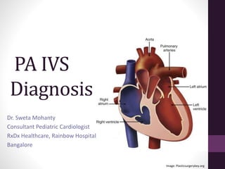
- 1. PA IVS Diagnosis Dr. Sweta Mohanty Consultant Pediatric Cardiologist RxDx Healthcare, Rainbow Hospital, Bangalore Image: Plasticsurgerykey.org
- 2. What is the diagnosis? Modified PLAX view
- 3. Diagnosis- PA IVS Is the Diagnosis Complete? Image: doctorlib Modified PLAX view
- 4. CASE EXAMPLE
- 5. History • 4 days old Term baby • Bluish discoloration noticed at 2 days • Progressive increase in cyanosis • Mild fast breathing noted Note: Cyanosis coincident with ductal closure Dyspnea with severe acidosis, reduced C.O. or pulmonary hypoplasia
- 7. Physical exam • S2 single • TR- PSM ± thrill at LLSB, MDM • PDA- murmur in 2nd or 3rd LICS • If TR severe or ASD restrictive hepatomegaly • If ASD restrictive compromising C.O. peripheral pulses feeble
- 8. Blood gas • Hypoxemia refractory to increased O2 • Hypocarbia from tachypnea • Metabolic acidosis in severe cases
- 9. CXR
- 10. Massive cardiomegaly in a newborn may be seen in all except: A] Ebstein’s anomaly and functional pulmonary atresia B] Pulmonary atresia and Ebstein’s anomaly C] ccTGA, severe left AVVR and functional aortic atresia D] Intrapericardial teratoma E] None of the above
- 12. CXR in PA IVS • Mildly enlarged heart with reduced pulmonary vascular markings & concave MPA segment Or • Gigantic cardiomegaly
- 14. Image: Thoracickey ECG QRS axis of +30 to +90 LV dominance, lack of RV forces RA enlargement ST-T abnormalities (subendocardial ischemia)
- 15. D.D. of Paucity of RV forces and LV dominance/ LVH on ECG
- 16. D.D. of Paucity of RV forces and LV dominance/ LVH on ECG •PA IVS •Tricuspid atresia •DILV
- 17. ECHOCARDIOGRAM
- 18. Role of Echo: To determine • Size of atrial communication • Size and morphology of tricuspid valve • RV morphology & inflow and outflow dimensions • Anatomical vs functional pulmonary atresia • Branch PA size and continuity • Presence of pulmonary blood flow from PDA • Coronary artery anatomy & connections with RV
- 20. Atrial septum Associations: • PFO or OS ASD with obligatory R to L shunt • If ASD restrictive, septum primum may become aneurysmal & herniate through mitral valve Subcostal view: IAS with aneurysmal flap, R-L shunt Image: pedcards.com
- 21. Atrial septum • Shunting at the atrial level is bidirectional but primarily R-to-L • Oxygenated pulmonary venous blood shunts L- to-R across TV perfuses the myocardium (via the RV-coronary fistulae) • Nonrestrictive flow is essential Image: Thoracickey
- 22. Coronary sinus Associations with PA IVS: • Stenosis/ atresia of coronary sinus ostium with decompression through unroofed coronary sinus • If atrial septum intact, coronary sinus-LA fenestration may be seen
- 23. TRICUSPID VALVE
- 24. Tricuspid valve 1) Extreme stenosis ≈ Underdeveloped RV Hypoplastic obstructive annulus Thick free margin, short dysplastic chordae, abnormal papillary muscles TV annulus hypoplastic, hypoplastic RV Image: doctorlib
- 25. Tricuspid valve 2) Profound regurgitation ≈ Large thin-walled RV Dilated annulus Ebstein-like anomaly/ Not displaced but dysplastic Image: doctorlib A4C view: Ebstein anomaly, severe TR
- 26. RIGHT VENTRICLE Image: Anesthesia and analgesia
- 27. Right ventricle • Heterogeneity in RV size, inlet, TV competency and RV function • Z value of tricuspid annulus correlates with RV cavity size • May be described as tripartite (with inlet, apical trabecular & outlet components) or underdeveloped Image: JACC
- 28. Myocardium • Ischemia, Fibrosis, Infarction, Rupture • Myocardial disarray, spongy myocardium • Endocardial fibroelastosis (inverse relation with ventriculo-coronary communications) Image: doctorlib.info
- 29. LVOT • May be obstructed by bulge in outlet septum in small and high-pressure RV • LVOTO worsens after Fontan Image: pedcards.com PSAX view: IVS bowing into LVOT
- 30. NATURE OF PULMONARY ATRESIA Image: doctorlib
- 31. Precordial SAX view with slight clockwise rotation Image: pedcards.com Membranous PA
- 32. Nature of pulmonary atresia Typically pulmonary valve morphology correlates with character of RV: • Well formed RV Membranous atresia likely: 3 semilunar cusps with complete fusion of commissures + well-formed infundibulum • Diminutive RV Muscular atresia likely: Primitive pulmonary valve + severely narrow/ atretic infundibulum
- 34. Pulmonary arteries • Usually MPA is present; pulmonary arteries are confluent & fed by left-sided PDA • If non-confluent PAs, bilateral PDAs or MAPCAs seen • LPA coarctation may occur at site of PDA insertion
- 36. Coronary arteries Associated abnormalities of coronaries: Subepicardial coronaries develop sinusoidal or fistulous connections with RV Absence of proximal aorto-coronary connection Coronary artery stenosis or interruption Coronary cameral fistula between RCA/ LMCA and RV
- 37. Ventriculocoronary connections & RV Ventriculocoronary connection correlates with: • Negative tricuspid valve Z value (Z score < -2.5) • RV classified as unipartite or bipartite Hypertensive RV with myointimal hyperplasia, background of glycosaminoglycans, endothelial irregularity, obliteration of lumen
- 38. Image: doctorlib A4C view of posterior RV and LV at level of Coronary sinus: Multiple coronary sinusoidal channels within RV myocardium
- 39. Ventriculocoronary connections • Ventriculocoronary connections may promote coronary stenosis • Normal aortic diastolic pressure may be insufficient to drive coronary blood flow if obstructive lesions present • If palliative measures like PGE1 or BT shunt further lower diastolic pressure, aortic diastolic pressure insufficient for coronary perfusion
- 40. Image: doctorlib PSAX view: Dilated proximal RCA with retrograde filling Ventriculocoronary connections • Myocardial perfusion sustained by retrograde coronary flow from hypertensive RV during systole • Systemic or suprasystemic RVSP Further coronary distortion • Any decrease in RV blood flow or RVSP coronary ischemia
- 41. Image: Giglia et al, Circulation Potential of adverse outcome after RV decompression: RV to coronary Fistula without coronary stenosis Potential RV steal phenomenon RV to coronary Fistula with proximal ± distal coronary stenosis Potential RV steal/ ischemia RV to coronary Fistula with Coronary occlusion/ atresia Potential Isolation and MI
- 42. This superior view demonstrates an atretic pulmonary artery (PA) with a LCA to RV fistula
- 43. Hypoplastic RV (HRV) with RCA to RV fistula. Ebstein-like malformation of the septal leaflet of TV
- 44. RV dependent coronary circulation (RVDCC) Coronary artery abnormalities requiring RV systolic events: • Absent aorto-coronary connections • Coronary artery interruption or stenosis • Profound coronary cameral steal or fistula
- 45. CARDIAC CATH
- 46. Role of Cardiac cath. • Coronary artery abnormalities (Color doppler cannot image obstructions to the coronary arteries with the precision of angiogram) • BAS if PFO restrictive • RV pressure- If low, suggests functional rather than anatomical PA Or global RV dysfunction • RV Angiogram- RV morphology & for ventriculocoronary connections • Aortography- PDA, pulmonary arteries
- 47. Tripartite RV Bipartite RV Monopartite RV Image: Circulation
- 48. Angiographic criteria for diagnosis of RVDCC Stenosis of ≥ 2 major coronary arteries (LMCA, LAD, circumflex or RCA) Or Atresia of the coronary ostia And Myocardium distal to the obstruction receiving blood supply via fistulous connections from RV Evidence of myocardial ischemia on surface ECG following catheter placement in RV is suspicious for RVDCC (Decompression of RV from catheter-related TR)
- 49. Angiographic diagnosis of RVDCC Angiographic techniques (Galindo et al): • RV angiogram • Aortogram • Aortogram with balloon occlusion of the aorta • Selective coronary angiograms
- 50. RV injection: Fistula to mid LAD with stenosis proximal to fistulous insertion LAD irregular proximally with additional proximal stenosis RCA fills from collaterals to LAD
- 51. Image: Sakurai, JTCVS, 2018 Lateral view Aortography: No coronaries arising from aorta Right ventriculogram: Proximal RCA, distal RCA & LCA arising from RV
- 53. In utero development of coronary a. abnormalities Lack of blood-egress from RV Elevated RV pressure Blood forced retrograde through thebesian veins Coronary sinusoids (enlarged thebesian veins) Fistulae (enlarged thebesian veins communicating with epicardial coronary arteries) Image: Thoracickey.com
- 54. In utero development of coronary a. abnormalities High pressure retrograde flow through epicardial coronaries Endothelial damage Coronary stenoses
- 55. Fetal echo findings • TV orifice proportional to RV size • TV often dysplastic TR or TS • Dilated RA common • Increased R-L shunt through PFO Dilated LV • Foraminal flap may be redundant Small, hypertrophied RV; small PE 28 weeks fetal echo Image: contemporaryobgyn.net
- 56. Fetal echo findings • MPA may be normal caliber • Ductal flow reversal essential finding Retrograde blood flow through the ductus Image: contemporaryobgyn.net 28 weeks fetal echo
- 57. Fetal echo associations Cardiac: • ASD • Tricuspid atresia • Proximal pulmonary artery atresia • RA dilatation • Aortic stenosis • Ebstein anomaly of the tricuspid leaflets Extracardiac: • None
- 58. Role of Fetal Echo Predictors for the postnatal surgical pathway: • Combination of z-scores of fetal cardiac measurements and tricuspid/mitral valve (TV/MV) ratios • Presence of coronary fistulae (usually predicts a single ventricle route) • RA pressure based on PFO (normal or restrictive), TV dimension and competency, ductus venosus Doppler (abnormal if end- diastolic flow absent or reversed)
- 59. Predictors of Biventricular repair • Fetal echo can predict biventricular circulation (at 26 weeks gestation) with 92% specificity • Best predictive scores for specific gestations: PV z-score (23 weeks) Median TV z-score (26 weeks) Combination of median PV z-score and TV/MV ratio (26 to 31 weeks) Combination of median TV z-score and TV/MV ratio (31 weeks)
- 60. Predictors of poor outcome • Fetal TV z score ≤ of −4 beyond 23 weeks of gestation • Fetal TV annulus of ≤ 5 mm beyond 30 weeks of gestation • RV:LV length or width < 0.5 • Absence of TR Peterson RE, et al.J Am Soc Echocardiogr. 2006.
- 61. Take-home messages Diagnosis of PA IVS should include: • 1) Adequacy of PDA to sustain adequate pulmonary blood flow; adequacy of ASD for decompression of systemic venous return to LA • 2) Size of RV; presence or absence of inlet, trabecular and/or infundibular portions • 3) Evaluation of RVOT, anatomic nature of pulmonary valve (muscular/ membranous) • 4) Any evidence of opening in a membranous pulmonary valve, esp. if presence of PR • 5) Evaluation of TV annulus diameter, any sign of Ebstein’s anomaly, related chordal attachment abnormalities • 6) Identification of coronary artery abnormalities and/or RVDCC
- 62. References • Nykanen DG. Pulmonary atresia and intact ventricular septum. In Ed. Allen HD, Driscoll DJ, Shaddy RE, Feltes TF. Moss and Adam’s Heart Disease in infants, children and adolescents. Seventh ed. Philadelphia, Lippincott Williams and Wilkins • Peterson RE, Levi DS, Williams RJ, et al. Echocardiographic predictors of outcome in fetuses with pulmonary atresia with intact ventricular septum.J Am Soc Echocardiogr. 2006;19:1393-1400. • Liu L, Wang H, Cui C, et al. Prenatal echocardiographic classification and prognostic evaluation strategy in fetal pulmonary atresia with intact ventricular septum. Medicine (Baltimore). 2019;98:e17492. • Giglia TM, Mandell VS, Connor AR, et al. Diagnosis and management of right ventricle-dependent coronary circulation in pulmonary atresia with intact ventricular septum. Circulation. 1992;86:1516-1528.
Hinweis der Redaktion
- PSAX view: Dilated LCA filling retrograde
- RAP score >3 predicts a biventricular repair
- Liu L, et al. Medicine 2019: In fetal PA/IVS, TV z-score >-3.28, TV/MV >0.71, RV/LV length >0.62, TID/CCD >33.95%, moderate and severe TR, and the absence of VCAC were associated with postnatal biventricular repair strategy