CEPHALOMETRIC ANALYSIS.ppt
•Als PPT, PDF herunterladen•
0 gefällt mir•211 views

for lecture of Undergraduates

Empfohlen
Weitere ähnliche Inhalte
Was ist angesagt?
Was ist angesagt? (20)
Ähnlich wie CEPHALOMETRIC ANALYSIS.ppt
Ähnlich wie CEPHALOMETRIC ANALYSIS.ppt (20)
Kürzlich hochgeladen
Kürzlich hochgeladen (20)
CEPHALOMETRIC ANALYSIS.ppt
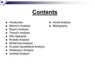
- 1. Contents Introduction Steiner’s Analysis Down’s Analysis Tweed’s Analysis Wits Apprasial Ricketts Analysis McNamara Analysis Di paolo Quadilateral Analysis Holdaway’s Analysis Jarabak Analysis Arnett Analysis Bibliography
- 2. Introduction :- The assessment of craniofacial dimensions is not a new skill in orthodontics. The earliest method was used to assess facial proportions from the artistic point of view. During the Renaissance, Durer analysed the human face, determined the ideal proportions & divide the face into four quadrants . Many centuries later, his method was applied to the analysis of cephalometric radiographs by de Coster & Moorees. Cephalometry (scientific measurement of the dimensions of the head ) was the first method to prove of value in orthodontics
- 3. The first X-ray picture of the skull in the standard lateral view were taken by Pacini & Carrera (1922). In the subsequent years, the various authors also produced the following type of radiographs for craniofacial measurements: MacGowen(1923), Simpson(1923), Comte(1927), Riesner(1929), & others. But none of them have given the accurate description of the method to take pictures. In 1931, Hofrath & Broadbent simultaneously & independently developed standarized method for the production of cephalometric radiographs, using special holders known as cephalostats.
- 4. This technique was developed in 1930s but gained wider acceptance for practical use during the last 3-4 decades The aims of the assessment tended to vary, ranging from studies on the facial growth, the location of malformations, aetiological studies to the assesment of treatment response, as a complement to status analysis in orthodontics. For clinical application, the methods designed to assist diagnosis are of particular interest. The many different diagnostic analysis may be differentiated in a number of ways.
- 5. RADIOGRAPHIC CEPHALOMETRIC TECHNIQUE The basic components of the equipment for producing a lateral cephalometric are: 1. An X-ray apparatus 2. An image receptor system 3. A cephalostat.
- 6. X-ray Apparatus:- comprises of X-rays tube, transformers, filters, collimators, and a coolant system, all encased in the machine’s housing. The three basic elements that generate the X- rays a. a cathode b. an anode c. the electrical power supply a. Cathode:- is a tungsten filament surrounded by a molybdenum. And serves as a source of electrons.
- 8. b. An anode :- It consist of a tungsten target embedded in a copper stem. The purpose of the target in an x ray tube is to convert the kinetic energy of the electrons generated from the filament in to x ray photons. Less than 1 % of the electron kinetic energy is converted to the x rays photons. The size of the focal spot , which determines image quality. The target face in the x ray tube is oriented at an angle of 15 to 200 to the cathode . The size or area of the effective focal spot created by the inclined target is between 1x1 mm2 and 1x2 mm2 .
- 9. the low- energy photons are filtered out by means of an aluminium filter The divergent x ray beam then passes through a lead diaphragm that fits over the opening of the machine housing and determine the beam’s size and shape. Only x ray with sufficient penetrating power are allowed to reach the patient c. the electric power supply :- the primary function of the power supply of an x ray machine are to 1. provide a low voltage current to heat the x ray tube filament by use of an step- down transformer 2. generate a high potential diff. between the anode and cathode by use of a high- voltage transformer
- 10. Image Receptor system ; Extra oral projection like lateral ceph. requires a complex image receptor system that consists of an extra oral film, intensifying screens, a cassette, a grid and soft tissue shield. Extra oral film is a screen film size ranging from 8x10 inches to 10x12 inches. Basic component of the film are an emulsion of silver halide crystals suspected in a gelatin frame work and a transparent blue- tinted cellulose acetate that serves as a base.
- 11. Cephalostat:- As described by Broadbent (1931). Patient’s head is fixed by 2 ear rods that are inserted into the ear holes so that the upper border of the ear holes rest on the upper part of the ear rods. Head is centered in the cephalostat, is oriented with the FHP parallel to floor and MSP vertical and parallel to the cassette. Standardized FHP is achieved by placing the infraorbital pointer at the patient’s orbit and then adjusting the head until the infra-orbital pointer and ear rods are at the same level. The upper part of face is supported by forehead clamp positioned at nasion.
- 12. The conventional use of 2 ear rods to stabilize the head in radiograph. Cephalometry is based on the assumption that the transmeatal axis of human is perpendicular to mid-sagittal plane. Actually, asymmetry is a general characteristic and the relationship of the left and right ears in their vertical and horizontal relation to each other which is frequently asymmetric. In these instances the insertion of ear-rods will obviously result in vertical and/or horizontal rotation of the head, which introduces a deficient and misleading image. So only the left ear-rods should be used in radiographic Cephalometry both for lateral and particular for the frontal projection.
- 15. Down’s Analysis It was given by William B Down in 1948 at the university of Illinois. Sample size :- 20 living individuals, ranging in age from 12-17 yrs & equally divided as to sex, & of Caucasian race. The Down’s analysis is divided into two parts:- 1. Skeletal pattern 2. Dental pattern
- 16. Skeletal pattern This include :- 1. Facial angle 2. Angle of convexity 3. A-B plane 4. Mandibular plane angle 5. Y axis
- 17. 87.80 Facial Angle :- N F H Plane Pog
- 18. Angle of Convexity :- N A Pog
- 20. 21.90 Mandibular Plane Angle:- F H Plane O P Go M
- 22. Dental pattern It includes :- 1. Cant of occlusal plane 2. Interincisal angle 3. Incisor-occlusal plane angle 4. Incisor mandibular plane angle 5. Protrusion of maxillary incisor
- 23. 9.30 F H Plane Cant of occlusal Plane:-
- 25. 14.50 Incisor occlusal plane angle:-
- 26. 1.40 Incisor mandibular plane angle:-
- 27. 2.7mm Upper incisor to A-Pog:- A Pog
- 28. The Polygon It was developed by Vorhies & Adams in 1951 which is also called as “wiggle” that represents the large group of cephalometric readings graphically
- 30. Steiner Analysis In the year 1950s, Cecil C. Steiner developed this analysis & is considered the first of the modern cephalometric analyses for two reasons: i) It displayed measurements in a way that emphasized not just the individual measurements but there interrelationship into a pattern . ii) It offered specific guides for use of cephalometric measurement in treatment planning The Steiner analysis is divided into three parts:- 1. Skeletal analysis 2. Dental analysis 3. Soft tissue analysis
- 31. Skeletal Analysis 1. S.N.A angle 2. S.N.B angle 3. A.N.B angle 4. Mandibular plane angle 5. Occlusal plane angle
- 39. Jenkins in 1955, elected to use the functional occlusal plane as a reference base for the measurement of the jaw disharmony. He established the “a” plane drawn through a point A at right angle to the occlusal plane, & then measured from the “a” plane to point B, gnathion, & the mandibular incisor edge.
- 40. Betty in 1975, ANB angle is not always an accurate method of establishing the actual amount of apical base divergence. He devised the AXD angle for measuring the apical base discrepancy, where point X is formed by projecting point A onto a perpendicular to SN line, & point D is located in the bony symphysis.
- 42. Dental Analysis This include:- 1. Upper incisor to N-A angle 2. Upper incisor to N-A linear 3. Lower incisor to N-B angle 4. Lower incisor to N-B linear 5. Inter Incisal angle
- 43. Upper incisor to N-A line:- 220 A N
- 44. Upper incisor to N-A line:- 4 mm N A
- 45. Lower incisor to N-B line :- 250 N B
- 46. Lower incisor to N-B line :- 4mm N B
- 48. Soft tissue Analysis S-line:- The lips in a well balanced faces, according to Steiner ,should touch a line extending from the soft tissue contour of the chin to the middle of an “S” formed by the lower border of the nose. This line is referred to as the S-line.
- 50. Steiner Stick It is also called as “chevrons”, a convenient shorthand for presenting the measurement . He calculated that what compromises in incisor positions would be necessary to achieve normal occlusion when the ANB angle is not ideal. This was a major step in applying cephalometric to routine treatment planning.
- 51. Steiner stick
- 52. Ricketts Analysis It was given by Robert Murray Ricketts in 1961 Purpose of analysis:- Characterize a particular type of condition Compare one individual to other Classify certain type of problems Communicate
- 53. Sample Size :-1000 consecutive patients Origin :-clinical cases with usual orthodontic race problems omitting surgical class III, traumatic TMJ cases & operated cleft palate patients Class I – 399 Class II Div 1- 367 Class II Div 2- 217 Class III – 17 Race :- white Sex :- 546 females ; 454 males of age group, 3-6 yrs – 61 7-10yrs – 497 11-14yrs- 343 15-18 yrs – 217 19-44 yrs – 33
- 54. Location of Xi point:- The location of the Xi point is keyed geometrically to the Frankfort horizontal & the pterygoid root vertical planes (PtV). The procedure follows :- 1. Locate FH plane & draw PtV plane perpendicular to the FH plane 2. Construct four planes tangent to points R1, R2 ,R3 & R4 on the border of the ramus
- 55. R1-deepest point on the anterior border of the ramus R2-located on the posterior border of the ramus R3-deepest point on the sigmoid notch, halfway between the superior & inferior curves R4-opposite R-3 on the inferior border of the mandible 3. The constructed plane form a rectangle enclosing the ramus. 4. Xi point is located at the centre of the rectangle at the intersection of the diagnosis.
- 57. Chin in Space This include :- 1. Facial axis 2. Facial (depth) angle 3. Mandibular plane
- 61. Convexity In this ;- 1. Convexity at point A It is measured from point A to the facial plane . The clinical norm at 9 yrs age is 2mm & decreases 10 every five yrs .high complexity implies a class II skeletal pattern. Negative complexity suggests a class III skeletal pattern .
- 62. N Pog A
- 63. Teeth This include :- 1. Lower incisor to A-Pog 2. Upper molar to PtV 3. Lower incisor inclination
- 64. 1mm A Pog Lower incisor to A-Pog line:-
- 65. Upper molar to Ptv:-
- 67. Profile Lower lip to E plane
- 68. Wits Appraisal Sample :- Size- 46 Race-Caucasian Sex 25 males;23 females Age – adults
- 69. AO BO
- 70. Tweed’s Triangle Sample :- Size - 95 The objectives are :- 1. The best balance & harmony of facial lines 2. Stability of denture after treatment 3. Healthy mouth tissues 4. An efficient chewing mechanism
- 71. 250 650 900 F H Plane
- 72. McNamara Analysis It was given by McNamara JA in the year 1984. The composite normative standards used in the McNamara were derived from 3 sourses:- Lateral cephalograms of the children comprising the Bolton standards Selected values from a group of untreated children from the Burlington Research Centre, & A sample of young adults from Ann Arobar ,Michigan, having good-to-excellent facial & dental configuration
- 73. Size – 111 young adults Race – caucasian Sex – male & female Age – average age of female is 26 yrs ; 8 months & average age of male is 30 yrs; 9 months
- 74. Relating the maxilla to the cranial base 1020 Soft tissue Evaluation
- 75. Hard tissue Evaluation:- F H Plane N A
- 76. Mandible to maxilla Effective length of maxilla (condyle to point A) Effective length of mandible (condyle to gnathion) Maxillo-mandibular differential Lower anterior facial height (ANS to menton) Mandibular plane angle Facial axis
- 79. Mandible to cranial base F H Plane N Pog
- 80. Upper incisor to point A vertical
- 81. Lower incisor to A-Po line
- 82. Airway Analysis Upper pharynx:-the upper pharyngeal wall width is measured from the posterior outline of the soft palate to the closest point on the pharyngeal wall. Lower pharynx:-it is measured from the intersection of the posterior border of the tongue & the inferior border of the mandible to the closest point on the posterior pharyngeal wall.
- 84. Di Paolo’s Quadilateral Analysis It was given by Di Paolo RJ in 1983. His sample size was 245 equal male and female, of 9- 15yr age group who had normal occlusion and were untreated orthodontic patients.
- 85. Skeletal Assessment This includes :- 1. Maxillary length 2. Mandibular length 3. AUFH (anterior upper facial height) 4. ALFH (anterior lower facial height) 5. PLFH (posterior lower facial height) 6. Facial (angle of facial convexity)
- 88. Dental Assessment This include :- 1. Pogonion line:. This is constructed by drawing a line tangent to pogonion and parallel to the anterior facial height. The most anterior point of the mandibular central incisor is then related perpendicular to the pogonion line. The average is 2 mm. anterior or posterior to the pogonion line. This measurement will indicate whether the chin is excessive or deficient in size.
- 89. 2. Point A line:-Maxillary incisor position is determined by drawing a line through point A parallel to the anterior lower facial height. A measurement is then made by drawing a perpendicular from this line to the most anterior point on the maxillary central incisor. The average measurement is 5 mm. plus or minus 1 mm. 3. Point B line :-Mandibular incisor position is determined by drawing a line through point B. This line is parallel to the anterior lower facial height. From this line, a measurement is made by drawing a perpendicular from this line to the most anterior point on the mandibular central incisor. The average measurement is 2 mm. plus or minus 1 mm.
- 91. Bibliography Radiographic cephalometry, Alexander jacobson pg no. 65-126, Quintessence publishing company Orthodontic cephalometry, Athanasios E Athanasiou pg no. 241-282, Mosby-Wolfe. Cephalometric radiography, Thomas Rakosi pg no. 7,34- 46 G William Arnett :Soft tissue cephalometric analysis Am J Ortho 1999;116:239-53.
- 92. Cecil C Steiner: Cephalometric for you & me Am J Ortho 1953 ;39 : 729-755 Charles H Tweed: The Frankfort-Mandibular Incisor Angle in Orthodontic Diagnosis, Treatment Planning & Prognosis AO 1954; 3: 121-169 Robert Murray Ricketts :Cephalometric Analysis & Synthesis Am J Ortho 1961; 31 :141-156 Rocco J Di Paolo :The quadrilateral analysis Am J Ortho 1984;86 :470-482 A Jacobson The Wits apprasial of jaw disharmony :Am J Ortho 1975;67 :125-138 Reed A Holdaway A soft-tissue cephalometric analysis & its use in orthodontic treatment planning Am J Ortho 1983;84:1-28