

Empfohlen
Weitere ähnliche Inhalte
Was ist angesagt?
Was ist angesagt? (20)
Ähnlich wie Operative Vaginal Delivery (Dr.Rozan)
Ähnlich wie Operative Vaginal Delivery (Dr.Rozan) (20)
Mehr von Rafi Rozan
Mehr von Rafi Rozan (11)
Kürzlich hochgeladen
Kürzlich hochgeladen (20)
Operative Vaginal Delivery (Dr.Rozan)
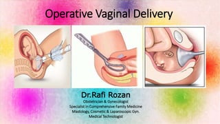
- 1. Operative Vaginal Delivery Dr.Rafi Rozan Obstetrician & Gynecologist Specialist in Comprehensive Family Medicine Mastology, Cosmetic & Laparoscopic Gyn. Medical Technologist
- 3. INTRODUCTION The era of modern operative obstetrics began with the invention of the forceps by Peter Chamberlen of England. Subsequently, over the years the ability to use forceps separated the obstetricians from the midwives. The use of forceps reached its acme in the United States as a result of the influence of DeLee, who in 1920 taught the importance of prophylactic forceps and episiotomy to protect against maternal and fetal injury. Dr.Rozan
- 4. In 1968 in New York City, 29.7% of births were forceps assisted, but by 1978 the incidence was down to 12.2%. At the same time, in the United States, cesarean birth rates rose from 5.5% in 1970 to 15.2% in 1978 and now is as high as 20–25%. 2015 – 3.1% OVD 0.56 Forceps 2.58% vacuum Dr.Rozan
- 5. Types of Episiotomy J Shape Lateral 8 o Clock MedioLateral Median Dr.Rozan
- 7. Types of Pelvis Caldwell-Molloy classification Acronym: GAAP Dr.Rozan
- 8. Rhombus of MichaelisPelvimetry External Dr.Rozan
- 9. Pelvimetry External External Conjugate of Baudelocque Biischial Spines Diameter Suprapubic Angle Dr.Rozan
- 11. The Bony Pelvis Ischial Spines OC = DC - 1.5 Dr.Rozan
- 12. Fetal Station Hodge Planes DeLee Planes ACOG Planes Dr.Rozan
- 13. Fetal Head Metopic Suture Coronal Suture Sagital Suture Lambdoid Suture Anterior Fontanel Mastoid Fontanel Posterior Fontanel Sphenoid Fontanel Dr.Rozan
- 15. Regions of the Fetal Head Dr.Rozan
- 16. Dr.Rozan
- 18. Classification & Criteria for Types of Forceps Deliveries Low Forceps Outlet Forceps Mid Forceps • Scalp is visible at the introitus without separating the labia • Fetal skull has reached the pelvic floor • Fetal head is at or on perineum • Sagittal suture is in anteroposterior diameter or right or left occiput anterior or posterior position • Rotation does not exceed 45 degrees • Leading point of the fetal skull is at station +2 cm or more and not on the pelvic floor • Without rotation: Rotation is 45 degrees or less (right or left occiput anterior to occiput anterior, or right or left occiput posterior to occiput posterior) • With rotation: Rotation is greater than 45 degrees • Station is above +2 cm but head is engaged Dr.Rozan
- 19. Indications for Operative Vaginal Delivery Nulliparous: 3 Hours • Suspicion of immediate or potential fetal compromise • Shortening of the second stage of labor for maternal benefit • Prolonged second stage of labor Multiparous: 2 Hours Epidural Anesthesia: 1 extra hour with RFT Acute Placental Abruption ValSalva Maneuver is Contraindicated Cardiac Abnormalities Neurological Disorder Muscular Disease Cystic Lung Disease Dr.Rozan
- 20. Contraindications • Extreme fetal prematurity • Fetal demineralizing disease (osteogenesis imperfect) • Fetal bleeding diathesis ( fetal hemophilia, neonatal alloimmune thrombocytopenia). • Unengaged head • Unknown fetal position. • Brow or face presentation. • Suspected fetal-pelvic disproportion. Relative contraindications to use of vacuum devices, but not forceps, include gestational age <34 weeks Dr.Rozan
- 21. Prerequisites for Operative Vaginal Delivery • Cervix fully dilated and retracted • Membranes ruptured • Engagement of the fetal head • Position, Presentation, Station, Ortientation and any Asynclitism must be know • Fetal weight estimation performed • Adequate anesthesia • Clinical Pelvimetry (Adequate pelvis to fetal size) • Maternal bladder has been emptied • Patient Consents to Procedure • Willingness to abandon trial of operative vaginal delivery and back-up plan in place in case of failure to deliver • This is an operative procedure, and it should be accorded the same respect and care for aseptic technique as any other operative procedure. Dr.Rozan
- 22. ANATOMY OF THE FORCEPS Handle Finger Guide Lock Shank Blades Cephalic Curve Pelvic Curve Heel Toe Fenestration Window Dr.Rozan
- 23. Some Types of Forceps Simpson forceps (1848) are the most commonly used among the types of forceps and has an elongated cephalic curve. These are used when there is substantial molding of the fetal head. Dr.Rozan
- 24. Elliot forceps (1860) are similar to Simpson forceps but with an adjustable pin in the end of the handles which can be drawn out as a means of regulating the lateral pressure on the handles when the instrument is positioned for use. They are used most often when there is minimal moulding. Dr.Rozan
- 25. Kielland forceps (1915, Norwegian) are distinguished by an extremely small pelvic curve and a sliding lock. The most common forceps used for rotation. The sliding lock is helpful in asynclitic Kielland forceps lack traction because they have almost no pelvic curve Dr.Rozan
- 26. Wrigley's forceps are used in low or outlet delivery and in cesarean section delivery where manual traction is proving difficult. The short length results in a lower chance of uterine rupture. Dr.Rozan
- 27. Piper's forceps have a perineal curve to allow application to the after- coming head in breech delivery. Dr.Rozan
- 28. Dr.Rozan
- 29. ANATOMY OF THE FORCEPS French lock English lock German lock Sliding Lock Pivot lock Dr.Rozan
- 30. Technique of forceps application The forceps must be applied so they fit the head accurately. They should lie evenly against the sides of the head, reaching from the parietal bones to and beyond the malar eminences covering symmetrically the spaces between the orbits and the ears. Parietal – Malar Ideal application Dr.Rozan
- 31. -With the criteria for forceps application met, the forceps can be applied. -The operator stands before the perineum with the forceps articulated and oriented to the position of the fetus' head. Holding the forceps articulated prevents confusion due to the crossing of the shanks. -For the left occipitoanterior (LOA) or direct OA positions, the left blade (posterior) is applied first. -The operator places his or her back to the maternal right thigh and holds the handle between the fingers, as in holding a pencil. -The shank is held perpendicular to the floor, the middle and index fingers are inserted into the vagina, and the thumb is applied to the heel of the blade. - The force necessary to insert the blade is exerted by the pressure of the thumb. - The left hand guides the handle in a wide arc until the blade is in place. This blade is then held in place by an assistant. Technique of forceps application Dr.Rozan
- 32. -The right blade is then inserted in a similar manner, with the operator's back to the patient's left thigh. -This blade is inserted more anteriorly in the vagina to avoid rotating the head further to the left. - Any adjustments to ensure a cephalic application should generally be made with the right blade. Technique of forceps application Dr.Rozan
- 34. Once the blades are applied, the accuracy of placement should be evaluated. Technique of forceps application 1. Lock articulation of the Forceps. Do not close the Handle 2. Ocipital Fontanel 1 finger beneath above the shank of the forceps 3. The shanks are perpendicular to the Sagital suture 4. One or less fingertip space at the heel of the blade 5. No maternal tissue has been grasped Dr.Rozan
- 35. The Pull … Technique of forceps application Pajot Saxtorph Maneuver Bill axis traction handle Scanzoni Maneuver OP Dr.Rozan Maternal Effort + Uterine Contraction + Devise Pull
- 36. Ritgen maneuver Is used to control delivery of the fetal head. It involves applying upward pressure from the coccygeal region to extend the head during actual delivery, thereby protecting the musculature of the perineum. Modified Ritgen Maneuver Dr.Rozan
- 37. Dr.Rozan
- 38. Dr.Rozan
- 39. Name: Jorge Odón Country: Argentina Occupation: Car Mechanic Dr.Rozan
- 40. Dr.Rozan
- 41. Odón's idea to Odón Device Dr.Rozan
- 46. 1. ACOG PB 154 2. Uptodate OVD 3. Williams Obstetrics 24 Ed 4. Martin JA, Hamilton BE, Osterman MJ, et al. Births: Final Data for 2015. Natl Vital Stat Rep 2017; 66:1. 5. Committee on Practice Bulletins—Obstetrics. ACOG Practice Bulletin No. 154 Summary: Operative Vaginal Delivery. Obstet Gynecol 2015; 126:1118. 6. Palatnik A, Grobman WA, Hellendag MG, et al. Predictors of Failed Operative Vaginal Delivery in a Contemporary Obstetric Cohort. Obstet Gynecol 2016; 127:501. 7. American College of Obstetricians and Gynecologists, Society for Maternal- Fetal Medicine. Obstetric care consensus no. 1: safe prevention of the primary cesarean delivery. Obstet Gynecol 2014; 123:693. Referenc es Dr.Rozan
- 47. Dr.Rozan
Hinweis der Redaktion
- The history of obstetrical forceps is long and, often, colorful. Sanskrit writings from approximately 1500 BC contain evidence of single and paired instruments; Egyptian, Greek, Roman, and Persian writings and pictures refer to forceps that were originally used for extraction following fetal demise to save the mother’s life DeLee's teaching was the impetus for the dramatic increase in the number of forceps deliveries performed in the United States from the 1930s through the 1950s. DeLee's has many contribution to the era of modern obstetric.. Namely the fetal station.
- However, due to the provocative studies by Friedman and colleagues and the increasing tendency for an adverse obstetric outcome to result in a malpractice suit, obstetric forceps delivery rates have fallen in the United States. OVD remains an important part of modern obstetrics and its knowledge and application is vital in every labor setting so that we can decrease CS and save a life if the needs arise.
- Knowing to do a Good Obstetric Explorartion is the Gold Key AP diameter = 11.5 to 12cm Oblique = 12.5 , Transverse = 13 cm, Inlet, mid, outlet and low
- The Rhombus of Michaelis, also known as the Michaelis-Raute or the quadrilateral of Michaelis, it is a rhombus-shaped contour that is sometimes visible on the lower back. The Rhombus of Michaelis is named after Gustav Adolf Michaelis, a 19th-century German obstetrician. 4 sides and angles
- Baudelocque, martin or Budin Pelvimeters. Bispinal = 24cm Distance between iliac crest= 28cm Distance between trocanters (Bitrocanter Diameter)= 32cm
- The concept of fetal station was initially described by DeLee in 1924 as the level of the presenting fetal part in the birth canal in relation to the ischial spines. Later, in 1988, ACOG first reported on a station classification system wherein the pelvis above and below the ischial spines is divided into fifths. These divisions are represented in centimeters; if the leading part of the fetus descends at a level between the spines, the station is designated as 0. Below the ischial spines, the presenting fetal part passes stations +1, +2, +3, +4, and +5 to delivery.
- Fontanels and sutures allows for molding of the fetal hear as it passes through the birth canal. In OVD it allows us to identify the Fetal Orientation thus being able to adequately and correctly place the Forceps.
- The regions of the fetal skull have been designated to aid in the description of the presenting Part felt at vaginal examination during labor.
- The sagittal suture is parallel to the transverse diameter of the pelvis- symmetricly (Selhiem Law). 2 unequal ovoid will align on there greater diameter axis reasons why the fetal head orients sideways to the transverse diameter of the Pelvis When the fetal head engages with some degree of tilt to the shoulder it results in asynclitism. Anterior asynclitism, in which the anterior part presents, is called Nägele obliquity. Posterior asynclitism, Litzman obliquity Bitemporal asynclitism, Roederer obliquity
- The American College of Obstetricians and Gynecologists' classification system for forceps deliveries is based on station and extent of rotation
- To reduce the rate of CS for failure to progress in the second stage, ACOG & SMFM recommend allowing 3H of pushing for nulliparous women and 2H of pushing for multiparous women before diagnosing arrest of labor, when maternal and fetal conditions permit. They did not provide specific criteria for the upper limit of the 2nd stage. Many obstetric providers allow an extra hour of pushing for women with epidural anesthesia when there is reassuring fetal testing. Use of forceps or vacuum is appropriate when expeditious delivery is indicated because of fetal compromise or probably imminent fetal compromise (eg, acute abruption)
- Instrumental delivery is contraindicated if the clinician or patient believes that the risk to mother or fetus is unacceptable. Examples include, but are not limited to
- Parietal Malar placement is important because It avoids over flexion or over extension of the fetal Head and as such preventing Maternal and fetal complications.
- Applying the left blade first has the advantage of not needing to cross the shanks in order to lock the forceps
- An alternative method of traction is to use a Bill axis traction handle and pull in the direction of the pointer. In exerting traction, it is well to keep in mind the bell-shaped curve of a uterine contraction as seen on a fetal monitor. The traction force should gradually increase, reaching its acme at about 30–40 seconds and then gradually relaxing. For the resident being taught, it is instructive to actually count off the seconds out loud so that they can appreciate the time involved. Between tractions, the handles are unlocked to relieve pressure on the fetus' head. Experts suggest 3 pulls are enough but its depends on the operator The OVD should be abandoned if no success in 15-20 minutes. When the baby he is crowned then Restrictive Episiotmy is done if indicated and the blades are removed starting with the Right one.
- Vacuum Assisted Delivery
- Forceps Assisted Delivery
- Argentinean Jorge Odón is a car mechanic by trade, and a tinkerer by nature. Recently, Odón watched a video about an easy method for removing a cork stuck in a wine bottle. And in the middle of the night it dawned on him that the same "trick" could be used during childbirth to help a baby that is stuck in the birth canal. Odón's children were fortunately born without complications, but his aunt suffered nerve damage during childbirth, so Odón was familiar with the potential complications. In an interview with the New York Times Odón explained that after seeing the wine bottle trick, it dawned on him that this could be used during childbirth.
- Worldwide, more than 13 million births each year face serious complications, and every day about 800 women die from preventable causes related to pregnancy and childbirth (about 300,000 annually). The use of forceps and other mechanical devices can aid significantly in the extraction of the fetus during second stage of labor.
- With the help of his wife, he constructed a prototype using his daughter's baby doll, a glass jar and a fabric bag. In time, and with several revisions of his design, Odón's idea — the Odón Device — won the endorsement of the World Health Organization (WHO), big-time donors, and a medical technology company that wants to develop it for production.
- Using the Odón Device, a lubricated plastic sleeve is slipped around the baby's head and inflated until it forms a grip. Doctors then pull on the bag until the baby emerges. According to Dr. Margaret Chan, director general of WHO, the Odón Device has the potential to save babies in poor countries, and reduce the number of emergency cesareans in rich ones. "The Odón Device, developed by WHO and now undergoing clinical trials, offers a low-cost simplified way to deliver babies, and protect mothers, when labour is prolonged. It promises to transfer life-saving capacity to rural health posts, which almost never have the facilities and staff to perform a C-section. If approved, the Odón Device will be the first simple new tool for assisted delivery since forceps and vacuum extractors were introduced centuries ago," Chan said in a speech to the 65th World Health Assembly.
- The Odon delivery is done in 5 Steps.