

Empfohlen
Weitere ähnliche Inhalte
Was ist angesagt?
Andere mochten auch
Andere mochten auch (20)
Ähnlich wie 23204946
Ähnlich wie 23204946 (20)
Mehr von radgirl
Kürzlich hochgeladen
Kürzlich hochgeladen (20)
23204946
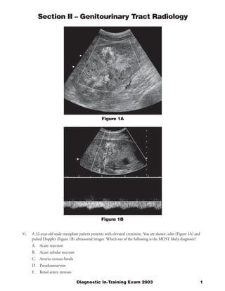
- 1. Section II – Genitourinary Tract Radiology Figure 1A Figure 1B 51. A 32-year-old male transplant patient presents with elevated creatinine. You are shown color (Figure 1A) and pulsed Doppler (Figure 1B) ultrasound images. Which one of the following is the MOST likely diagnosis? A. Acute rejection B. Acute tubular necrosis C. Arterio-venous fistula D. Pseudoaneurysm E. Renal artery stenosis Diagnostic In-Training Exam 2003 1
- 2. Section II – Genitourinary Tract Radiology Question #51 Findings: The Color Doppler image demonstrates a vascular cavity with inflow, outflow, and spectral broadening. On pulsed Doppler, this area has a low resistance arterial/venous waveform. Rationales: A) Incorrect. In normal transplant kidneys, the resistive index (RI=peak systolic velocity minus diastolic velocity/ peak systolic velocity) is usually 0.7 and below. In rejection, RI’s tend to be elevated, usually above 0.8. Elevated RI’s are not specific, and can be seen with acute tubular necrosis. In this case, the Doppler waveform demonstrates increased diastolic velocity, therefore, the RI is low. This would be unusual for rejection. B) Incorrect. See the rationale for 1A. The Doppler waveforms for rejection and ATN may be indistinguishable. C) Correct. AV fistulas are usually the result of graft biopsies, as was the situation in this case. At Doppler interrogation, they demonstrate increased velocity with low resistance, and spectral broadening. D) Incorrect. Graft pseudoaneurysms are also usually a result of biopsies. They are characterized by to-and-fro flow by color and pulsed Doppler. E) Incorrect. Stenosis of the graft anastomosis is characterized by a high velocity jet (>2.0 m/s in many cases) at the area of narrowing, and distal dampening. This is manifested as a tardus parvus waveform (prolonged acceleration time), and spectral broadening 2 American College of Radiology
- 3. Section II – Genitourinary Tract Radiology Figure 2 52. A 19-year-old woman is involved in a high speed motor vehicle accident. You are shown a contrast enhanced CT scan (Figure 2). Which one of the following is the MOST likely diagnosis? A. Renal contusions B. Contrast reaction C. Hypotension D. Renal arterial injuries E. Ureteral transections Diagnostic In-Training Exam 2003 3
- 4. Section II – Genitourinary Tract Radiology Question #52 Findings: Non-enhancement of both kidneys after blunt abdominal trauma with blood in the perirenal and pararenal spaces. Rationales: A) Incorrect. Renal contusions are characterized by small intrarenal hematomas and areas of decreased function due to edema and increased intrarenal pressure. In this case, both kidneys demonstrate a global lack of any function. B) Incorrect. Contrast reactions resulting in hypotension demonstrate a persistent nephrogram on delayed images. In this case, there is no appreciable uptake of contrast material into either kidney. C) Incorrect. This would appear similar to B D) Correct. This is a typical appearance for a bilateral renal arterial injury, most of which are caused by a tear in the intima of the renal artery with subsequent thrombosis. Most cases of renal arterial injury are unilateral. There is essentially no enhancement of either kidney after the administration of intravenous contrast material. In some cases, particularly when the diagnosis is delayed, rim enhancement of peripheral cortex from capsular collaterals can be seen. E) Incorrect. Ureteral transection can be diagnosed by CT when extravasation of contrast material from the ureter is seen on delayed images. 4 American College of Radiology
- 5. Section II – Genitourinary Tract Radiology Figure 3 53. You are shown a single image from an intravenous contrast-enhanced CT of the pelvis (Figure 3) on a 61- year-old man with a history of prostate cancer. Which one of the following is the MOST likely diagnosis? A. Uretero-vesical junction calculus B. Transitional cell carcinoma of the bladder C. Simple ureterocele D. Ectopic ureterocele E. Malakoplakia Diagnostic In-Training Exam 2003 5
- 6. Section II – Genitourinary Tract Radiology Question #53 Findings: This CT image with intravenous contrast material demonstrates a bulbous, contrast filled structure originating from the uretero-vesical junction and protruding into the bladder. Rationales: A) Incorrect. Ureteropelvic junction obstructions occur at the renal pelvis, proximal ureter junction, clearly more proximally than in this case. On contrast enhanced delayed CT scans, a dilated renal pelvis and renal collecting system is characteristic. Less contrast material would be expected to be seen in the ipsilateral ureter due to the proximal obstruction. B) Incorrect. Transitional cell carcinoma of the bladder typically appears as an irregular urothelial-based filling defect. In this case, the ureterocele is very smooth, and no filling defect is seen within the contrast filled ureterocele lumen. C) Correct. This is a typical appearance for a simple ureterocele. Important features of this diagnosis include a thin-walled contrast filled cavity, and a smooth ovoid appearance. D) Incorrect. Ectopic ureteroceles tend to occur in more inferior and medial locations than simple ureteroceles. By comparing the position of the ureterocele to the contralateral ureteral orifice, it is apparent that this ureterocele originates in the expected position as the uretero-vesical junction, and is thus most likely to be a simple ureterocele. E) Incorrect. Malakoplakia is an unusual condition of the urinary tract associated with urinary tract infection, and characterized by the presence of soft raised plaques, usually in the urinary bladder. These are indistinguishable from other causes of bladder masses, and a tissue diagnosis is usually necessary 6 American College of Radiology
- 7. Section II – Genitourinary Tract Radiology Figure 4 54. A 15-year-old African-American girl with sickle cell trait presents with gross hematuria (Figure 4). What is the MOST likely diagnosis? A. Lymphoma B. Medullary renal carcinoma C. Renal cell carcinoma D. Global renal infarct E. Acute pyelonephritis Diagnostic In-Training Exam 2003 7
- 8. Section II – Genitourinary Tract Radiology Question #54 Findings: This single contrast enhanced CT section shows a heterogeneous mass in the central right kidney causing enlargement of the kidney, but sparing the cortex. There is periaortic adenopathy, which displaces the inferior vena cava anteriorly. Rationales: A) Incorrect. Renal involvement by lymphoma is more often associated with non-Hodgkin’s lymphoma than with Hodgkin’s disease and it is commonly bilateral. Most patients are asymptomatic and the lesion is detected on a follow-up CT. The common CT appearance of renal lymphoma is that of multiple soft-tissue nodules. Other patterns include direct invasion from adjacent lymph nodes, a solitary mass, and nephromegaly due to diffuse parenchymal infiltration. Intrarenal lymphoma is hypo attenuating relative to the surrounding renal parenchyma and shows minimal enhancement. Secondary findings such as splenomegaly and widespread lymphadenopathy are common. Although lymphoma could be an explanation for the renal mass in this patient, the clinical history makes this not the best diagnosis.. B) Correct. Renal medullary carcinoma is usually found in your black patients with sickle cell trait. This is an aggressive neoplasm with a relentlessly progressive course. Spread, as evidenced by adenopathy in this case, is typical when the tumor is first diagnosed. The tumor arises in the region of the renal medulla and often expands centrally and enlarges the kidney. C) Incorrect. Renal cell carcinoma can certainly look like this, and if the clinical setting were in an elderly patient, would be a likely diagnosis, as would transitional cell carcinoma extending from the collecting system into the renal parenchyma. D) Incorrect. In acute global infarction, the kidney is of normal size and lacks contrast enhancement. The unenhanced parenchyma usually appears homogeneous. A faint rim of cortical (i.e., a cortical rim sign) due to perfusion by capsular collateral vessels may be seen. Although the peripheral enhancement in this case could be seen in global infarction, the renal enlargement argues against the diagnosis. E) Incorrect. Acute pyelonephritis, if fulminant, could conceivably be this diffuse, though it would be extremely unusual to see this degree of adenopathy. Again, the presenting symptoms do not support the diagnosis of infection. 8 American College of Radiology
- 9. Section II – Genitourinary Tract Radiology Figure 5 55. A 60-year-old man presented with suspect aortic injury but a normal aortic arch arteriogram. You are shown an unenhanced CT scan obtained 48 hours later (Figure 5). What is the MOST likely diagnosis? A. Renal vein thrombosis B. Arterial occlusion C. Pyelonephritis D. Renal neoplasm E. Acute tubular necrosis Diagnostic In-Training Exam 2003 9
- 10. Section II – Genitourinary Tract Radiology Question #55 Findings: CT scan is performed without IV contrast and thus, there is no contrast in aorta and hepatic vessels. However, contrast enhancement is demonstrated within kidneys, which has pattern of corticomedullary phase enhancement. Although this appearance can normally be seen when scanning within 70 seconds of start of the contrast bolus, it would not be expected to persist 48 hours after contrast administration. The CT findings are therefore consistent with bilateral renal failure. Rationales: A) Incorrect. CT findings of renal vein thrombosis include renal enlargement (usually unilateral), a prolonged corticomedullary phase of enhancement, and impaired contrast excretion because of edema resulting from the obstructed venous drainage. Renal vein thrombosis could theoretically result in the pattern seen in this patient but bilateral disease is extremely rare. A more likely cause of bilateral renal failure with a corticomedullary pattern of enhancement is acute tubular necrosis. B) Incorrect. Acute arterial occlusion may be a global or segmental event. In acute global infarction, the kidney is of normal size and lacks contrast enhancement, although a faint rim of cortical enhancement (i.e., a cortical rim sign) may be seen because of perfusion by capsular collateral vessels. Segmental infarction appears as a peripheral, wedge-shaped, or triangular area of diminished enhancement. Bilateral arterial occlusion would not result in corticomedullary enhancement. C) Incorrect. Acute pyelonephritis is a bacterial infection of the kidney. Typically, it has a patchy distribution, but in severe cases, the entire kidney can be involved. Contrast-enhanced CT scans usually demonstrate ill-defined, wedge-shaped zones of diminished attenuation. Contrast enhancement may be seen in areas of inflammation on scans obtained several hours after contrast administration, but enhancing parenchyma would not be expected 48 hours after contrast administration. In addition, pyelonephritis would be an unlikely cause of symmetric bilateral renal failure. D) Incorrect. Renal neoplasm would be an unlikely cause of symmetric bilateral renal failure. E) Correct. Acute tubular necrosis refers to a nephrotoxic or ischemic injury to the renal tubules accompanied by clinical manifestations of acute renal failure. It is the most common cause of acute renal failure and often results in a persistent corticomedullary phase of renal enhancement sometime lasting for days. Acute tubular necrosis is the best answer. 10 American College of Radiology