SlideShare ist ein Scribd-Unternehmen logo
1 von 10
Downloaden Sie, um offline zu lesen
Section III – Thoracic Radiology (Cardiac)


                                                  Figure 1A


                                                  Figure 1B

96.   You are shown a frontal chest radiograph (Figure 1A) and an axial 2-D time-of-flight MRI (Figure 1B) of a
      35-year-old patient with pulmonary arterial hypertension and cyanosis. The patient had surgery as a child.
      Which one of the following lesions was MOST likely repaired?
      A. Transposition of the great arteries
      B.   Patent ductus arteriosus
      C. Tetralogy of Fallot
      D. Aorto-pulmonary window
      E.   Truncus arteriosus

                                      Diagnostic In-Training Exam 2002                                             1
Section III – Thoracic Radiology (Cardiac)
Question #96
Findings: The radiograph demonstrates enlarged central pulmonary arteries compatible with pulmonary arterial
hypertension. The central pulmonary arteries also have calcified walls. In addition, there is deformity of the left
upper ribs compatible with a prior thoracotomy. The MR confirms the presence of enlarged central pulmonary
arteries and decreased signal in the central branches suggesting slow flow from elevated resistance. There is also an
anastomosis between the left pulmonary artery and the descending aorta.

Rationales:
A) Incorrect. Repair of D-transposition of the Great Arteries usually takes place through a median sternotomy not
   a posterior thoracotomy as this case. Transposition repair can be done by an atrial switch operation (Mustard,
   Senning) in which the systemic venous and pulmonary venous blood are re-routed appropriately, and more
   recently by the arterial switch or Jatene operation in which the great arteries themselves are relocated to their
   respective ventricles. The surgical connection between the descending aorta and pulmonary artery in the test
   case cannot be explained on the basis of any of these operations, therefore excluding transposition as an option.
B) Incorrect. Repair of a patent ductus arteriosus requires ligation of the ductus. This is done through a left
   thoracotomy. An anastomosis between the pulmonary artery and aorta is not required. Moreover, pulmonary
   hypertension is not expected after surgical repair. Thus, although the rib changes can be seen after PDA repair,
   the pulmonary artery to aorta conduit is not consistent with this type of repair.
C) Correct. Surgical palliation of TOF usually requires anastomosis between the obstructed right heart and
   systemic circulation before definitive repair is performed. The Potts-Smith operation was performed in this
   patient. In this palliative operation, the descending aorta and left pulmonary artery are anastomosed in a side-
   by-side fashion through a thoracotomy. Unfortunately, a common complication of this procedure is the
   development of pulmonary arterial hypertension because of increased blood flow to the lungs if the size of the
   anastomosis is not carefully constructed. The findings of pulmonary hypertension, a left thoracotomy and the
   surgical anastomosis between the systemic and pulmonary circulations support the diagnosis of tetralogy of
   Fallot. More recently, definitive repair of tetralogy of Fallot is performed early in life when feasible, thus
   obviating temporizing palliating procedures. The Potts-Smith operation has essentially been abandoned.
D) Incorrect. An aorto-pulmonary window is a congenital defect in which there is a broad-based connection
   between the proximal aorta and the pulmonary artery. This condition can result in pulmonary artery
   hypertension if surgical repair is not done. However, the increased pulmonary flow characteristic of this
   anomaly prior to surgery usually regresses completely or nearly so after surgery. Repair requires closing the
   abnormal communication generally through an sternal splitting anterior approach. In the test patient, the left
   posterior thoracotomy as well as the anastomosis between the descending aorta and pulmonary artery argue
   against the diagnosis of aorto-pulmonary window.
E) Incorrect. Prior to surgical repair, the shunt in truncus arteriosus is at the level of the ascending aorta and
   pulmonary artery. Repair requires separation of the pulmonary arteries from the aortic segment and then
   reconnection to the right ventricle, usually with an aortic homograft conduit. The conduit may eventually
   calcify. Normal vascularity is expected after surgery. In the test patient, the pulmonary hypertension and the
   shunt at the level of the descending, rather than the ascending, aorta make truncus arteriosus untenable. In
   addition, surgical repair of truncus is performed through a sternal splitting anterior approach, not through a
   thoracotomy.

Citations:
Potts WJ, Smith S, Gibson S. Anastomosis of the aorta to a pulmonary artery. JAMA 132:627, 1946
Piccoli GP, Dickinson DF, Musumeci F, Hamilton DI. A changing policy for the surgical treatment of tetralogy of Fallot: Early and
late results in 235 consecutive patients. Ann Thorac Surg 33:365, 1982
RL Soulen, RM Donner, and M Capitanio. Postoperative evaluation of complex congenital heart disease by magnetic resonance
imaging. RadioGraphics 1987 7: 975-1000


2                                           American College of Radiology
Section III – Thoracic Radiology (Cardiac)


                                                  Figure 2A


                                                  Figure 2B

97.   You are shown a plain radiograph (Figure 2A) and a right ventriculogram (Figure 2B) of a 2-day-old infant
      with tachypnea and cyanosis. Which one of the following is the MOST likely diagnosis?
      A. Tetralogy of Fallot
      B.   Truncus arteriosus
      C. Pulmonary atresia and intact ventricular septum
      D. Ebstein’s anomaly of tricuspid valve
      E.   D-transpostion of the great vessels


                                   Diagnostic In-Training Exam 2002                                               3
Section III – Thoracic Radiology (Cardiac)
Question #97
Findings: The chest radiograph demonstrates decreased pulmonary vascularity with a normal heart size and slight
elevation of the cardiac apex. The location of the aortic arch is not readily visible on this radiograph. The right
ventriculogram reveals opacification of the aorta, indicating a ventricular septal defect shunting right-to-left and
hypoplasia of the right ventricular outflow tract. Other findings include competent tricuspid and mitral valves. The
findings are consistent with Tetralogy of Fallot.

Rationales:
A. Correct. Tetralogy of Fallot is the most common cardiac cause of neonatal cyanosis. Anatomic features of
   Tetralogy of Fallot include a VSD, pulmonic stenosis, overriding aorta, and right ventricular hypertrophy. If
   there is severe obstruction of the right ventricular outflow tract, right to left shunting occurs across the VSD.
   Infants with severe obstruction usually present soon after birth with cyanosis and dyspnea. The infundibular
   stenosis results in decreased pulmonary vascularity on chest radiographs. The cardiac size is normal with an
   uplifted apex as a result of the right ventricular hypertrophy. The right heart obstruction, concomitant
   hypoplasia of the right ventricular outflow tract and atretic pulmonary valve in this patient is characteristic of
   severe tetralogy of Fallot also known as pseudotruncus arteriosus.
B. Incorrect. Truncus arteriosus is a cyanotic cardiac anomaly characterized by a single arterial trunk arising from
   the heart and giving origin to the pulmonary arteries and ascending aorta. There is a single truncal valve which
   may have 3 to 6 semilunar leaflets. Invariably, there is a large VSD below the valve leaflets. Pulmonary
   vascularity is markedly increased because blood flows from the aorta to the pulmonary circulation. The cardiac
   size is enlarged. The decreased pulmonary vascularity, normal heart size and 2 separate semilunar valves
   demonstrated on the ventriculogram essentially exclude the diagnosis of truncus.
C. Incorrect. In patients with pulmonary atresia and intact ventricular septum (also referred to as trilogy of Fallot)
   there is no communication between the pulmonary outflow tract and the right ventricle. The right ventricle is
   usually hypoplastic. There is also obligatory right to left shunting across an atrial septal defect. The absence of
   antegrade flow through the pulmonary valve results in diminished pulmonary vascularity on chest radiography.
   The heart is usually normal size in the immediate postnatal period. The chest radiographic findings in the test
   patient are compatible with pulmonary atresia and intact ventricular septum. However, the presence of a
   ventricular septal defect on the right ventriculogram excludes the diagnosis.
D. Incorrect. Patients with Ebstein’s anomaly have a redundant tricuspid valve which is adherent to the right
   ventricular wall distal to the annulus of the tricuspid valve. This results in tricuspid regurgitation and
   subsequent right atrial enlargement. The right ventricle is decreased in size and is slow to empty, leading to
   decreased blood flow into the pulmonary arteries and right to left shunting across a patent foramen ovale or
   atrial septal defect. Hence, affected infants are usually cyanotic. The combination of decreased vascularity, a
   normal heart size and a competent tricuspid valve on the ventriculogram in the test patient makes Ebstein
   anomaly an unlikely diagnosis.
E. Incorrect. D transposition of the great vessels (TGV) occurs when the aorta and main pulmonary artery arise
   from the morphologic right ventricle and morphologic left ventricle, respectively, creating two circulations in
   parallel. Desaturated venous blood flows from the right atrium into the right ventricle and then into the aorta,
   resulting in cyanosis. Radiographic findings include a narrow mediastinum, mild cardiomegaly and prominent
   pulmonary vascularity. The normal heart size and decreased vascularity in the test case would be unusual in a
   patient with d-TGV.

Citations:
Van Praagh R, Van Praagh S, Nebesar RA et al: Tetralogy of Fallot. Underdevelopment of the pulmonary infundibulum and its
sequelae. Am J Cardiol 26:25-33, 1970
Adams FH, Emmanoulides GC, Riemenschneider TA. In Heart Disease in Infants, Children, and Adolescents. Fourth Edition
Williams, Wilkins Baltimore 1989


4                                         American College of Radiology
Section III – Thoracic Radiology (Cardiac)


                                                  Figure 3A


                                                   Figure 3B

98.   This 63-year-old patient presented with chest pain. You are shown two levels (Figure 3A and Figure 3B) of a
      contrast enhanced CT scan. Which one of the following is the MOST likely diagnosis?
      A. Arrhythmogenic right ventricular dysplasia
      B.   Congenital partial absence of the pericardium
      C. Left ventricular aneurysm
      D. Pericardial cyst
      E.   Sinus of Valsalva aneurysm

                                  Diagnostic In-Training Exam 2002                                                  5
Section III – Thoracic Radiology (Cardiac)
Question #98
Findings: The CT scan demonstrates a large mass that fills part of the anterior mediastinum in front of the
pulmonary outflow tract (A). This mass contains calcification. Image (B) obtained at a lower level shows that the
mass is in continuity with the left ventricle. In addition, the left ventricular wall is thin in the anteroseptal portion
with subendocardial mural thrombus formation and peripheral calcification. All of these findings in combination
are diagnostic of a large left ventricular aneurysm that extends superiorly into the anterior mediastinum

Rationales:
A. Incorrect. Arrhythmogenic right ventricular dysplasia is a condition in which fibrofatty replacement of the
   ventricular wall can lead to outpouchings in the wall of the right ventricle. Patients have frequent ventricular
   arrhythmias, and sudden death has been described. A large calcified mass as seen in this case would be atypical.
   In addition, the mass in the test patient arises from the left ventricle and the right ventricle appears normal,
   thus excluding such a diagnosis.
B. Incorrect. Congenital absence of the pericardium can be total or partial. The partial form is the most common,
   usually involving the portion of the pericardium along the left heart border near the left atrial appendage.
   Patients can present clinically with chest pain, and strangulation of the left atrial appendage has been described.
   CT shows a protrusion of the left ventricle through the area of absent pericardium. The thickness of the
   ventricular wall is normal. The findings in the test patient of a thinned ventricular wall and a calcified mass in
   continuity with the anterior left ventricular wall excludes absence of the pericardium as a diagnostic alternative.
C. Correct. There is thinning of the left ventricular wall over its entire anteroseptal aspect. Mural thrombus is
   seen on the inside of the thin ventricular wall. In addition, a large aneurysm has formed which has extended
   superiorly in front of the right ventricular outflow tract. This aneurysm has partly calcified over time. The
   findings are diagnostic of a left ventricular aneurysm.
D. Incorrect. Pericardial cyst is a congenital entity related to the celomic cavity, in which a fluid containing
   structure forms adjacent to and not in communication with the pericardial cavity. Most are located in the right
   cardiophrenic angle. Heart chambers are not involved in the condition. The typical CT findings of pericardial
   cyst are those of a water attenuation mass adjacent to the cardiac border. A soft tissue attenuation mass with
   areas of calcification and the left ventricular involvement shown in the test case exclude a pericardial cyst.
E. Incorrect. Sinus of Valsalva aneurysm results from a deficiency between the aortic media and fibrous annulus
   of the aortic valve resulting in distension and eventual aneurysm formation. If the aneurysm becomes large
   enough, it can rupture into the right atrial or right ventricular cavity. The test case demonstrates predominantly
   a left ventricular process excluding this diagnosis of sinus of Valsalva aneurysm.

Citations:
Adams FH, Emmanoulides GC, Riemenschneider TA. In Heart Disease in Infants, Children, and Adolescents. Fourth Edition
Williams, Wilkins Baltimore 1989
Dahnert Wolfgang. Radiology Review Manual. Lippincott Williams and Wilkins 4th Edition 1999


6                                        American College of Radiology
Section III – Thoracic Radiology (Cardiac)


                                                  Figure 4A


                                                  Figure 4B

99.   An afebrile 3-week-old infant is referred because of tachypnea. A chest radiograph (Figure 4A) was obtained.
      This was followed by an electrocardiogram, which was abnormal, as well as an aortogram (Figure 4B). Which
      one of the following is the MOST likely diagnosis?
      A. Hypoplastic left heart syndrome
      B.   Bland-White-Garland syndrome
      C. Kawasaki arteritis
      D. Single coronary artery
      E.   Aortic insufficiency

                                  Diagnostic In-Training Exam 2002                                               7
Section III – Thoracic Radiology (Cardiac)
Question #99
Findings: The frontal chest radiograph shows cardiomegaly, normal pulmonary vascularity and retrocardiac
atelectasis. A left sided obstructive lesion should be suspected. Aortogram shows a normal aortic valve and
ascending aorta. However, the left coronary artery fills in a retrograde fashion by collaterals from the right coronary
artery. No connection is seen between the proximal left coronary system and the aorta. On the other hand, it
appears that the proximal left coronary artery drains to the region of the main pulmonary artery. Also note that
the left anterior descending and circumflex branches come together as a common trunk.

Rationales:
A. Incorrect. The term hypoplastic left heart syndrome refers to a spectrum of anomalies characterized by a poorly
   developed left ventricle and ascending aorta. There is often underdevelopment of the mitral valve and left
   atrium. The resultant pathology is that there is no forward flow of blood from the left ventricle and so
   congestive heart failure is common soon after birth. Chest radiographs usually demonstrate cardiomegaly,
   increased vascularity and pulmonary edema. The normal pulmonary vascularity in the test radiographs and
   normal size aortic valve and ascending aorta on the aortogram make hypoplastic left heart unlikely.
B. Correct. Bland-White –Garland Syndrome is a rare condition in which one of the coronary arteries (usually
   the left) originates from the pulmonary artery. Shortly after birth as the pulmonary artery resistance normally
   decreases, the myocardial perfusion from the anomalous coronary is compromised, resulting in ischemia and
   transmural infarction Patients present with congestive heart failure as a result of the ischemic insult. ECG
   usually suggests the diagnosis.
C. Incorrect. Kawasaki arteritis is an acute febrile vasculitis of childhood. The illness occurs in young children,
   approximately 80% are under 5 years of age. Rarely, it affects infants or adolescents. Kawasaki disease causes
   a severe vasculitis of all blood vessels, with predilection for the coronary arteries. Acutely, patients present with
   high spiking fevers. Cardiomegaly and normal vascularity can be seen on chest radiographs. Aneurysms of the
   coronary arteries are common, but anomalous coronary origin is not a feature of this disease. The age of the
   test patient and the abnormal coronary artery anatomy makes Kawasaki disease unlikely.
D. Incorrect. Rarely, infants are born with a single coronary artery. Most often, there is a single left branch which
   courses between the aorta and pulmonary artery . The anomalous artery can be “pinched” between the two
   vessels, resulting in ischemia and sudden death. In the test patient, there are two coronary arteries, excluding
   the diagnosis of a single artery.
E. Incorrect. In the test patient, the left anterior descending (LAD) branch of the coronary artery and the
   circumflex branch join together as a common trunk, thus excluding the diagnosis of an anomalous origin.
   Anomalous origin of the LAD is particularly common in patients with tetralogy of Fallot, occurring in 5 to
   9% of this population This anatomic variant can have important surgical implications at total repair.

Citation:
Choh JH, Levinsky L, Srinivasan V, Idbeis B, Subramanian S. Anomalous origin of the left coronary artery from the pulmonary
trunk: Its clinical spectrum and current surgical management. Thorac and Cardiovasc Surg 28:239-242, 1980.


8                                         American College of Radiology
Section III – Thoracic Radiology (Cardiac)


                                               Figure 5A


                                                 Figure 5B

100. You are shown an axial T1-weighted turbo spin-echo (Figure 5A) and coronal gradient echo image (Figure 5B)
     of a 13-year-old girl with a diastolic murmur in the left precordial space. Which one of the following is the
     MOST likely diagnosis?
      A. Rheumatic aortic valvular disease
      B.   Bicuspid aortic valve
      C. Aortic dissection
      D. Williams syndrome
      E.   Takayasu’s arteritis

                                   Diagnostic In-Training Exam 2002                                             9
Section III – Thoracic Radiology (Cardiac)
Question #100 (question was not scored in the exam)
Both sequences demonstrate an enlarged aortic root that measures approximately 4.5 cm in diameter. In addition,
there is thickening of the walls of the ascending aorta and pulmonary arteries. The gradient echo image
demonstrates a mild degree of aortic valvular insufficiency. The findings are consistent with an arteritis, such as
Takayasu’s arteritis.

Rationales:
A. Incorrect. Rheumatic fever is an inflammatory condition that affects primarily the heart. The inflammatory
   process can involve the heart valves producing scarring that can lead to stenosis as well as insufficiency. The
   walls of the great vessels are not affected by the condition, thus excluding the diagnosis in the test case.
B. Incorrect. Bicuspid aortic valve is a common congenital defect affecting the aortic valve, occurring in 1-2% of
   the population. Due to hemodynamic alteration of the flow through the valve, fibrosis and eventual
   calcification ensue producing stenosis. Although the enlargement of the aortic root from post-stenotic dilatation
   as well as the mild degree of valvular regurgitation could be explained on the basis of a bicuspid aortic valve, the
   thickening of the aortic and pulmonary arteries excludes the diagnosis.
C. Incorrect. Aortic dissection is associated with an intramural hematoma which produces widening of the aorta.
   True wall thickening does not occur. A late sequela can be aortic insufficiency. Although the finding of aortic
   valvular insufficiency in the test case is consistent with dissection, the presence of wall thickening is atypical for
   dissection. Moreover, the patient’s age is unusual for this diagnosis.
D. Incorrect. Williams syndrome is an autosomal dominant condition that is characterized by aortitis, mental
   retardation and elfin-like facies. Although thickening of the wall of the ascending aorta and pulmonary artery
   can be a feature of the disease, there is usually narrowing of the lumen of the ascending aorta causing
   supravalvular stenosis that can be diffuse at times. The aortic root dilatation seen in the test case virtually
   excludes Williams syndrome.
E. Correct. Takayasu arteritis is a granulomatous inflammation of the aorta, its branches and the pulmonary
   artery. It usually affects persons younger than 50 years of age. An acute stage that is characterized by a
   granulomatous infiltrate of elastic fibers of media of arterial walls is followed by a chronic fibrotic period that
   eventually leads to fibrosis of the media and adventitia. Takayasu arteritis is divided into several types. Type IV
   is the so called dilated type, which affects aorta and its branches as well as the pulmonary arteries.

Citations:
Danhert Wolfgang in Radiology Review Manual 4th Edition Lippincott-Williams Wilkins 1999


10                                       American College of Radiology

Weitere ähnliche Inhalte

Was ist angesagt?

Aortic Disssection. Dr Nikrish Hegde.
Aortic Disssection. Dr Nikrish Hegde.Aortic Disssection. Dr Nikrish Hegde.
Aortic Disssection. Dr Nikrish Hegde.
DrNikrish Hegde
 
Ct pulmonary angiogram
Ct pulmonary angiogramCt pulmonary angiogram
Ct pulmonary angiogram
muhammed Yasar
 

Was ist angesagt? (20)

Imaging of aortic pathologies
Imaging of aortic pathologies Imaging of aortic pathologies
Imaging of aortic pathologies
 
Spectrum Of Ct Findings In Rupture And Impendinging Rupture Of AAA
Spectrum Of Ct Findings In Rupture And Impendinging Rupture Of AAASpectrum Of Ct Findings In Rupture And Impendinging Rupture Of AAA
Spectrum Of Ct Findings In Rupture And Impendinging Rupture Of AAA
 
Aortic aneurysms and dissection 2016
Aortic aneurysms and dissection 2016Aortic aneurysms and dissection 2016
Aortic aneurysms and dissection 2016
 
Normal variants of heart structures
Normal variants of heart structuresNormal variants of heart structures
Normal variants of heart structures
 
Presentation1, x ray film reading of the chest.
Presentation1, x ray film reading of the chest.Presentation1, x ray film reading of the chest.
Presentation1, x ray film reading of the chest.
 
Aortic Disssection. Dr Nikrish Hegde.
Aortic Disssection. Dr Nikrish Hegde.Aortic Disssection. Dr Nikrish Hegde.
Aortic Disssection. Dr Nikrish Hegde.
 
Image of Thoracic Aortic Disease
Image of Thoracic Aortic DiseaseImage of Thoracic Aortic Disease
Image of Thoracic Aortic Disease
 
Full story pulomnary embolism imaging Dr Ahmed Esawy
Full story pulomnary embolism imaging Dr Ahmed EsawyFull story pulomnary embolism imaging Dr Ahmed Esawy
Full story pulomnary embolism imaging Dr Ahmed Esawy
 
Aortic dissection 01
Aortic dissection 01Aortic dissection 01
Aortic dissection 01
 
Aortic aneurysm imaging
Aortic aneurysm imagingAortic aneurysm imaging
Aortic aneurysm imaging
 
Aortic aneurysm
Aortic aneurysmAortic aneurysm
Aortic aneurysm
 
Ct pulmonary angiogram
Ct pulmonary angiogramCt pulmonary angiogram
Ct pulmonary angiogram
 
Aortic aneurysm imaging
Aortic aneurysm imagingAortic aneurysm imaging
Aortic aneurysm imaging
 
23204949
2320494923204949
23204949
 
Pulmonary embolism imaging
Pulmonary embolism imagingPulmonary embolism imaging
Pulmonary embolism imaging
 
Diseases of the thoracic aorta
Diseases of the thoracic aortaDiseases of the thoracic aorta
Diseases of the thoracic aorta
 
Acute Aortic Syndrome
Acute Aortic SyndromeAcute Aortic Syndrome
Acute Aortic Syndrome
 
23204992
2320499223204992
23204992
 
Aortitis ppt
Aortitis pptAortitis ppt
Aortitis ppt
 
Transposition of the great arteries
Transposition of the great arteriesTransposition of the great arteries
Transposition of the great arteries
 

Andere mochten auch

What is I2 Final-Approved
What is I2 Final-ApprovedWhat is I2 Final-Approved
What is I2 Final-Approved
Duane Blackburn
 
sketch
sketchsketch
sketch
blitzz
 
Delegation & Empowerment
Delegation & EmpowermentDelegation & Empowerment
Delegation & Empowerment
kunna
 
DiffCalculus: August 27, 2012
DiffCalculus: August 27, 2012DiffCalculus: August 27, 2012
DiffCalculus: August 27, 2012
Carlos Vázquez
 
Fuengirola.ayer.hoy
Fuengirola.ayer.hoyFuengirola.ayer.hoy
Fuengirola.ayer.hoy
iesboliches
 

Andere mochten auch (20)

Baie Des Cochons
Baie Des CochonsBaie Des Cochons
Baie Des Cochons
 
What is I2 Final-Approved
What is I2 Final-ApprovedWhat is I2 Final-Approved
What is I2 Final-Approved
 
Maria Seredyszyn Hames , School And Students
Maria Seredyszyn Hames , School And StudentsMaria Seredyszyn Hames , School And Students
Maria Seredyszyn Hames , School And Students
 
Koncernpresentation
KoncernpresentationKoncernpresentation
Koncernpresentation
 
Urcus Spre Manastire
Urcus Spre ManastireUrcus Spre Manastire
Urcus Spre Manastire
 
Llistat llibres
Llistat llibresLlistat llibres
Llistat llibres
 
Diplomas 2009 - 2010
Diplomas 2009 - 2010Diplomas 2009 - 2010
Diplomas 2009 - 2010
 
Chipul Lui Iisus In Pictura Lui Ocampo
Chipul Lui Iisus  In Pictura Lui OcampoChipul Lui Iisus  In Pictura Lui Ocampo
Chipul Lui Iisus In Pictura Lui Ocampo
 
Wsi Franchise Webinar Presentation
Wsi Franchise Webinar PresentationWsi Franchise Webinar Presentation
Wsi Franchise Webinar Presentation
 
Ab430trainingoverview
Ab430trainingoverviewAb430trainingoverview
Ab430trainingoverview
 
sketch
sketchsketch
sketch
 
Pursutpp
PursutppPursutpp
Pursutpp
 
Året som gick
Året som gickÅret som gick
Året som gick
 
Delegation & Empowerment
Delegation & EmpowermentDelegation & Empowerment
Delegation & Empowerment
 
DiffCalculus: August 27, 2012
DiffCalculus: August 27, 2012DiffCalculus: August 27, 2012
DiffCalculus: August 27, 2012
 
Tugas
TugasTugas
Tugas
 
Banking CH 17
Banking CH 17Banking CH 17
Banking CH 17
 
Fuengirola.ayer.hoy
Fuengirola.ayer.hoyFuengirola.ayer.hoy
Fuengirola.ayer.hoy
 
KMap
KMapKMap
KMap
 
The National Biometrics Challenge (2011)
The National Biometrics Challenge (2011)The National Biometrics Challenge (2011)
The National Biometrics Challenge (2011)
 

Ähnlich wie 23204931

Faisal al atawi study2
Faisal al atawi study2Faisal al atawi study2
Faisal al atawi study2
alatawi2
 
Pulmonary Arteriovenous Malformation Presenting As An Enlarging Lung Mass And...
Pulmonary Arteriovenous Malformation Presenting As An Enlarging Lung Mass And...Pulmonary Arteriovenous Malformation Presenting As An Enlarging Lung Mass And...
Pulmonary Arteriovenous Malformation Presenting As An Enlarging Lung Mass And...
Bassel Ericsoussi, MD
 
Cc no adulto II
Cc no adulto IICc no adulto II
Cc no adulto II
gisa_legal
 
ICM case based discussions
ICM case based discussionsICM case based discussions
ICM case based discussions
pbsherren
 
Complete transposition of the great arteries (D-TGA.pptx
Complete transposition of the great arteries (D-TGA.pptxComplete transposition of the great arteries (D-TGA.pptx
Complete transposition of the great arteries (D-TGA.pptx
DrPNatarajan2
 
Seminar congenital cardiac disorders (pda,TA and AP Window)
Seminar congenital cardiac disorders (pda,TA and AP Window)Seminar congenital cardiac disorders (pda,TA and AP Window)
Seminar congenital cardiac disorders (pda,TA and AP Window)
Uma Binoy
 
A rara associação de drenagem anômala total de veias pulmonares e cor triatri...
A rara associação de drenagem anômala total de veias pulmonares e cor triatri...A rara associação de drenagem anômala total de veias pulmonares e cor triatri...
A rara associação de drenagem anômala total de veias pulmonares e cor triatri...
gisa_legal
 
surgery.Congenital heart disease.(dr.aram)
surgery.Congenital heart disease.(dr.aram)surgery.Congenital heart disease.(dr.aram)
surgery.Congenital heart disease.(dr.aram)
student
 

Ähnlich wie 23204931 (20)

Faisal al atawi study2
Faisal al atawi study2Faisal al atawi study2
Faisal al atawi study2
 
Dysplastic tricuspid valve may 2021
Dysplastic tricuspid valve  may 2021Dysplastic tricuspid valve  may 2021
Dysplastic tricuspid valve may 2021
 
EHJ-CR-Slide-Set (1).pptx
EHJ-CR-Slide-Set (1).pptxEHJ-CR-Slide-Set (1).pptx
EHJ-CR-Slide-Set (1).pptx
 
Pulmonary Arteriovenous Malformation Presenting As An Enlarging Lung Mass And...
Pulmonary Arteriovenous Malformation Presenting As An Enlarging Lung Mass And...Pulmonary Arteriovenous Malformation Presenting As An Enlarging Lung Mass And...
Pulmonary Arteriovenous Malformation Presenting As An Enlarging Lung Mass And...
 
Coarctation of aorta
Coarctation of aortaCoarctation of aorta
Coarctation of aorta
 
Cc no adulto II
Cc no adulto IICc no adulto II
Cc no adulto II
 
Mitral valve disease
Mitral valve disease Mitral valve disease
Mitral valve disease
 
Tricuspid pulmonary valves
Tricuspid  pulmonary valvesTricuspid  pulmonary valves
Tricuspid pulmonary valves
 
ICM case based discussions
ICM case based discussionsICM case based discussions
ICM case based discussions
 
Loops Around the Heart – A Giant Snakelike Right Coronary Artery Ectasia with...
Loops Around the Heart – A Giant Snakelike Right Coronary Artery Ectasia with...Loops Around the Heart – A Giant Snakelike Right Coronary Artery Ectasia with...
Loops Around the Heart – A Giant Snakelike Right Coronary Artery Ectasia with...
 
Cyanotic heart diseases
Cyanotic heart diseasesCyanotic heart diseases
Cyanotic heart diseases
 
Pediatric Cardiology (1).pptx
Pediatric Cardiology (1).pptxPediatric Cardiology (1).pptx
Pediatric Cardiology (1).pptx
 
Congenital heart diseases
Congenital heart diseasesCongenital heart diseases
Congenital heart diseases
 
Complete transposition of the great arteries (D-TGA.pptx
Complete transposition of the great arteries (D-TGA.pptxComplete transposition of the great arteries (D-TGA.pptx
Complete transposition of the great arteries (D-TGA.pptx
 
Coarctation of aorta
Coarctation of aortaCoarctation of aorta
Coarctation of aorta
 
Seminar congenital cardiac disorders (pda,TA and AP Window)
Seminar congenital cardiac disorders (pda,TA and AP Window)Seminar congenital cardiac disorders (pda,TA and AP Window)
Seminar congenital cardiac disorders (pda,TA and AP Window)
 
A rara associação de drenagem anômala total de veias pulmonares e cor triatri...
A rara associação de drenagem anômala total de veias pulmonares e cor triatri...A rara associação de drenagem anômala total de veias pulmonares e cor triatri...
A rara associação de drenagem anômala total de veias pulmonares e cor triatri...
 
23204989
2320498923204989
23204989
 
Emergency cadiopulmonary imaging
Emergency cadiopulmonary imagingEmergency cadiopulmonary imaging
Emergency cadiopulmonary imaging
 
surgery.Congenital heart disease.(dr.aram)
surgery.Congenital heart disease.(dr.aram)surgery.Congenital heart disease.(dr.aram)
surgery.Congenital heart disease.(dr.aram)
 

Mehr von radgirl (20)

23205065
2320506523205065
23205065
 
23205062
2320506223205062
23205062
 
23205059
2320505923205059
23205059
 
23205056
2320505623205056
23205056
 
23205042
2320504223205042
23205042
 
23205052
2320505223205052
23205052
 
23205045
2320504523205045
23205045
 
23205048
2320504823205048
23205048
 
23205039
2320503923205039
23205039
 
23205036
2320503623205036
23205036
 
23205032
2320503223205032
23205032
 
23205029
2320502923205029
23205029
 
23205025
2320502523205025
23205025
 
23205022
2320502223205022
23205022
 
23205019
2320501923205019
23205019
 
23205016
2320501623205016
23205016
 
23205011
2320501123205011
23205011
 
23204998
2320499823204998
23204998
 
23205008
2320500823205008
23205008
 
23205004
2320500423205004
23205004
 

Kürzlich hochgeladen

🌹Attapur⬅️ Vip Call Girls Hyderabad 📱9352852248 Book Well Trand Call Girls In...
🌹Attapur⬅️ Vip Call Girls Hyderabad 📱9352852248 Book Well Trand Call Girls In...🌹Attapur⬅️ Vip Call Girls Hyderabad 📱9352852248 Book Well Trand Call Girls In...
🌹Attapur⬅️ Vip Call Girls Hyderabad 📱9352852248 Book Well Trand Call Girls In...
Call Girls In Delhi Whatsup 9873940964 Enjoy Unlimited Pleasure
 

Kürzlich hochgeladen (20)

Saket * Call Girls in Delhi - Phone 9711199012 Escorts Service at 6k to 50k a...
Saket * Call Girls in Delhi - Phone 9711199012 Escorts Service at 6k to 50k a...Saket * Call Girls in Delhi - Phone 9711199012 Escorts Service at 6k to 50k a...
Saket * Call Girls in Delhi - Phone 9711199012 Escorts Service at 6k to 50k a...
 
Night 7k to 12k Navi Mumbai Call Girl Photo 👉 BOOK NOW 9833363713 👈 ♀️ night ...
Night 7k to 12k Navi Mumbai Call Girl Photo 👉 BOOK NOW 9833363713 👈 ♀️ night ...Night 7k to 12k Navi Mumbai Call Girl Photo 👉 BOOK NOW 9833363713 👈 ♀️ night ...
Night 7k to 12k Navi Mumbai Call Girl Photo 👉 BOOK NOW 9833363713 👈 ♀️ night ...
 
Best Rate (Patna ) Call Girls Patna ⟟ 8617370543 ⟟ High Class Call Girl In 5 ...
Best Rate (Patna ) Call Girls Patna ⟟ 8617370543 ⟟ High Class Call Girl In 5 ...Best Rate (Patna ) Call Girls Patna ⟟ 8617370543 ⟟ High Class Call Girl In 5 ...
Best Rate (Patna ) Call Girls Patna ⟟ 8617370543 ⟟ High Class Call Girl In 5 ...
 
Most Beautiful Call Girl in Bangalore Contact on Whatsapp
Most Beautiful Call Girl in Bangalore Contact on WhatsappMost Beautiful Call Girl in Bangalore Contact on Whatsapp
Most Beautiful Call Girl in Bangalore Contact on Whatsapp
 
Call Girls Service Jaipur {9521753030} ❤️VVIP RIDDHI Call Girl in Jaipur Raja...
Call Girls Service Jaipur {9521753030} ❤️VVIP RIDDHI Call Girl in Jaipur Raja...Call Girls Service Jaipur {9521753030} ❤️VVIP RIDDHI Call Girl in Jaipur Raja...
Call Girls Service Jaipur {9521753030} ❤️VVIP RIDDHI Call Girl in Jaipur Raja...
 
Top Quality Call Girl Service Kalyanpur 6378878445 Available Call Girls Any Time
Top Quality Call Girl Service Kalyanpur 6378878445 Available Call Girls Any TimeTop Quality Call Girl Service Kalyanpur 6378878445 Available Call Girls Any Time
Top Quality Call Girl Service Kalyanpur 6378878445 Available Call Girls Any Time
 
Call Girls in Delhi Triveni Complex Escort Service(🔝))/WhatsApp 97111⇛47426
Call Girls in Delhi Triveni Complex Escort Service(🔝))/WhatsApp 97111⇛47426Call Girls in Delhi Triveni Complex Escort Service(🔝))/WhatsApp 97111⇛47426
Call Girls in Delhi Triveni Complex Escort Service(🔝))/WhatsApp 97111⇛47426
 
Andheri East ) Call Girls in Mumbai Phone No 9004268417 Elite Escort Service ...
Andheri East ) Call Girls in Mumbai Phone No 9004268417 Elite Escort Service ...Andheri East ) Call Girls in Mumbai Phone No 9004268417 Elite Escort Service ...
Andheri East ) Call Girls in Mumbai Phone No 9004268417 Elite Escort Service ...
 
🌹Attapur⬅️ Vip Call Girls Hyderabad 📱9352852248 Book Well Trand Call Girls In...
🌹Attapur⬅️ Vip Call Girls Hyderabad 📱9352852248 Book Well Trand Call Girls In...🌹Attapur⬅️ Vip Call Girls Hyderabad 📱9352852248 Book Well Trand Call Girls In...
🌹Attapur⬅️ Vip Call Girls Hyderabad 📱9352852248 Book Well Trand Call Girls In...
 
Jogeshwari ! Call Girls Service Mumbai - 450+ Call Girl Cash Payment 90042684...
Jogeshwari ! Call Girls Service Mumbai - 450+ Call Girl Cash Payment 90042684...Jogeshwari ! Call Girls Service Mumbai - 450+ Call Girl Cash Payment 90042684...
Jogeshwari ! Call Girls Service Mumbai - 450+ Call Girl Cash Payment 90042684...
 
Coimbatore Call Girls in Coimbatore 7427069034 genuine Escort Service Girl 10...
Coimbatore Call Girls in Coimbatore 7427069034 genuine Escort Service Girl 10...Coimbatore Call Girls in Coimbatore 7427069034 genuine Escort Service Girl 10...
Coimbatore Call Girls in Coimbatore 7427069034 genuine Escort Service Girl 10...
 
Pondicherry Call Girls Book Now 9630942363 Top Class Pondicherry Escort Servi...
Pondicherry Call Girls Book Now 9630942363 Top Class Pondicherry Escort Servi...Pondicherry Call Girls Book Now 9630942363 Top Class Pondicherry Escort Servi...
Pondicherry Call Girls Book Now 9630942363 Top Class Pondicherry Escort Servi...
 
Manyata Tech Park ( Call Girls ) Bangalore ✔ 6297143586 ✔ Hot Model With Sexy...
Manyata Tech Park ( Call Girls ) Bangalore ✔ 6297143586 ✔ Hot Model With Sexy...Manyata Tech Park ( Call Girls ) Bangalore ✔ 6297143586 ✔ Hot Model With Sexy...
Manyata Tech Park ( Call Girls ) Bangalore ✔ 6297143586 ✔ Hot Model With Sexy...
 
Model Call Girls In Chennai WhatsApp Booking 7427069034 call girl service 24 ...
Model Call Girls In Chennai WhatsApp Booking 7427069034 call girl service 24 ...Model Call Girls In Chennai WhatsApp Booking 7427069034 call girl service 24 ...
Model Call Girls In Chennai WhatsApp Booking 7427069034 call girl service 24 ...
 
8980367676 Call Girls In Ahmedabad Escort Service Available 24×7 In Ahmedabad
8980367676 Call Girls In Ahmedabad Escort Service Available 24×7 In Ahmedabad8980367676 Call Girls In Ahmedabad Escort Service Available 24×7 In Ahmedabad
8980367676 Call Girls In Ahmedabad Escort Service Available 24×7 In Ahmedabad
 
Premium Call Girls In Jaipur {8445551418} ❤️VVIP SEEMA Call Girl in Jaipur Ra...
Premium Call Girls In Jaipur {8445551418} ❤️VVIP SEEMA Call Girl in Jaipur Ra...Premium Call Girls In Jaipur {8445551418} ❤️VVIP SEEMA Call Girl in Jaipur Ra...
Premium Call Girls In Jaipur {8445551418} ❤️VVIP SEEMA Call Girl in Jaipur Ra...
 
💕SONAM KUMAR💕Premium Call Girls Jaipur ↘️9257276172 ↙️One Night Stand With Lo...
💕SONAM KUMAR💕Premium Call Girls Jaipur ↘️9257276172 ↙️One Night Stand With Lo...💕SONAM KUMAR💕Premium Call Girls Jaipur ↘️9257276172 ↙️One Night Stand With Lo...
💕SONAM KUMAR💕Premium Call Girls Jaipur ↘️9257276172 ↙️One Night Stand With Lo...
 
Night 7k to 12k Chennai City Center Call Girls 👉👉 7427069034⭐⭐ 100% Genuine E...
Night 7k to 12k Chennai City Center Call Girls 👉👉 7427069034⭐⭐ 100% Genuine E...Night 7k to 12k Chennai City Center Call Girls 👉👉 7427069034⭐⭐ 100% Genuine E...
Night 7k to 12k Chennai City Center Call Girls 👉👉 7427069034⭐⭐ 100% Genuine E...
 
Independent Call Girls In Jaipur { 8445551418 } ✔ ANIKA MEHTA ✔ Get High Prof...
Independent Call Girls In Jaipur { 8445551418 } ✔ ANIKA MEHTA ✔ Get High Prof...Independent Call Girls In Jaipur { 8445551418 } ✔ ANIKA MEHTA ✔ Get High Prof...
Independent Call Girls In Jaipur { 8445551418 } ✔ ANIKA MEHTA ✔ Get High Prof...
 
Russian Call Girls Service Jaipur {8445551418} ❤️PALLAVI VIP Jaipur Call Gir...
Russian Call Girls Service  Jaipur {8445551418} ❤️PALLAVI VIP Jaipur Call Gir...Russian Call Girls Service  Jaipur {8445551418} ❤️PALLAVI VIP Jaipur Call Gir...
Russian Call Girls Service Jaipur {8445551418} ❤️PALLAVI VIP Jaipur Call Gir...
 

23204931

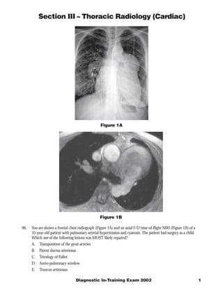
  • 1. Section III – Thoracic Radiology (Cardiac) Figure 1A Figure 1B 96. You are shown a frontal chest radiograph (Figure 1A) and an axial 2-D time-of-flight MRI (Figure 1B) of a 35-year-old patient with pulmonary arterial hypertension and cyanosis. The patient had surgery as a child. Which one of the following lesions was MOST likely repaired? A. Transposition of the great arteries B. Patent ductus arteriosus C. Tetralogy of Fallot D. Aorto-pulmonary window E. Truncus arteriosus Diagnostic In-Training Exam 2002 1
  • 2. Section III – Thoracic Radiology (Cardiac) Question #96 Findings: The radiograph demonstrates enlarged central pulmonary arteries compatible with pulmonary arterial hypertension. The central pulmonary arteries also have calcified walls. In addition, there is deformity of the left upper ribs compatible with a prior thoracotomy. The MR confirms the presence of enlarged central pulmonary arteries and decreased signal in the central branches suggesting slow flow from elevated resistance. There is also an anastomosis between the left pulmonary artery and the descending aorta. Rationales: A) Incorrect. Repair of D-transposition of the Great Arteries usually takes place through a median sternotomy not a posterior thoracotomy as this case. Transposition repair can be done by an atrial switch operation (Mustard, Senning) in which the systemic venous and pulmonary venous blood are re-routed appropriately, and more recently by the arterial switch or Jatene operation in which the great arteries themselves are relocated to their respective ventricles. The surgical connection between the descending aorta and pulmonary artery in the test case cannot be explained on the basis of any of these operations, therefore excluding transposition as an option. B) Incorrect. Repair of a patent ductus arteriosus requires ligation of the ductus. This is done through a left thoracotomy. An anastomosis between the pulmonary artery and aorta is not required. Moreover, pulmonary hypertension is not expected after surgical repair. Thus, although the rib changes can be seen after PDA repair, the pulmonary artery to aorta conduit is not consistent with this type of repair. C) Correct. Surgical palliation of TOF usually requires anastomosis between the obstructed right heart and systemic circulation before definitive repair is performed. The Potts-Smith operation was performed in this patient. In this palliative operation, the descending aorta and left pulmonary artery are anastomosed in a side- by-side fashion through a thoracotomy. Unfortunately, a common complication of this procedure is the development of pulmonary arterial hypertension because of increased blood flow to the lungs if the size of the anastomosis is not carefully constructed. The findings of pulmonary hypertension, a left thoracotomy and the surgical anastomosis between the systemic and pulmonary circulations support the diagnosis of tetralogy of Fallot. More recently, definitive repair of tetralogy of Fallot is performed early in life when feasible, thus obviating temporizing palliating procedures. The Potts-Smith operation has essentially been abandoned. D) Incorrect. An aorto-pulmonary window is a congenital defect in which there is a broad-based connection between the proximal aorta and the pulmonary artery. This condition can result in pulmonary artery hypertension if surgical repair is not done. However, the increased pulmonary flow characteristic of this anomaly prior to surgery usually regresses completely or nearly so after surgery. Repair requires closing the abnormal communication generally through an sternal splitting anterior approach. In the test patient, the left posterior thoracotomy as well as the anastomosis between the descending aorta and pulmonary artery argue against the diagnosis of aorto-pulmonary window. E) Incorrect. Prior to surgical repair, the shunt in truncus arteriosus is at the level of the ascending aorta and pulmonary artery. Repair requires separation of the pulmonary arteries from the aortic segment and then reconnection to the right ventricle, usually with an aortic homograft conduit. The conduit may eventually calcify. Normal vascularity is expected after surgery. In the test patient, the pulmonary hypertension and the shunt at the level of the descending, rather than the ascending, aorta make truncus arteriosus untenable. In addition, surgical repair of truncus is performed through a sternal splitting anterior approach, not through a thoracotomy. Citations: Potts WJ, Smith S, Gibson S. Anastomosis of the aorta to a pulmonary artery. JAMA 132:627, 1946 Piccoli GP, Dickinson DF, Musumeci F, Hamilton DI. A changing policy for the surgical treatment of tetralogy of Fallot: Early and late results in 235 consecutive patients. Ann Thorac Surg 33:365, 1982 RL Soulen, RM Donner, and M Capitanio. Postoperative evaluation of complex congenital heart disease by magnetic resonance imaging. RadioGraphics 1987 7: 975-1000 2 American College of Radiology
  • 3. Section III – Thoracic Radiology (Cardiac) Figure 2A Figure 2B 97. You are shown a plain radiograph (Figure 2A) and a right ventriculogram (Figure 2B) of a 2-day-old infant with tachypnea and cyanosis. Which one of the following is the MOST likely diagnosis? A. Tetralogy of Fallot B. Truncus arteriosus C. Pulmonary atresia and intact ventricular septum D. Ebstein’s anomaly of tricuspid valve E. D-transpostion of the great vessels Diagnostic In-Training Exam 2002 3
  • 4. Section III – Thoracic Radiology (Cardiac) Question #97 Findings: The chest radiograph demonstrates decreased pulmonary vascularity with a normal heart size and slight elevation of the cardiac apex. The location of the aortic arch is not readily visible on this radiograph. The right ventriculogram reveals opacification of the aorta, indicating a ventricular septal defect shunting right-to-left and hypoplasia of the right ventricular outflow tract. Other findings include competent tricuspid and mitral valves. The findings are consistent with Tetralogy of Fallot. Rationales: A. Correct. Tetralogy of Fallot is the most common cardiac cause of neonatal cyanosis. Anatomic features of Tetralogy of Fallot include a VSD, pulmonic stenosis, overriding aorta, and right ventricular hypertrophy. If there is severe obstruction of the right ventricular outflow tract, right to left shunting occurs across the VSD. Infants with severe obstruction usually present soon after birth with cyanosis and dyspnea. The infundibular stenosis results in decreased pulmonary vascularity on chest radiographs. The cardiac size is normal with an uplifted apex as a result of the right ventricular hypertrophy. The right heart obstruction, concomitant hypoplasia of the right ventricular outflow tract and atretic pulmonary valve in this patient is characteristic of severe tetralogy of Fallot also known as pseudotruncus arteriosus. B. Incorrect. Truncus arteriosus is a cyanotic cardiac anomaly characterized by a single arterial trunk arising from the heart and giving origin to the pulmonary arteries and ascending aorta. There is a single truncal valve which may have 3 to 6 semilunar leaflets. Invariably, there is a large VSD below the valve leaflets. Pulmonary vascularity is markedly increased because blood flows from the aorta to the pulmonary circulation. The cardiac size is enlarged. The decreased pulmonary vascularity, normal heart size and 2 separate semilunar valves demonstrated on the ventriculogram essentially exclude the diagnosis of truncus. C. Incorrect. In patients with pulmonary atresia and intact ventricular septum (also referred to as trilogy of Fallot) there is no communication between the pulmonary outflow tract and the right ventricle. The right ventricle is usually hypoplastic. There is also obligatory right to left shunting across an atrial septal defect. The absence of antegrade flow through the pulmonary valve results in diminished pulmonary vascularity on chest radiography. The heart is usually normal size in the immediate postnatal period. The chest radiographic findings in the test patient are compatible with pulmonary atresia and intact ventricular septum. However, the presence of a ventricular septal defect on the right ventriculogram excludes the diagnosis. D. Incorrect. Patients with Ebstein’s anomaly have a redundant tricuspid valve which is adherent to the right ventricular wall distal to the annulus of the tricuspid valve. This results in tricuspid regurgitation and subsequent right atrial enlargement. The right ventricle is decreased in size and is slow to empty, leading to decreased blood flow into the pulmonary arteries and right to left shunting across a patent foramen ovale or atrial septal defect. Hence, affected infants are usually cyanotic. The combination of decreased vascularity, a normal heart size and a competent tricuspid valve on the ventriculogram in the test patient makes Ebstein anomaly an unlikely diagnosis. E. Incorrect. D transposition of the great vessels (TGV) occurs when the aorta and main pulmonary artery arise from the morphologic right ventricle and morphologic left ventricle, respectively, creating two circulations in parallel. Desaturated venous blood flows from the right atrium into the right ventricle and then into the aorta, resulting in cyanosis. Radiographic findings include a narrow mediastinum, mild cardiomegaly and prominent pulmonary vascularity. The normal heart size and decreased vascularity in the test case would be unusual in a patient with d-TGV. Citations: Van Praagh R, Van Praagh S, Nebesar RA et al: Tetralogy of Fallot. Underdevelopment of the pulmonary infundibulum and its sequelae. Am J Cardiol 26:25-33, 1970 Adams FH, Emmanoulides GC, Riemenschneider TA. In Heart Disease in Infants, Children, and Adolescents. Fourth Edition Williams, Wilkins Baltimore 1989 4 American College of Radiology
  • 5. Section III – Thoracic Radiology (Cardiac) Figure 3A Figure 3B 98. This 63-year-old patient presented with chest pain. You are shown two levels (Figure 3A and Figure 3B) of a contrast enhanced CT scan. Which one of the following is the MOST likely diagnosis? A. Arrhythmogenic right ventricular dysplasia B. Congenital partial absence of the pericardium C. Left ventricular aneurysm D. Pericardial cyst E. Sinus of Valsalva aneurysm Diagnostic In-Training Exam 2002 5
  • 6. Section III – Thoracic Radiology (Cardiac) Question #98 Findings: The CT scan demonstrates a large mass that fills part of the anterior mediastinum in front of the pulmonary outflow tract (A). This mass contains calcification. Image (B) obtained at a lower level shows that the mass is in continuity with the left ventricle. In addition, the left ventricular wall is thin in the anteroseptal portion with subendocardial mural thrombus formation and peripheral calcification. All of these findings in combination are diagnostic of a large left ventricular aneurysm that extends superiorly into the anterior mediastinum Rationales: A. Incorrect. Arrhythmogenic right ventricular dysplasia is a condition in which fibrofatty replacement of the ventricular wall can lead to outpouchings in the wall of the right ventricle. Patients have frequent ventricular arrhythmias, and sudden death has been described. A large calcified mass as seen in this case would be atypical. In addition, the mass in the test patient arises from the left ventricle and the right ventricle appears normal, thus excluding such a diagnosis. B. Incorrect. Congenital absence of the pericardium can be total or partial. The partial form is the most common, usually involving the portion of the pericardium along the left heart border near the left atrial appendage. Patients can present clinically with chest pain, and strangulation of the left atrial appendage has been described. CT shows a protrusion of the left ventricle through the area of absent pericardium. The thickness of the ventricular wall is normal. The findings in the test patient of a thinned ventricular wall and a calcified mass in continuity with the anterior left ventricular wall excludes absence of the pericardium as a diagnostic alternative. C. Correct. There is thinning of the left ventricular wall over its entire anteroseptal aspect. Mural thrombus is seen on the inside of the thin ventricular wall. In addition, a large aneurysm has formed which has extended superiorly in front of the right ventricular outflow tract. This aneurysm has partly calcified over time. The findings are diagnostic of a left ventricular aneurysm. D. Incorrect. Pericardial cyst is a congenital entity related to the celomic cavity, in which a fluid containing structure forms adjacent to and not in communication with the pericardial cavity. Most are located in the right cardiophrenic angle. Heart chambers are not involved in the condition. The typical CT findings of pericardial cyst are those of a water attenuation mass adjacent to the cardiac border. A soft tissue attenuation mass with areas of calcification and the left ventricular involvement shown in the test case exclude a pericardial cyst. E. Incorrect. Sinus of Valsalva aneurysm results from a deficiency between the aortic media and fibrous annulus of the aortic valve resulting in distension and eventual aneurysm formation. If the aneurysm becomes large enough, it can rupture into the right atrial or right ventricular cavity. The test case demonstrates predominantly a left ventricular process excluding this diagnosis of sinus of Valsalva aneurysm. Citations: Adams FH, Emmanoulides GC, Riemenschneider TA. In Heart Disease in Infants, Children, and Adolescents. Fourth Edition Williams, Wilkins Baltimore 1989 Dahnert Wolfgang. Radiology Review Manual. Lippincott Williams and Wilkins 4th Edition 1999 6 American College of Radiology
  • 7. Section III – Thoracic Radiology (Cardiac) Figure 4A Figure 4B 99. An afebrile 3-week-old infant is referred because of tachypnea. A chest radiograph (Figure 4A) was obtained. This was followed by an electrocardiogram, which was abnormal, as well as an aortogram (Figure 4B). Which one of the following is the MOST likely diagnosis? A. Hypoplastic left heart syndrome B. Bland-White-Garland syndrome C. Kawasaki arteritis D. Single coronary artery E. Aortic insufficiency Diagnostic In-Training Exam 2002 7
  • 8. Section III – Thoracic Radiology (Cardiac) Question #99 Findings: The frontal chest radiograph shows cardiomegaly, normal pulmonary vascularity and retrocardiac atelectasis. A left sided obstructive lesion should be suspected. Aortogram shows a normal aortic valve and ascending aorta. However, the left coronary artery fills in a retrograde fashion by collaterals from the right coronary artery. No connection is seen between the proximal left coronary system and the aorta. On the other hand, it appears that the proximal left coronary artery drains to the region of the main pulmonary artery. Also note that the left anterior descending and circumflex branches come together as a common trunk. Rationales: A. Incorrect. The term hypoplastic left heart syndrome refers to a spectrum of anomalies characterized by a poorly developed left ventricle and ascending aorta. There is often underdevelopment of the mitral valve and left atrium. The resultant pathology is that there is no forward flow of blood from the left ventricle and so congestive heart failure is common soon after birth. Chest radiographs usually demonstrate cardiomegaly, increased vascularity and pulmonary edema. The normal pulmonary vascularity in the test radiographs and normal size aortic valve and ascending aorta on the aortogram make hypoplastic left heart unlikely. B. Correct. Bland-White –Garland Syndrome is a rare condition in which one of the coronary arteries (usually the left) originates from the pulmonary artery. Shortly after birth as the pulmonary artery resistance normally decreases, the myocardial perfusion from the anomalous coronary is compromised, resulting in ischemia and transmural infarction Patients present with congestive heart failure as a result of the ischemic insult. ECG usually suggests the diagnosis. C. Incorrect. Kawasaki arteritis is an acute febrile vasculitis of childhood. The illness occurs in young children, approximately 80% are under 5 years of age. Rarely, it affects infants or adolescents. Kawasaki disease causes a severe vasculitis of all blood vessels, with predilection for the coronary arteries. Acutely, patients present with high spiking fevers. Cardiomegaly and normal vascularity can be seen on chest radiographs. Aneurysms of the coronary arteries are common, but anomalous coronary origin is not a feature of this disease. The age of the test patient and the abnormal coronary artery anatomy makes Kawasaki disease unlikely. D. Incorrect. Rarely, infants are born with a single coronary artery. Most often, there is a single left branch which courses between the aorta and pulmonary artery . The anomalous artery can be “pinched” between the two vessels, resulting in ischemia and sudden death. In the test patient, there are two coronary arteries, excluding the diagnosis of a single artery. E. Incorrect. In the test patient, the left anterior descending (LAD) branch of the coronary artery and the circumflex branch join together as a common trunk, thus excluding the diagnosis of an anomalous origin. Anomalous origin of the LAD is particularly common in patients with tetralogy of Fallot, occurring in 5 to 9% of this population This anatomic variant can have important surgical implications at total repair. Citation: Choh JH, Levinsky L, Srinivasan V, Idbeis B, Subramanian S. Anomalous origin of the left coronary artery from the pulmonary trunk: Its clinical spectrum and current surgical management. Thorac and Cardiovasc Surg 28:239-242, 1980. 8 American College of Radiology
  • 9. Section III – Thoracic Radiology (Cardiac) Figure 5A Figure 5B 100. You are shown an axial T1-weighted turbo spin-echo (Figure 5A) and coronal gradient echo image (Figure 5B) of a 13-year-old girl with a diastolic murmur in the left precordial space. Which one of the following is the MOST likely diagnosis? A. Rheumatic aortic valvular disease B. Bicuspid aortic valve C. Aortic dissection D. Williams syndrome E. Takayasu’s arteritis Diagnostic In-Training Exam 2002 9
  • 10. Section III – Thoracic Radiology (Cardiac) Question #100 (question was not scored in the exam) Both sequences demonstrate an enlarged aortic root that measures approximately 4.5 cm in diameter. In addition, there is thickening of the walls of the ascending aorta and pulmonary arteries. The gradient echo image demonstrates a mild degree of aortic valvular insufficiency. The findings are consistent with an arteritis, such as Takayasu’s arteritis. Rationales: A. Incorrect. Rheumatic fever is an inflammatory condition that affects primarily the heart. The inflammatory process can involve the heart valves producing scarring that can lead to stenosis as well as insufficiency. The walls of the great vessels are not affected by the condition, thus excluding the diagnosis in the test case. B. Incorrect. Bicuspid aortic valve is a common congenital defect affecting the aortic valve, occurring in 1-2% of the population. Due to hemodynamic alteration of the flow through the valve, fibrosis and eventual calcification ensue producing stenosis. Although the enlargement of the aortic root from post-stenotic dilatation as well as the mild degree of valvular regurgitation could be explained on the basis of a bicuspid aortic valve, the thickening of the aortic and pulmonary arteries excludes the diagnosis. C. Incorrect. Aortic dissection is associated with an intramural hematoma which produces widening of the aorta. True wall thickening does not occur. A late sequela can be aortic insufficiency. Although the finding of aortic valvular insufficiency in the test case is consistent with dissection, the presence of wall thickening is atypical for dissection. Moreover, the patient’s age is unusual for this diagnosis. D. Incorrect. Williams syndrome is an autosomal dominant condition that is characterized by aortitis, mental retardation and elfin-like facies. Although thickening of the wall of the ascending aorta and pulmonary artery can be a feature of the disease, there is usually narrowing of the lumen of the ascending aorta causing supravalvular stenosis that can be diffuse at times. The aortic root dilatation seen in the test case virtually excludes Williams syndrome. E. Correct. Takayasu arteritis is a granulomatous inflammation of the aorta, its branches and the pulmonary artery. It usually affects persons younger than 50 years of age. An acute stage that is characterized by a granulomatous infiltrate of elastic fibers of media of arterial walls is followed by a chronic fibrotic period that eventually leads to fibrosis of the media and adventitia. Takayasu arteritis is divided into several types. Type IV is the so called dilated type, which affects aorta and its branches as well as the pulmonary arteries. Citations: Danhert Wolfgang in Radiology Review Manual 4th Edition Lippincott-Williams Wilkins 1999 10 American College of Radiology