Tuberculosis of knee
•Als PPTX, PDF herunterladen•
141 gefällt mir•26,723 views

knee tuberculosis, a simple presentation

Empfohlen
Weitere ähnliche Inhalte
Was ist angesagt?
Was ist angesagt? (20)
Ähnlich wie Tuberculosis of knee
Ähnlich wie Tuberculosis of knee (20)
Mehr von Ard Nepid
Kürzlich hochgeladen
Kürzlich hochgeladen (20)
Tuberculosis of knee
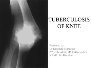
- 1. TUBERCULOSIS OF KNEE Presented by: Dr Dipendra Maharjan 2nd yr Resident, MS Orthopaedics NAMS, Bir Hospital
- 2. Introduction • Skeletal Tuberculosis – Ancient disease – 2% of all Tuberculosis cases – The most common form of skeletal TB is Pott’s disease – The next most common form of musculoskeletal TB is tuberculous arthritis
- 3. • The knee joint – the largest joint – the largest intra-articular space – third common site for osteoarticular tuberculosis – accounts for nearly 10% of all skeletal tuberculous lesions
- 4. Pathology • Initial focus – hematogenous dissemination in the synovium – in the subchondral bone – juxta-articular osseous focus. • The synovial lesion may for many months remain purely as tubercular synovitis. • The synovial membrane gets congested, edematous and studded with tubercles.
- 5. • The naked eye examination – pinkish-blue or pinkish-gray appearance. • The synovial lining becomes hypertrophied and thickened with granulation tissue. • The joint fluid in the initial stages is increased, serous, opalescent, turbid ellowish and may contain fibrinous flakes.
- 6. • In advanced stage of the disease – tuberculous process becomes osteoarticular, – the tuberculouse granulation tissue like the pannus erodes the articular margins, destroys the bones – involves the cruicate ligaments, periarticular tissues, capsule and ligaments.
- 7. • In cases which start as osseous lesions, there may be tuberculous abscess in – subchondral bone – epiphyseal bone – metaphyseal region • Abscess in the epiphyses and metaphyses may sometimes be seen traversing the epiphyseal cartilage plate
- 8. Clinical Features • The onset and course is insidious • The knee shows – Swelling • warm • patellar tap is present due to synovial effusion • the thickened synovium – filling up all parapatellar fossa appreciated earliest in medial parapatellar fossa.
- 9. • Tenderness is present to pressure is most marked at the synovial reflection and along the joint line. • When the arthritis has set – movements are grossly restricted, – painful – accompanied by muscle • regional lymphadenopathy.
- 10. • Quadricep muscle shows gross wasting • In the neglected case, – triple deformity • Once the flexion deformity established – tensor fasciae lata exantuates the deformity. • In long case – Posterior capsule of the knee joint gets contracted
- 11. • synovial stage – Generalized osteoporosis and – increased soft tissue swelling caused by • synovial effusion, • thickened synovium and capsule. • As the arthritis sets – loss of definition of articular surfaces, – marginal erosions, – diminution of the joint space and – destruction of the bones forming the joint. Roentgonograms
- 12. • In advance stage, – gross destruction and deformation of bone ends, – osteolytic cavities, – tubercular sequestra and – triple deformity may be seen
- 13. right knee demonstrating extreme valgus deformity and joint destruction.
- 14. Differential Diagnosis • Monoarticular affections – rheumatic arthritis (in children) – chronic traumatic synovitis due to chronic internal derangement of knee (e.g. • meniscal tears, loose bodies, • osteochondritis dissecans, • chondromalaciapatellae, • discoid semilunar cartilage etc) • Rheumatoid arthritis (in adults), • subacute pyogenic arthritis/synovitis, • hemarthrosis, • dysenteric arthritis, • villonodular synovitis, • synovial chondromatosus, • synovioma, • foreign body granuloma.
- 15. Prognosis • lee et al 1995, Hoffman et al 2002 – With the modern methods of management the functional results are directly related to the extent of disease at the onset of antitubercular drugs • In the stage of synovitis, – non operative or operative – complete healing – excellent range of movements.
- 16. In advance arthritis with subluxation severe restriction of motion is inevitable, arthrodesis (in adults) in functioning position (5 to 10 degree of flexion) is one of the option of treatment.
- 17. Treatment • Non operative treatment with antitubercular drugs is employed in – tubercular synovitis – children. • Traction is applied to – prevent (for correct) flexion and subluxation deformity and to – keep the joint surfaces distracted. • In addition to the systemic drugs, the joint may be aspirated
- 18. • With the quiescence of acute local signs, gently active and assisted knee bending should be. • • Usually after 12 weeks of treatment the patient may be permitted ambulation with suitable orthosis and crutches. • After 6 to 12 months of treatment, in cases with favorable response, the crutches or orthosis may be discarded. • Unprotected weight bearing is usually permitted 9 to 12 months after the start of treatment.
- 19. • In children with arthritis the deformity and subluxation is corrected/minimized by – employing double traction or – rarely by corrective plasters. • Arthrodesis of the grossly destroyed knee in children should be deferred till the completion of growth potentioal fo the distal femur and proximal tibia.
- 20. Operative Treatment • In the synovial stage – arthrotomy and synovectomy should be carried out. • In early arthritis, – synovectomy, – removal of loose/rice bodies, debris, pannus, loose articular cartilage and – careful curettage of osseous juxtaarticular foci • Postoperatively triple drug therapy, – traction, – intermittent active and assisted exercises, – suitable brace ambulation should be continued
- 21. • In adults with advanced arthritis or in cases which resulted in painful fibrous ankylosis during the process of healing, the knee joint may be treated by arthrodesis. • This option provides – painless stable knee, – prevents recrudescence, – corrects deformity and the – patients can do long hours of standing and walking. However it imposes a lot of restrictions in sitting, using public transport and many other social activities.
- 22. Arthroplasty • Besser (1980), wray and roy (1987) performed arthroplasty inadvertently in the preoperatively unsuspected cases of tuberculosis of the knee. • Kim(1988) reported good results after total knee arthroplasty in selected cases of old healed tuberculosis of knee. • Gale and harding (1991) reported a short term result of total knee arthroplasty in the presence of active disease.
- 23. • Indication for arthroplasty for a healed disease may be more justified for the knee than for any other joint • At present replacement arthroplasty of knee is being offered to selected patients. Most of the authors suggest this operation at least 5 to 10 years after the last evidence of activity of infection (Eskola et al 1988, kim 1988, gale and harding 1991) • Mandatory coverage by modern antitubercular drugs for about 5 months after replacement surgery is advised.
- 24. Thank You!
- 25. References • Tuberculosis of skeletal system by M.S. Tuli
Hinweis der Redaktion
- The pannus may erode the margins of the articular cartilage, grow between the articular cartilage and the subchondral bone thus detaching the cartilage from the bone and may grow over the articular cartilage as a sheet of granulation tissue.
- the metaphyseal region usually in children leading to various degree of destruction of bone. Abscess in the epiphyses and metaphyses may sometimes be seen traversing the epiphyseal cartilage plate giving an appearance of la lesion sitting astride the physis.
- The onset and course is insidious without usual systemic and local feature of tuberculous disease. The knee shows swelling, filling up all parapatellar fossar appreciated earliest in medial parapatellar fossa, suprapatellar pouch and even popliteal fossa. The swelling is warm, patellar tap is present if the swelling is predominantly due to the synovial effusion, the thickened synovium gives a boggy (doughy or semielastic) feel and can be rolled between the fingers and the underlying femur. It is best palpated on the medial side of the knee because vastus medialis remains muscular up to its insertion to patella and gets wasted early.
- Tenderness is present to pressure is most marked at the synovial reflection and aling the joint line. In the synovial disease, for a long time, there may only terminal restriction of movements. When the arthritis has set in the movements are grossly restricted, painful and accompanied by muscle spasm (particularly of hamstring) Quadricep muscle shows gross wasting regional lymphadenopathy.
- In the synovial stage if the disease is not responding favorably or the diagnosis is uncertain even after semi-invasive procedure arthrotomy and synovectomy should be carried out. In early arthritis, in addition to synovectomy removal of losse/rice bodies, debris, pannus, loose articular cartilage and carefyl curettage of osseous juxtaarticular foci should be carried out. Postoperatively triple drug therapy, traction, intermitten active and assisted exercises, suitable brace abmbulation should be continued
- In adults with advanced arthritis or in cases which resulted in painful fibrous ankylosis during the process of healing, the knee joint may be treated by arthrodesis. This option provides a painless stable knee, prevents recrudexcence, corrects deformity and the patients can do long hours of standing and walking. However it imposes a lot of restrictions in sitting, using public transport and many other social activities.