Weitere ähnliche Inhalte Ähnlich wie Hemodynamic assessment of partial mechanical circulatory support: data derived from computed tomography angiographic images and computational fluid dynamics (20) Mehr von Paul Schoenhagen (16) Kürzlich hochgeladen (20) 1. © Cardiovascular Diagnosis and Therapy. All rights reserved. Cardiovasc Diagn Ther 2015;5(2):160-165www.thecdt.org
Introduction
Left ventricular assist devices (LVAD) with full mechanical
support and left-ventricular unloading are limited to end-
stage heart-failure patients. In contrast, partial mechanical
circulatory support devices are intended for patients at
an earlier stage of heart failure. The Circulite Synergy
Micro-pump was the first of such a device (1-3) that has
been applied clinically and has been demonstrated to be
associated with significantly improved hemodynamics in a
study of 27 New York Heart Association class IIIB and early
class IV patients (2).
Conceptually, the support flow created by the Circulite
Synergy Micro-pump with its inflow graft connected to
the left atrium travels from the anastomosis site of the
outflow graft at the right subclavian artery through the
innominate artery (flow reversal) to the aorta. Alterations
in hemodynamics with this new concept are not well
understood. Direct measurements are only available from
Doppler ultrasound techniques as the device itself is a
contraindication to other methods such as phase contrast
magnetic resonance imaging. Non-invasive Doppler
techniques are limited to selected arterial segments and
Technical Notes
Hemodynamic assessment of partial mechanical circulatory
support: data derived from computed tomography angiographic
images and computational fluid dynamics
Christof Karmonik1
, Sasan Partovi2
, Fabian Rengier3,4
, Hagen Meredig3
, Mina Berty Farag5
, Matthias
Müller-Eschner3,4
, Rawa Arif5
, Aron-Frederik Popov6
, Hans-Ulrich Kauczor3
, Matthias Karck5
, Arjang
Ruhparwar5
1
Magnetic Resonance Imaging Core, Houston Methodist Research Institute, Houston, Texas, USA; 2
Department of Radiology, University Hospitals
Case Medical Center, Case Western Reserve University, Cleveland, Ohio, USA; 3
Department of Diagnostic and Interventional Radiology, University
Hospital Heidelberg, Heidelberg, Germany; 4
Department of Radiology E010, German Cancer Research Center (DKFZ), Heidelberg, Germany;
5
Department of Cardiac Surgery, University Hospital Heidelberg, Heidelberg, Germany; 6
Department of Cardiothoracic Transplantation and
Mechanical Support, Royal Brompton and Harefield NHS Trust, Harefield Hospital, London, UK
Correspondence to: Sasan Partovi, MD. Department of Radiology, University Hospitals Case Medical Center, Case Western Reserve University, 11100
Euclid Ave, Cleveland, Ohio 44106, USA. Email: sasan.partovi@case.edu or sasan.partovi@uhhospitals.org.
Abstract: Partial mechanical circulatory support represents a new concept for the treatment of advanced
heart failure. The Circulite Synergy Micro Pump®
, where the inflow cannula is connected to the left atrium
and the outflow cannula to the right subclavian artery, was one of the first devices to introduce this concept
to the clinic. Using computational fluid dynamics (CFD) simulations, hemodynamics in the aortic tree
was visualized and quantified from computed tomography angiographic (CTA) images in two patients. A
realistic computational model was created by integrating flow information from the native heart and from
the Circulite device. Diastolic flow augmentation in the descending aorta but competing/antagonizing
flow patterns in the proximal innominate artery was observed. Velocity time curves in the ascending aorta
correlated well with those in the left common carotid, the left subclavian and the descending aorta but poorly
with the one in the innominate. Our results demonstrate that CFD may be useful in providing a better
understanding of the main flow patterns in mechanical circulatory support devices.
Keywords: Heart failure; partial mechanical circulatory support; computational fluid dynamics (CFD); flow
Submitted Jan 10, 2015. Accepted for publication Feb 09, 2015.
doi: 10.3978/j.issn.2223-3652.2015.03.03
View this article at: http://dx.doi.org/10.3978/j.issn.2223-3652.2015.03.03
2. 161Cardiovascular Diagnosis and Therapy, Vol 5, No 2 April 2015
© Cardiovascular Diagnosis and Therapy. All rights reserved. Cardiovasc Diagn Ther 2015;5(2):160-165www.thecdt.org
a complete three-dimensional picture of the entire flow
field during the cardiac cycle is not easily obtained. An
alternative to direct measurements is the indirect calculation
of hemodynamics using computational fluid dynamics
(CFD). In this approach, the geometry of the arterial
tree and the inflow and outflow conditions of the arterial
segment of interest are used to solve the Navier Stokes
partial differential equation which yields a complete three-
dimensional description of the blood flow velocity for the
entire cardiac cycle.
Here, we demonstrate the use of CFD based on
computed tomography angiographic (CTA) images and
Doppler ultrasound measurements obtained from two
patients. We aimed to demonstrate the interaction between
flow created by the device and the pulsatile native cardiac
outflow at different time points in the cardiac cycle.
Materials and methods
Patient information
Permission of the Internal Review Board was obtained for
this retrospective study. Two female patients, both 63 years
old, with ischemic cardiomyopathy post Circulite Synergy
Micro-pump implantation underwent a CTA examination
using a SOMATOM Definition Flash CT scanner (Siemens,
Erlangen, Germany) and retrospective ECG-gating within
2 months after device implantation. The dose length
products of the two CTA studies were 1,147 and 1,943 mGycm,
respectively.
Computational fluid dynamics (CFD) technique
The lumen of the thoracic aorta, the origins of the supra-
aortic vessels and the right subclavian artery including the
outflow anastomosis site were manually segmented. From
this segmentation, a 3D surface reconstruction of the
luminal boundary was created (Paraview, Kitware Inc.). The
native cardiac outflow velocity waveform was reconstructed
from measurements obtained with transthoracic Doppler
(TTD) echocardiography and used as inflow waveform
in the CFD simulations as was the (constant) outflow
velocity of the Circulite Synergy Micro-pump (Figure 1 and
Table 1). The 3D surface reconstruction of the segmented
lumen was imported into Star-CCM+ (version 8.02.008)
and computational polyhedral meshes were created (case
1: 121,740 elements; case 2: 112,897 elements). Blood
was modeled as a Newtonian fluid with a constant density
of 1,050 kg/m3
and a constant viscosity of 0.004 Pa·s. A
laminar flow model was assumed (no inclusion of turbulence
effects). To allow for the decay of initial transients in the
computational simulation, three cardiac cycles were and
results are reported from the third cardiac cycle with a
method described previously (4).
Post-processing of simulation results
Total velocities, velocities in the inferior-superior direction
and pressures were temporally averaged in the proximal
innominate artery, the left common carotid artery and
the left subclavian artery. Velocity time curves in these
arteries were compared with the one of the ascending
aorta using the Pearson correlation coefficient as means
to reveal modifications of the hemodynamics. Pseudo-
color representations of velocity magnitudes, pressures
and wall shear stresses (WSS) during systole and diastole
were created to illustrate overall hemodynamics in the
computational model.
Results
Case 1
Pressures in the distal subclavian artery immediately
proximal to the anastomosis site exhibited elevated
pressures during the entire cardiac cycle compared to
the remainder of the arteries, where high pressures
were only observed during systole (Figure 1A). During
diastole, prominent retrograde flow from the outflow
graft anastomosis site entering the aorta was observed
(Figure 1B). This flow was reversed during peak systole
in the proximal innominate and subclavian arteries due
to increased native cardiac output, causing a “collision”
of both flows, leading to unordered and stagnant flow. In
addition, a region of recirculating or slow flow formed in
the proximal subclavian artery at this time, which traveled
more proximal during beginning and end systole. For case 1,
the retrograde average velocity in the innominate was
0.03 m/s during diastole which, was reversed during systole
with an average of 0.08 m/s. The average total velocity
in the left innominate was considerably lower than the
velocities in the other two supra-aortic vessels but pressures
where the highest of all three (Figure 2). A low correlation
of 0.31 between the velocity time curve in the ascending
aorta and the innominate artery further points to disturbed
flow in this artery.
3. 162 Karmonik et al. CFD in partial mechanical circulatory support
© Cardiovascular Diagnosis and Therapy. All rights reserved. Cardiovasc Diagn Ther 2015;5(2):160-165www.thecdt.org
The blood flow of this case is illustrated in the Figures 3
and 4.
Case 2
A similar flow pattern was observed for case 2 as described
for case 1 with a reversal of retrograde average velocity
from 0.18 m/s during diastole and of 0.12 at systole
(Figure 1D). Average and maximum WSS values exhibited
a maximum at the anastomosis site but also a minimum on
the anterior wall of the ascending aorta (Figure 1C). Also for
this case, average total velocity in the innominate artery was
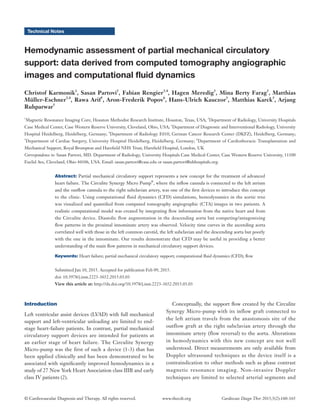
Figure 1 (A) Pressure distribution on the wall of the computational model for case 1 during diastole (left) and systole (right). While elevated
pressures in systole exist in the ascending aorta and the innominate, these are only found at the anastomosis site during diastole; (B) flow
patterns for case 1 (visualized by the velocity magnitude) demonstrate a change in the overall flow pattern, which is dominated during
diastole by the Circulite device and during systole by the native flow from the heart; (C) WSS maximum and average during the cardiac
cycle both exhibit a minimum at the anterior wall of the ascending aorta but also a maximum at the anastomosis site; (D) as for case 1, also
the flow patterns for case 2 (visualized by the velocity magnitude) demonstrate a change in the overall flow pattern, which is dominated
during diastole by the Circulite device and during systole by the native flow from the heart. Red boxes mark the regions where velocities and
pressures were quantified. WSS, wall shear stresses.
Table 1 Hemodynamics data for both cases
Items Case 1 Case 2
Outflow graft (m/s) 0.843 0.929
RR interval (msec) 500 900
EF (%) 21 17
SV (mL) 65.5 51.1
EDV (mL) 311.4 306.9
ESV (mL) 245.9 255.8
Cardiac outflow (L/min) 7.4 2.3
EF, ejection fraction; SV, stroke volume; EDV, end diastolic
volume; ESV, end systolic volume.
A C
B D
Case 1 Case 2
4. 163Cardiovascular Diagnosis and Therapy, Vol 5, No 2 April 2015
© Cardiovascular Diagnosis and Therapy. All rights reserved. Cardiovasc Diagn Ther 2015;5(2):160-165www.thecdt.org
Figure 2 (A) Top panel: velocity time curves in the ascending aorta (asc), the innominate artery (innom), the left carotid artery (lcarotid),
the left subclavian artery (lsubcl) and the descending aorta (desc) for both cases. The deviation of shape for the innominate artery can be
appreciated for both cases, likely related to the described hemodynamics. Middle panel: velocity time curves for flow in inferior-superior
direction. Flow reversal during cardiac cycle in the innominate is clearly visible pointing to competing effects of flow created by the
Circulate device and by the native heart. Lower panel: the upper triangular matrix displays the correlation coefficients between the velocity
time curves shown in A while the lower triangular matrix graphically visualizes these correlation coefficients (blue: positive, red: negative).
Low positive correlation for case 1 and strong negative correlation for case 2 quantifies the difference in shapes shown in the upper panel; (B)
average values of velocities, velocities in inferior-superior direction (IS velocity) and pressure for both cases.
Case 1 Case 2
0.31 0.95 0.94 0.75 –0.88 0.86 0.86 0.96
–0.82 –0.85 –0.88
1.00 0.80
0.80
0.34 0.42 0.71
0.99 0.77
0.83
asc
innom
lcarotid
lsubcl
desc
innom
lcarotid
lsubcl
desc
asc
innom
lcarotid
lsubcl
desc
1.5
1
0.5
0
0.8
0.6
0.4
0.2
0
–0.2
–0.4
–0.6
–0.8
–1
–1.2
2
1.5
1
0.5
0
–0.5
–1
–1.5
–2
2
1.5
1
0.5
0
1 3 5 7 9 11
1 2 3 4 5 6 7 8 9 10 11
innom
lcarotid
lsubcl
desc
asc asc
innom innom
lcarotid lcarotid
lsubcl lsubcl
desc desc
Case 1 velocity IS pressure Case 2 velocity IS pressure
velocity velocity
asc
innom
lcarotid
lsubcl
desc
asc
innom
lcarotid
lsubcl
desc
0.397
0.192
0.335
0.324
0.515
0.501
0.398
0.464
0.553
0.669
0.015
0.136
0.119
–0.496
–0.142
0.317
0.402
–0.672
479
371
297
309
853
552
624
593
A
B
asc
asc
5. 164 Karmonik et al. CFD in partial mechanical circulatory support
© Cardiovascular Diagnosis and Therapy. All rights reserved. Cardiovasc Diagn Ther 2015;5(2):160-165www.thecdt.org
lowest for the innominate and flow direction on average was
actually retrograde (Figure 2).
Discussion
To access efficacy of new partial mechanical circulatory
support devices, a better understanding of the induced
flow alterations is warranted. Direct measurements in vivo
are difficult to obtain for the entire artery tree. As an
alternative, we have demonstrated that CFD in combination
with clinical CTA images together with patient-specific
inflow boundary conditions, is capable of reproducing flow
feature in the aorta and adjacent arteries. While blood
velocity was increased in the aorta distal to the origin of the
innominate artery during diastole, flow during systole was
not affected as the Circulite device and the native cardiac
output worked against each other thereby creating an area
of recirculating flow in the distal innominate artery partially
extending into the subclavian artery. Clinical studies
evaluating the effectiveness of the Circulite Synergy Micro-
pump have reported encouraging results, demonstrating
that the device improved hemodynamics in heart failure
patients of class IIIB and IV and appeared to interrupt
and partially reverse the progressive hemodynamic
deterioration typical of end-stage heart failure (2). In a
study of 54 chronic heart failure patients, older patients
(≥70 years) implanted with Synergy had smaller body
sizes and worse renal function than younger patients.
Both groups experienced similar hemodynamic benefits
and functional improvements, though peak VO2 was less
improved in the elderly (3).
Our results demonstrate increased support of aortic
flow during most of the cardiac cycle, but ineffectiveness
of the device during peak systole. As a consequence of the
flow reversal in the innominate artery, a region of low and
oscillating wall shear stress WSS is created. This particular
hemodynamic condition has previously been identified to
be promote atherosclerosis by inducing endothelial cell
apoptosis via a mechanism that involves the induction of
GATA4, FZD5 and BMP2; molecules that have a well-
defined role in embryonic development (7). A recent
microRNA study reported alterations in miR-regulated
differential gene expressions with varying hemodynamic
forces. Several microRNAs were identified mediating a
protective role with high WSS and expression of miR-21,
miR-92a, and miR-663 activated with low WSS resulted
in a pathological EC phenotype (8). A synchronization of
partial mechanical support devices to the cardiac cycle of the
individual may avoid this potential adverse hemodynamic
condition and potentially improve outcome.
Figure 3 Illustrates flow reversal from retrograde flow pre and post
systole to antegrade flow at systole at the origin of the innominate
artery and the immediate distal aortic section for case 1 (5).
Higher flow velocities are indicated by yellow, orange and red
colors. A region of slow and stagnant flow can be appreciated in
the anterior ascending aorta during systole. Also during systole, the
lowest blood flow velocities can be found at this location. These
observed alterations in hemodynamics are a direct consequence of
competing flows from the partial support device and native cardiac
output.
Available online: http://www.asvide.com/articles/470
Figure 4 Shows the entire model in similar fashion as supplement
#1 (6). Inflow from the partial support device is indicated by high
velocities (yellow and red colors) on the left. Interacting flows of
opposite direction meet at the proximal section of the innominate
artery resulting in a ‘traveling’ band of slow velocities (dark blue
region). Flow patterns are complex pre and post systole and deviate
considerably from the pattern at systole where native flow from the
heart dominates.
Available online: http://www.asvide.com/articles/471
▲
▲
6. 165Cardiovascular Diagnosis and Therapy, Vol 5, No 2 April 2015
© Cardiovascular Diagnosis and Therapy. All rights reserved. Cardiovasc Diagn Ther 2015;5(2):160-165www.thecdt.org
As with any model, our CFD results present a simplification
of reality in which only the most pertinent features were
considered. Aortic wall motion effects were not included
nor were fluid structure interactions of the aortic wall with
surrounding parenchyma. Consequently, our results have
to be considered to reproduce major flow patterns, which
may be refined by including the features discussed above.
Nevertheless, they help to understand the general concept
of the altered hemodynamics: antagonistic flows between
the heart and the supporting device during systole interfere
in a destructive pattern. As no device-specific assumptions
were included in the CFD simulation, similar flow patterns
may be anticipated for other such kind of devices unless
alternative algorithms of pump activity are implemented.
In contrast to patients with conventional continuous flow
LVADs, patients with partial mechanical circulatory support
still have a considerable output of the native heart and
antagonistic flows between the device and the heart should
play a major role when devising new concepts. Moreover
a periodic rotation of the impeller with acceleration and
deceleration of the rotor allows for fixed speed while
superimposing periodic episodes of pulsatility.
A larger study including more cases may help to better
understand the interactions of device-induced flow and
native cardiac output and may therefore be of help in
optimizing the design of new devices operating with this
principle.
Acknowledgements
Fabian Rengier and Matthias Müller-Eschner received
support from the German Research Foundation (DFG)
within project R03, SFB/TRR 125 “Cognition-Guided
Surgery”.
Disclosure: The authors declare no conflict of interest.
References
1. Meyns B, Ector J, Rega F, et al. First human use of partial
left ventricular heart support with the Circulite synergy
micro-pump as a bridge to cardiac transplantation. Eur
Heart J 2008;29:2582.
2. Meyns BP, Simon A, Klotz S, et al. Clinical benefits of
partial circulatory support in New York Heart Association
Class IIIB and Early Class IV patients. Eur J Cardiothorac
Surg 2011;39:693-8.
3. Barbone A, Pini D, Rega F, et al. Circulatory support in
elderly chronic heart failure patients using the CircuLite®
Synergy® system. Eur J Cardiothorac Surg 2013;44:207-
12; discussion 212.
4. Karmonik C, Partovi S, Loebe M, et al. Influence of
LVAD cannula outflow tract location on hemodynamics in
the ascending aorta: a patient-specific computational fluid
dynamics approach. ASAIO J 2012;58:562-7.
5. Karmonik C, Partovi S, Rengier F, et al. Illustrate flow reversal
from retrograde flow pre and post systole to antegrade flow
at systole at the origin of the innominate artery and the
immediate distal aortic section for case 1. Asvide 2015;2:016.
Available online: http://www.asvide.com/articles/470
6. Karmonik C, Partovi S, Rengier F, et al. Show the entire
model in similar fashion as supplement #1. Asvide 2015;2:017.
Available online: http://www.asvide.com/articles/471
7. Mahmoud M, Kim R, De Luca A, et al. 189 Disturbed
flow promotes endothelial cell injury via the induction of
developmental genes. Heart 2014;100 Suppl 3:A105.
8. Neth P, Nazari-Jahantigh M, Schober A, et al. MicroRNAs
in flow-dependent vascular remodelling. Cardiovasc Res
2013;99:294-303.
Cite this article as: Karmonik C, Partovi S, Rengier F,
Meredig H, Farag MB, Müller-Eschner M, Arif R, Popov
AF, Kauczor HU, Karck M, Ruhparwar A. Hemodynamic
assessment of partial mechanical circulatory support: data
derived from computed tomography angiographic images
and computational fluid dynamics. Cardiovasc Diagn Ther
2015;5(2):160-165. doi: 10.3978/j.issn.2223-3652.2015.03.03