
Empfohlen
Weitere ähnliche Inhalte
Was ist angesagt?
Was ist angesagt? (20)
Ähnlich wie Sectional fixed orthodontic appliance
Ähnlich wie Sectional fixed orthodontic appliance (20)
Mehr von Maher Fouda
Mehr von Maher Fouda (20)
Kürzlich hochgeladen
Kürzlich hochgeladen (20)
Sectional fixed orthodontic appliance
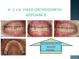
- 1. Lectured by Prof . MAHER FOUDA
- 2. The basic 2X4 appliance design is as follows: bands cemented on both upper first permanent molars. brackets bonded onto the erupted maxillary incisors. continuous archwires to provide/maintain good arch form, as well as control of anterior teeth. supporting stainless steel tubing placed in the long archwire spans between the molars and incisors.
- 3. Archwire
- 5. A nine-year-old girl was referred by her dentist regarding both upper central incisors, which were in crossbite. She presented with a Class I incisor relationship on a Skeletal I base in the mixed dentition. The upper labial segment was spaced with the lower being well aligned. She had a premature contact on the central incisors with a resultant 2 mm anterior displacement on full closure.
- 7. Bands were place on both upper first molars and brackets were bonded to all the upper incisors with an initial aligning wire of 0.016 inch nickel titanium being placed. At the next visit, 5 weeks later, the overjet had been corrected. A 0.016 inch stainless steel wire was then placed with power chain for a further 4 weeks to close any residual space and the patient was debonded .Total treatment time was 9 weeks. No retainer was indicated and the result was stable 4 months later.
- 8. Intra-oral photographs after 6 weeks of treatment. Incisor relationship is now corrected
- 13. An 8 year-old boy was referred by his GDP regarding the delayed eruption of the upper left central incisor due to the presence of supernumerary tooth. He presented with a Class I incisor relationship on a Skeletal I base with well-aligned upper and lower arches .The supernumerary was removed and the central incisor bonded to a gold chain using a closed technique under a general anaesthetic.
- 15. Brackets were bonded to the three erupted incisors and bands were cemented onto both upper first molars with an initial aligning wire of 0.016 inch nickel titanium. The wire sequence progressed through a 0.01860.025 inch nickel titanium to a 0.01960.025 inch stainless steel working arch wire. This was then used as a base wire coupled with a piggyback 0.016 inch nickel titanium wire applying traction to the unerupted central incisor via the gold chain.
- 17. Once the incisor was through a bracket was placed and 0.016 inch nickel titanium archwire fully engaged. The archwire was then stepped up to a 0.018 inch stainless steel wire with powerchain to close any residual spacing prior to debond. The incisor was self-retaining. Total active orthodontic treatment time was 10 months
- 20. A 7 year-old boy was initially referred by his GDP for the removal of two supernumerary teeth present in the upper midline. He presented in the early mixed dentition. The supernumerary teeth were removed and the patient reviewed 1 year later when he presented with a very irregular and rotated upper incisors .A course of 264 appliance therapy was prescribed.
- 22. Initially, all four incisors were bonded with bands place on both upper first permanent molars and an initial aligning wire of 0.012 inch nickel titanium due to the severe rotations associated with the upper incisors. Progression was via a 0.016 inch nickel titanium achwire to a 0.018 × 0.025 inch nickel titanium archwire with a working archwire of 0.019 × 0.025 inch stainless steel.
- 23. Powerchain was used for a single visit to close any remaining anterior spacing. Once a positive overjet and overbite were established the appliance was removed, and a palatally-bonded retainer cemented. Total active orthodontic treatment time was thirteen months.
- 25. Intra-oral photographs after alignment of incisors
- 26. Post-treatment photographs. Palatal bonded retainer
- 29. An 8 year-old girl was referred by her GDP who was concerned about both upper central incisors being in crossbite. She presented with a Class III incisor relationship on a Skeletal III base in the mixed dentition with an anterior and displacement to the left of the mandible after initial contact.
- 31. All four upper incisors were bonded, the upper first molars banded with a soldered quadhelix, which was activated, and an initial aligning wire of 0.016 inch nickel titanium placed .Rapid correction of the incisor relationship occurred and the patient was debonded after 5 months of treatment. She was kept under review with the occlusion remaining stable 3 years later.
- 33. Pre- treatment
- 36. Correction of anterior crossbite with fixed appliance.
- 41. The boy, 8 years and 9 months had a crossbite of all four upper incisors. In case history his mother stated she had a frontal crossbite as a young girl, and her cousin was operated for the mandibular prognathism. There was the crossbite of central incisors in the boy's deciduous dentition.
- 42. Class I in molars and canines, crossbite of 21 +12, flattened upper frontal segment, spaces in both upper and lower frontal teeth .The orthopantomographic radiograph shows the teeth are present in the range of second permanent molars. Patient crossbite befor treatment
- 43. Cephalogram was assessed: Skeletal Class III according to WITS, Skeletal Class I according to ANB angle, neutral growth rotation, interincisal angle of 129°, 1+1 to NS angle is 104°, 1-1 to ML angle is 95°.
- 44. The treatment started with bands on teeth 6+6, 6-6, transpalatal arch Burstone of steel 0.032" x 0.032" as the anchorage, and with attachment of brackets on teeth 21 +12, and 21 -12. In the maxilla we used an arch of TMA wire, diameter of 0.016" x 0.022", utility type; lower arch was of stainless steel, diameter of 0.016". At the same time we made bite blocks for the teeth V+V of photocomposite to eliminate incisors from articulation. After a month, Class III elastics were added. Positive overjet of 21 +12 was reached 6 weeks after the beginning of treatment. At that time the bite blocks were removed . Overbite achieved
- 45. For retention a stainless steel arch, diameter of 0.017" x 0.025" was used. Upper and lower fixed appliances were removed 4 months after treatment beginning (Fig. 4a-c). Currently, the patient is in retention phase already for 19 months, he has upper and lower retainer, incisors are still in positive overjet. After removal of fixed appliance
- 48. A 7-year-old female presented with the upper four deciduous incisors in crossbite . Glass ionomer cement was added to the occlusal surfaces of the mandibular second deciduous molars. Two months later, with the crossbite corrected, the cement was removed . The patient’s profile also improved significantly.
- 49. Pretreatment
- 52. A 10-year-old male presented with the upper central incisors in crossbite .The lower first permanent molars were built up with glass ionomer cement .Three months later, the crossbite was resolved .The upper lateral incisors erupted in a normal relationship to the lower arch, while the lower anterior crowding resolved itself as the occlusion was unlocked .
- 53. 10-year-old male patient with upper central incisors in crossbite before treatment.
- 54. Glass ionomer cement bonded to occlusal surfaces of lower first permanent molars.
- 55. Case 2. Correction of crossbite in three months.
- 56. Case 2. Upper lateral incisors erupting in proper positions as lower anterior crowding resolves Spontaneously.
- 57. A 7-year-old male presented with a crossbite of the upper left central incisor and gingival recession of the lower left central incisor (Fig. 9). Two months after a glass ionomer build-up of the lower molars, the crossbite was resolved, and the perio - dontal condition of the lower incisor had improved significantly (Fig. 10). Three months later, the gingival contours of the lower incisor were almost normal (Fig. 11).
- 59. Correction of crossbite in two months.
- 62. Physiological spacing (ugly duckling) stage. A transient anterior open bite can be associated with eruption of the incisors as they approach the occlusal plane and this invariably improves with time. The maxillary central incisors can also be quite distally inclined when they first erupt, which produces a midline diastema between them. This physiological spacing or ‘ugly duckling’ stage is thought to be due to the combined effect of the maxillary incisor apices being initially quite close together in the anterior maxilla as the incisors erupt and lateral pressure from the erupting maxillary lateral incisors and canines
- 63. 7 year 9 year 14 year
- 64. As these teeth erupt this pressure is transferred from the apical region of the maxillary incisors more coronally, improving their inclination and usually closing the diastema.
- 70. Pretreatment periapical x-ray of maxillary central incisors
- 72. Final intraoral photogragh Final periapical radiogragh of maxillary central incisors
- 78. 1- presence or absence of an anterior mandibular displacement. 2-possible damage that has or might occur to the dentition through excessive tooth wear, or to the supporting periodontal structures. 3- prevention of establishment of the developing Malocclusion. 4-space availability – this may be rectified by the early removal of both the upper deciduous canines. 5-the position of the developing permanent canines in relation to the roots of the lateral incisors. 6-the depth of the overbite
- 79. The magnitude of the crossbite —does it involve a single tooth or an entire segment? Is there a displacement associated with the crossbite? How significant is the skeletal component and will it be possible to compensate for this discrepancy with tooth movement only?
- 80. If expansion is indicated at an early stage, then this can be carried out easily and simultaneously by adding a quadhelix to the 2x4 appliance.
- 82. previous history of trauma. early extraction of deciduous teeth allowing closure of eruption space or formation of fibrous gingival tissue. retained deciduous teeth. supernumerary teeth. Odontomes.
- 83. The major advantages in carrying out this treatment with a 2x4 appliance are the ease with which space opening can be controlled with a fixed appliance, and also that the force magnitude and vector can be controlled much more precisely than with a removable appliance.
- 84. Minimal discomfort. Reduces need for patient co-operation. Increase control of tooth movements. Movement possible in all three planes of space.
- 85. Appliance rarely worn full time. Appliance damage/lost appliances. Difficulty in speech/eating. Gagging. Decalcification/caries. Gingivitis/palatal hyperplasia/fungal infections. Incorrect activation produces unhelpful changes. Allow only tipping of teeth.
- 86. Treatment carried out in this mixed dentition stage may take as little as a couple of weeks,15 but in the more difficult cases can take longer. In the majority of cases, however, the end result can be more effectively and efficiently achieved than if a removable appliance was used.
- 87. واهلها مصر احفظ اللهم