1. PROBABILITY MAPPING
Keywords: myocardial infarction, imaging, decision support system, infarction core area and border zone
Background
As the population ages, the incidence and prevalence of cardiovascular diseases CVD increases
and the need for early detection and intervention will be exacerbated. In addition, diagnostic
imaging provides cardiologists with detailed anatomical (CT, MRI, US), metabolic and
functional (nuclear imaging, MRI) information upon which an accurate diagnosis can be made.
Currently no single imaging test is superior to all the others and a variety of imaging modalities
may be used to diagnose or monitor CVD.
Description
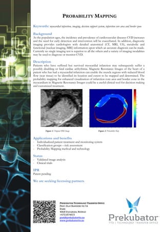
Patients who have suffered but survived myocardial infarction may subsequently suffer a
possibly disabling or fatal cardiac arrhythmia. Magnetic Resonance Images of the heart of a
patient who has had a myocardial infarction can enable the muscle regions with reduced blood
flow (scar tissue) to be identified its location and extent to be mapped and determined. The
probability mapping for enhanced visualization of infarction core area and border zone in the
myocardium in Magnetic Resonance Images could be a useful clinical tool for decision making
and customized treatment.
Figure 1: Original MRI Image Figure 2: Probability Map
Applications and benefits
- Individualized patient treatment and monitoring system
- Classification groups – risk assessment
- Probability Mapping method and technology
Status
- Validated image analysis
- Clinical trials
IPR
Patent pending
We are seeking licensing partners.