
Empfohlen
Weitere ähnliche Inhalte
Was ist angesagt?
Was ist angesagt? (20)
Ähnlich wie Assignment on Circulation of blood
Ähnlich wie Assignment on Circulation of blood (20)
Kürzlich hochgeladen
Kürzlich hochgeladen (20)
Assignment on Circulation of blood
- 2. Content Topic Page 01 Circulatory System 04 02 Cardiovascular System 05 - Cardiovascular System Anatomy - The Heart and Circulation 03 Blood Vessel 10 - Arteries and Arterioles -Capillaries -Veins and Venules 04 Coronary Circulation 13 05 Hepatic Portal Circulation 15 06 The Path of Blood through the Human Body 15 07 Pulmonary circulation 16 08 Systemic circulation 17 09 Capillary exchange 18 10 References 19 2
- 3. Circulatory system The circulatory system is an organ system that permits blood and lymph circulation to transport nutrients, oxygen, carbon dioxide, hormones, blood cells, etc. to and from cells in the body to nourish it and help to fight diseases, stabilize body temperature and pH, and to maintain homeostasis. This system may be seen strictly as a blood distribution network, but some consider the circulatory system as composed of the cardiovascular system, which distributes blood, and the lymphatic system, which returns excess filtered blood plasma from the interstitial fluid (between cells) as lymph. While humans, as well as other vertebrates, have a closed cardiovascular system (meaning that the blood never leaves the network of arteries, veins and capillaries), some invertebrate groups have an open cardiovascular system. The more primitive, diploblastic animal phyla lack circulatory systems. The lymphatic system, on the other hand, is an open system providing an accessory route for excess interstitial fluid to get returned to the blood. Figure 1: Sectional view of Human heart 3
- 4. Two types of fluids move through the circulatory system: blood and lymph. Lymph is essentially recycled blood plasma after it has been filtered from the blood cells and returned to the lymphatic system. The blood, heart and blood vessels form the cardiovascular system. The essential components of the human cardiovascular system are the heart, blood, and blood vessels. It includes: the pulmonary circulation, a "loop" through the lungs where blood is oxygenated; and the systemic circulation, a "loop" through the rest of the body to provide oxygenated blood. An average adult contains five to six quarts (roughly 4.7 to 5.7 liters) of blood, accounting for approximately 7% of their total body weight. Blood consists of plasma, red blood cells, white blood cells, and platelets. Also, the digestive system works with the circulatory system to provide the nutrients the system needs to keep the heart pumping. Cardiovascular System The cardiovascular system consists of the heart, blood vessels, and the approximately 5 liters of blood that the blood vessels transport. Figure 2: Cardiovascular System 4
- 5. Responsible for transporting oxygen, nutrients, hormones, and cellular waste products throughout the body, the cardiovascular system is powered by the body’s hardest-working organ — the heart, which is only about the size of a closed fist. Even at rest, the average heart easily pumps over 5 liters of blood throughout the body every minute. Cardiovascular System Anatomy The Heart and Circulation The heart is a muscular pumping organ located medial to the lungs along the body’s midline in the thoracic region. The bottom tip of the heart, known as its apex, is turned to the left, so that about 2/3 of the heart is located on the body’s left side with the other 1/3 on right. The top of the heart, known as the heart’s base, connects to the great blood vessels of the body: the aorta, vena cava, pulmonary trunk, and pulmonary veins. Figure 3: Blood Circulation Through Heart 5
- 6. Circulatory Loops There are 2 primary circulatory loops in the human body: the pulmonary circulation loop and the systemic circulation loop. Pulmonary circulation transports deoxygenated blood from the right side of the heart to the lungs, where the blood picks up oxygen and returns to the left side of the heart. The pumping chambers of the heart that support the pulmonary circulation loop are the right atrium and right ventricle. Systemic circulation carries highly oxygenated blood from the left side of the heart to all of the tissues of the body (with the exception of the heart and lungs). Systemic circulation removes wastes from body tissues and returns deoxygenated blood to the right side of the heart. The left atrium and left ventricle of the heart are the pumping chambers for the systemic circulation loop. Figure 4: Represents The circulation 6
- 7. The primary function of the heart is to pump blood through blood vessels to the body's cells. Imagine a simple machine like a water pump working for perhaps 70 or more years without attention and without stopping. Impossible? Yet this is exactly what the heart can do in our bodies. The heart is really a muscular bag surrounding four hollow compartments, with a thin wall of muscle separating the left hand side from the right hand side. The muscles in the heart are very strong because they have to work harder than any of the other muscles in our body, pushing the blood to our head and feet continuously. The blood flow around our body is called our circulation. The heart connects the two major portions of the circulation's continuous circuit, the systemic circulation and the pulmonary circulation. The blood vessels in the pulmonary circulation carry the blood through the lungs to pick up oxygen and get rid of carbon dioxide, while the blood vessels in the systemic circulation carry the blood throughout the rest of our body. The heart actually has two separate sides, one designed to pump deoxygenated blood into the pulmonary circulation where the blood becomes oxygenated, and one designed to pump the oxygenated blood into the systemic circulation where the blood flows throughout the body. Each side of the heart has two chambers or compartments. The top chamber on each side is called the atrium. The right atrium receives incoming deoxygenated blood from the body and the left atrium receives incoming oxygenated blood from the lungs. The thin-walled atrium on each side bulges as it fills with blood, and as the lower heart muscle relaxes, the atrium contracts and squeezes the blood into a second chamber, the thick muscular ventricle. The ventricle is the pumping chamber that, with each muscular contraction, pushes the blood forcefully out and into the lungs (right ventricle) and the rest of the body (left ventricle). The atrium and ventricle on each side of the heart are separated by tissue flaps called valves. The structure of these valves prevents blood from flowing backward into the atrium as the ventricle squeezes blood out. The valve on the right side, between the atrium and the ventricle, is called the tricuspid valve. The valve on the left side, between the atrium and the ventricle, is called the bicuspid or mitral valve. There are two other important valves that help to keep the blood Rowing in the proper direction. These two valves are located at the two points where blood 7
- 8. exits the heart. The pulmonary valve is located between the right ventricle and the pulmonary artery that carries the deoxygenated blood from the heart to the lungs, and the aortic valve is located between the left ventricle and the aorta, the major artery that carries the oxygenated blood from the heart to the rest of the body. The arteries are the blood vessels that transport blood out of the heart under high pressure to the tissues. The arterioles are the last small branch of the arterial system through which blood is released into the capillaries. The capillaries are very small, thin-walled blood vessels where the exchange of gases, nutrients, and waste takes place between the cells and the blood. Blood flows with almost no resistance in the larger blood vessels, but in the arterioles and capillaries, considerable resistance to flow does occur because these vessels are so small in diameter that the blood must squeeze all its contents through them. The venules collect blood from the capillaries and gradually feed into progressively larger veins. The veins transport the blood from the tissues back to the heart. The walls of the veins are thin and very elastic and can fold or expand to act as a reservoir for extra blood, if required by the needs of the body. Figure 5: The path of a typical RBC through the heart. 8
- 9. Let us follow a single red blood cell (RBC) through one full cycle along the circulatory pathway. Remember that RBCs carry oxygen throughout the body. Since the blood travels endlessly, an arbitrary choice must be made of a starting point to describe the RBC's route. We will begin at the point where the RBC has delivered its oxygen to a cell in need and is on its return back to the heart. 1. Once the deoxygenated red blood cell (RBC) returns to the heart, it enters either through the superior vana cava or the inferior vena cava. The superior vena cava returns deoxygenated blood from the upper part of the body to the heart. The inferior vena cava returns deoxygenated blood from the lower part of the body to the heart. These large veins lead into the right atrium. 2. The RBC passes through the tricuspid valve into the right ventricle. 3. The RBC is then pumped through the pulmonary valve into the pulmonary artery and on to the lungs. There the RBC gives off carbon dioxide and picks up oxygen. 4. The RBC returns to the heart through a pulmonary vein, enters the left atrium, passes through the mitral valve, and flows into the left ventricle. 5. The left ventricle pumps the fully oxygenated RBC through the aortic valve, into the aorta, the body's main artery, and out to the body. 6. From the aorta, the RBC flows into one of the many arteries of the body, through the arterioles, and then to the capillaries, where the RBC will deliver oxygen and nutrients to the cells and remove wastes and carbon dioxide. Next it moves through the venules, veins, and on to the vena cava in a deoxygenated state, and returns to the heart, only to begin its repetitive journey once again. This whole process has taken approximately 20 seconds! That single RBC will travel about 950 miles (more than 1500 kilometers) in its brief 4-month lifetime! Blood Vessels Blood vessels are the body’s highways that allow blood to flow quickly and efficiently from the heart to every region of the body and back again. The size of blood vessels corresponds with the amount of blood that passes through the vessel. All blood vessels contain a hollow area called the lumen through which blood is 9
- 10. able to flow. Around the lumen is the wall of the vessel, which may be thin in the case of capillaries or very thick in the case of arteries. All blood vessels are lined with a thin layer of simple squamous epithelium known as the endothelium that keeps blood cells inside of the blood vessels and prevents clots from forming. The endothelium lines the entire circulatory system, all the way to the interior of the heart, where it is called the endocardium. Figure 6: Three types of blood vessels There are three major types of blood vessels: arteries, capillaries and veins. Blood vessels are often named after either the region of the body through which they carry blood or for nearby structures. For example, the brachiocephalic artery 10
- 11. carries blood into the brachial (arm) and cephalic (head) regions. One of its branches, the subclavian artery, runs under the clavicle; hence the name subclavian. The subclavian artery runs into the axillary region where it becomes known as the axillary artery. Arteries and Arterioles: Arteries are blood vessels that carry blood away from the heart. Blood carried by arteries is usually highly oxygenated, having just left the lungs on its way to the body’s tissues. The pulmonary trunk and arteries of the pulmonary circulation loop provide an exception to this rule – these arteries carry deoxygenated blood from the heart to the lungs to be oxygenated. Arteries face high levels of blood pressure as they carry blood being pushed from the heart under great force. To withstand this pressure, the walls of the arteries are thicker, more elastic, and more muscular than those of other vessels. The largest arteries of the body contain a high percentage of elastic tissue that allows them to stretch and accommodate the pressure of the heart. Smaller arteries are more muscular in the structure of their walls. The smooth muscles of the arterial walls of these smaller arteries contract or expand to regulate the flow of blood through their lumen. In this way, the body controls how much blood flows to different parts of the body under varying circumstances. The regulation of blood flow also affects blood pressure, as smaller arteries give blood less area to flow through and therefore increases the pressure of the blood on arterial walls. Arterioles are narrower arteries that branch off from the ends of arteries and carry blood to capillaries. They face much lower blood pressures than arteries due to their greater number, decreased blood volume, and distance from the direct pressure of the heart. Thus arteriole walls are much thinner than those of arteries. Arterioles, like arteries, are able to use smooth muscle to control their aperture and regulate blood flow and blood pressure. Capillaries: Capillaries are the smallest and thinnest of the blood vessels in the body and also the most common. They can be found running throughout almost every tissue of the body and border the edges of the body’s avascular tissues. Capillaries connect to arterioles on one end and venules on the other. 11
- 12. Capillaries carry blood very close to the cells of the tissues of the body in order to exchange gases, nutrients, and waste products. The walls of capillaries consist of only a thin layer of endothelium so that there is the minimum amount of structure possible between the blood and the tissues. The endothelium acts as a filter to keep blood cells inside of the vessels while allowing liquids, dissolved gases, and other chemicals to diffuse along their concentration gradients into or out of tissues. Precapillary sphincters are bands of smooth muscle found at the arteriole ends of capillaries. These sphincters regulate blood flow into the capillaries. Since there is a limited supply of blood, and not all tissues have the same energy and oxygen requirements, the precapillary sphincters reduce blood flow to inactive tissues and allow free flow into active tissues. Veins and Venules: Veins are the large return vessels of the body and act as the blood return counterparts of arteries. Because the arteries, arterioles, and capillaries absorb most of the force of the heart’s contractions, veins and venules are subjected to very low blood pressures. This lack of pressure allows the walls of veins to be much thinner, less elastic, and less muscular than the walls of arteries. Veins rely on gravity, inertia, and the force of skeletal muscle contractions to help push blood back to the heart. To facilitate the movement of blood, some veins contain many one-way valves that prevent blood from flowing away from the heart. As skeletal muscles in the body contract, they squeeze nearby veins and push blood through valves closer to the heart. When the muscle relaxes, the valve traps the blood until another contraction pushes the blood closer to the heart. Venules are similar to arterioles as they are small vessels that connect capillaries, but unlike arterioles, venules connect to veins instead of arteries. Venules pick up blood from many capillaries and deposit it into larger veins for transport back to the heart. Coronary Circulation The heart has its own set of blood vessels that provide the myocardium with the oxygen and nutrients necessary to pump blood throughout the body. The left and right coronary arteries branch off from the aorta and provide blood to the left and 12
- 13. right sides of the heart. The coronary sinus is a vein on the posterior side of the heart that returns deoxygenated blood from the myocardium to the vena cava. Figure 7: Coronary Circulation Figure 8: Hepatic Circulation 13
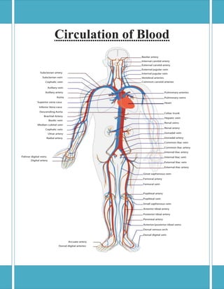
- 14. Hepatic Portal Circulation The veins of the stomach and intestines perform a unique function: instead of carrying blood directly back to the heart, they carry blood to the liver through the hepatic portal vein. Blood leaving the digestive organs is rich in nutrients and other chemicals absorbed from food. The liver removes toxins, stores sugars, and processes the products of digestion before they reach the other body tissues. Blood from the liver then returns to the heart through the inferior vena cava. The Path of Blood through the Human Body: When a heart contracts and forces blood into the blood vessels, there is a certain path that the blood follows through the body. Figure 9: Blood circulation through human body 14
- 15. The blood moves through pulmonary circulation and then continues on through systemic circulation. Pulmonary and systemic are the two circuits in the two-circuit system of higher animals with closed circulatory systems. Humans and other mammals have two-circuit circulatory systems: one circuit is for pulmonary circulation (circulation to the lungs; pulmo = lungs), and the other circuit is for systemic circulation (the rest of the body). As each atrium and ventricle contract, blood is pumped into certain major blood vessels, and from there, continues through the circulatory system. Pulmonary circulation Blood that is lacking oxygen is said to be deoxygenated. This blood has just exchanged oxygen for carbon dioxide across cell membranes, and now contains mostly carbon dioxide. Deoxygenated blood enters the right atrium through the superior vena cava and the inferior vena cava. Superior means higher, and inferior means lower, so the superior vena cava is at the top of the right atrium, and the inferior vena cava enters the bottom of the right atrium. From the right atrium, the deoxygenated blood drains into the right ventricle through the right atrioventricular (AV) valve, which is so named because it is between the atrium and the ventricle. This valve is also referred to as the tricuspid valve because it has three flaps in its structure. When the ventricles contract, the AV valve closes off the opening between the ventricle and the atrium so that blood does not flow back up into the atrium. As the right ventricle contracts, it forces the deoxygenated blood through the pulmonary semilunar valve and into the pulmonary artery. Semilunar means halfmoon and refers to the shape of the valve. Note that this is the only artery in the body that contains deoxygenated blood; all other arteries contain oxygenated blood. The semilunar valve keeps blood from flowing back into the right ventricle once it is in the pulmonary artery. The pulmonary artery carries the blood that is very low in oxygen to the lungs, where it becomes oxygenated. 15
- 16. Systemic circulation Freshly oxygenated blood returns to the heart via the pulmonary veins. Note that these are the only veins in the body that contain oxygenated blood; all other veins contain deoxygenated blood. The pulmonary veins enter the left atrium. When the left atrium relaxes, the oxygenated blood drains into the left ventricle through the left AV valve. This valve is also called the bicuspid valve because it has only two flaps in its structure. Now the heart really squeezes. As the left ventricle contracts, the oxygenated blood is pumped into the main artery of the body — the aorta. To get to the aorta, blood passes through the aortic semilunar valve, which serves to keep blood flowing from the aorta back into the left ventricle. The aorta branches into other arteries, which then branch into smaller arterioles. The arterioles meet up with capillaries, which are the blood vessels where oxygen is exchanged for carbon dioxide. Figure 10: Pulmonary and Systemic Circulation 16
- 17. Capillary exchange Capillaries bridge the smallest of the arteries and the smallest of the veins. Near the arterial end, the capillaries allow materials essential for maintaining the health of cells to diffuse out (water, glucose, oxygen, and amino acids). To maintain the health of cells, it is also necessary for the capillaries to transport wastes and carbon dioxide to places in the body that can dispose of them. The waste products enter near the venous end of the capillary. Water diffuses in and out of capillaries to maintain blood volume, which adjusts to achieve homeostasis. Capillaries are only as thick as one cell, so the contents within the cells of the capillaries can easily pass out of the capillary by diffusing through the capillary membrane. And, because the capillary membrane abuts the membrane of other cells all over the body, the capillary’s contents can easily continue through the abutting cell’s membrane and get inside the adjoining cell. The process of capillary exchange is how oxygen leaves red blood cells in the bloodstream and gets into all the other cells of the body. Capillary exchange also allows nutrients to diffuse out of the bloodstream and into other cells. At the same time, the other cells expel waste products that then enter the capillaries, and carbon dioxide diffuses out of the body’s cells and into the capillaries. 17
- 18. References: Kent. G.C’.Jr (1954). Comparative anatomy of the Vertebrates. Kotpal, R.L. (2000). Modern Textbook of Zoology, Vertebrates. http://en.wikipedia.org/wiki/Circulatory_system http://www.innerbody.com/anatomy/cardiovascular-male http://www.nsbri.org/humanphysspace/focus2/heartcirculation.html 6. http://www.dummies.com/how-to/content/the-path-of-bloodthrough-the-human-body.html 1. 2. 3. 4. 5. 18