SlideShare ist ein Scribd-Unternehmen logo
1 von 17
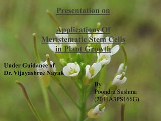
Presentation on 
Applications Of 
Meristematic Stem Cells 
in Plant Growth 
By 
Poondru Sushma 
(2011A3PS166G) 
Under Guidance of 
Dr. Vijayashree Nayak
Meristematic Cells 
•Meristem cells are unspecialized cells found in plants, similar 
to stem cells found in animals. 
•Meristem cells form much of the cell growth with in a plant. 
•This growth is broken down into upward and outward. 
 Upward and downward growth is the result of fast growing apical 
meristem cells 
 Out ward growth is the result of somewhat slower lateral meristem cells. 
• The term meristem was first used in 1858 by Karl Wilhelm 
von Nägeli (1817–1891) in his book Beiträge zur 
Wissenschaftlichen Botanik. It is derived from the Greek 
word merizein, meaning to divide, in recognition of its 
inherent function.
Apical Meristem 
Organization of an 
apical meristem 
(growing tip) 
1 - Central zone 
2 - Peripheral zone 
3 - Medullary (i.e. 
central) meristem 
4 - Medullary 
tissue
Apical Meristems 
Primary Meristems 
Protoderm Procambium Ground 
Meristem 
lies around the 
outside of the stem 
and develops into 
the epidermis 
lies just inside of the 
protoderm and 
develops into 
primary xylem and 
primary phloem. It 
also produces 
the vascular 
cambium, a 
secondary meristem 
It develops in to 
the cortex and the pith. 
It produces the cork 
cambium, another 
secondary meristem.
Apical Meristems 
At the meristem summit, there is a small group of slowly 
dividing cells, which is commonly called the central zone. 
Cells of this zone have a stem cell function and are essential 
for meristem maintenance. 
Apical meristems are composed of several layers. The number 
of layers varies according to plant type. In general the 
outermost layer is called the tunica while the innermost layers 
are the corpus. 
In monocots, the tunica determines the physical characteristics 
of the leaf edge and margin. In dicots, layer two of the corpus 
determines the characteristics of the edge of the leaf. The 
corpus and tunica play a critical part of the plant physical 
appearance as all plant cells are formed from the meristems.
Shoot Apical Meristem 
The shoot apical meristem is the site of most of the 
embryogenesis in flowering plants. Primordia of leaves, 
sepals, petals, stamens and ovaries are initiated here at the 
rate of one every time interval, called a plastochron. It is 
where the first indications that flower development has 
been evoked are manifested. 
The shoot apical meristem consists of 4 distinct cell 
groups: 
*Stem cells 
*The immediate daughter cells of the stem cells 
*A subjacent organizing Centre 
*Founder cells for organ initiation in surrounding regions
Shoot Apical Meristem 
3 interacting CLAVATA genes are required to regulate the size of the stem 
cell reservoir in the shoot apical meristem by controlling the rate of cell division. 
CLV1 and CLV2 are predicted to form a receptor complex (of the LRR receptor 
like kinase family) to which CLV3 is a ligand. 
Another important gene in plant meristem maintenance is WUSCHEL (shortened 
to WUS), which is a target of CLV signaling. WUS is expressed in the cells below 
the stem cells of the meristem and its presence prevents the differentiation of the 
stem cells.
Meristem structure 
• SAMs are usually dome shaped and have a layered structure described as 
a tunica and corpus. 
• The tunica consists usually of 2 cell layers (often only 1 in monocots) where 
cell division occurs primarily in the anticlinal plane (perpendicular to the 
surface) and the surface to remain as monolayers. 
• The cells in the L3 layer divide in random orientations to form a multilayered 
structure referred to as the rib meristem (RM) or the organizing center (OC), 
which provides stem cell-promoting cues.
Meristem structure 
• SAMs also show cytological zonations independent of the layered structure. 
• Central zone that stains lightly with histological stains. The central zone 
includes both tunica and corpus cells. 
• The peripheral zone is a ring of densely staining cells that surround the 
central zone and also includes both tunica and corpus. 
• Below the central zone is an area called the rib meristem. 
• Several approaches have shown a correlation in the mitotic activity with the 
zonation. 
• Mitotic activity is most prevalent in the peripheral region as shown by 
incorporation of tritiated thymidine which reflects DNA replication and 
therefore mitotic activity. 
• The central zone is the region of the apical initials. These divide infrequently 
and occasionally contribute cells to the rapidly dividing peripheral zone. 
Because most genetic mistakes are generated during mitosis, this is probably a 
mechanism to minimize the occurrence of genetic mistakes in the permanent 
cell population of the meristem.
Initiation and maintenance of meristems 
• Initiation of the shoot meristem in the embryo requires the action of 
several genes that were identified in genetic screens for mutants that 
lack a SAM. 
• One factor is a pair of genes called CUP-SHAPED COTYLEDONS 
(CUC)1 and 2. These are duplicate factors. That is that there are two 
different genes that function redundantly and both have to be mutated 
to see a mutant phenotype. 
• The phenotype consists of both cotyledons forming in a continuous 
fused cup-shaped structure with no SAM. Occasionally, one can get a 
shoot to form in tissue culture and then growth is fairly normal up to 
flowering. This indicates that the CUC genes are required for SAM 
initiation but not maintenance. 
• Mutations in the SHOOT MERISTEMLESS (STM) also result in 
embryos without a SAM. Occasionally, in weak stm alleles, 
adventitious shoots will form but the shoots progressively lose their 
meristem indicating that STM is required for both the initiation and 
maintenance of SAMs. STM encodes a homeodomain transcription 
factor.
Initiation and maintenance of meristems 
• Maintenance of shoot apical meristems requires regulating the balance 
between cell proliferation and differentiation. 
• Several genes have been identified that regulate this balance. As 
mentioned, STM is required to maintain the SAM and in mutants the 
SAM dwindles away because of either too rapid cell differentiation or 
insufficient cell proliferation. 
• Accordingly, the proposed function of STM is to either promote 
proliferation (i.e., promote the meristematic state) or to inhibit 
differentiation. Consistent with this, ectopic expression of STM-like 
genes, such as the maize KNOTTED gene, in leaves causes the 
formation of ectopic meristems. 
• Another gene called WUSHEL (WUS) has a similar function (ie, 
similar mutant phenotype) and also encodes a homeodomain 
transcription factor. 
• Three genes called CLAVATA (CLV)1,2 and 3 have the opposite effect 
in that mutations cause an over proliferation of cells in the SAM (the 
SAM gets too big). Therefore the function of these genes is to restrict 
cell proliferation or to promote cell differentiation.
Initiation and maintenance of meristems 
• CLV1 is a receptor kinase and CLV2 is another receptor-like protein. These 2 
are proposed to form a heterodimeric receptor complex. 
• CLV3 is a small protein that is predicted to be secreted and is hypothesized to 
function as the activating signal ligand for the CLV1/2 receptor complex. 
• CLV1 is expressed mostly in the corpus region of the SAM and CLV3 is 
expressed predominantly in the tunica region of the SAM, highlighting the 
interaction among layers of the SAM. 
• Because cells in both the tunica and corpus proliferate abnormally 
in clv1 mutants, it is concluded that CLV1 acts non-autonomously. 
• It is possible that activation of the CLV1 receptor results in the production of a 
secondary signal that diffuses throughout the meristem. 
• The balance between the activity of the proliferation promoting 
(differentiation inhibiting) and the proliferation inhibiting (differentiation 
promoting) pathways is required for the long-term function of the SAM.
Initiation and maintenance of meristems 
• The CLV and STM pathways are antagonistic to each other 
because clv and stm mutants partially suppress one another. 
• This also indicates that these represent independent pathways 
because each still functions in the absence of the other. 
• On the other hand, the WUSHEL gene acts downstream of CLV 
because clv / wus double mutants look just like wus. 
• The CLV pathway acts to inhibit the expression of WUS which 
acts to promote cell proliferation. In clv mutants there is nothing 
to restrict WUS, resulting in overproliferation. However, if wus is 
mutant, then it doesn’t matter whether CLV is around to restrict it 
or not.
WUSHEL 
• Cell–cell communication between distinct cell types within stem cell 
niches is critical for stem cell maintenance in both plants and animals, 
although they differ in their niche architecture and cell behaviors. 
• In the shoot apical meristem (SAM) stem cell niche not all stem cells 
make contact with the niche, they also do not exhibit oriented and 
asymmetric cell divisions to regulate stem cell numbers. 
• For example, the Arabidopsis SAM stem cell niche is a collection of ∼500 
cells located at the growing tip of each shoot. 
• The CZ of the SAM harbors stem cells. The stem cell progeny that are 
displaced into the adjacent peripheral zone (PZ) proliferate before 
differentiating. 
• Visually, the SAM stem cell niche is a multilayered structure consisting of 
three clonally distinct layers of cells, and stem cells are found in each of 
these layers. 
• WUSCHEL (WUS), a homeodomain-containing transcription factor, is 
both necessary and sufficient for stem cell specification. 
• WUS RNA is found in a few cells of the RM/OC located just beneath the 
CZ .
WUSHEL 
• Restriction of WUS transcription to cells of the OC is critical for maintaining a 
constant number of stem cells, and this is mediated by the CLAVATA (CLV) 
signaling pathway. 
• CLAVATA3 (CLV3), expressed in the CZ, encodes a small peptide that is 
secreted into the extracellular space and binds to CLAVATA1 (CLV1), a 
leucine-rich repeat receptor kinase predominantly expressed in cells of the 
RM. 
• Activation of CLV1 and related receptor kinases has been shown to mediate 
repression of WUS transcription through a signaling cascade that is not well 
understood. 
• WUS, which is expressed in cells of the RM/OC, not only specifies stem cell 
fate in overlying cells of the CZ, but also activates its own negative regulator, 
CLV3, in a non-cell-autonomous manner. 
• Thus, the WUS–CLV feedback system forms a self-correcting mechanism for 
maintaining a constant number of stem cells and the SAM size.
WUSHEL 
*WUS protein migrates from OC/niche into adjacent cells 
*WUS protein migration is required for shoot meristem 
function 
*WUS binds to CLV3 regulatory regions to activate its 
transcription
Bio finalppt

Weitere ähnliche Inhalte

Was ist angesagt?

Dicot and monocot leaf anatomy
Dicot and monocot leaf anatomyDicot and monocot leaf anatomy
Dicot and monocot leaf anatomyRajinapp
 
Tunica carpus theory
Tunica carpus theoryTunica carpus theory
Tunica carpus theoryFatima Fizan
 
Evolution of sporophyte in bryotphytes
Evolution of sporophyte in bryotphytesEvolution of sporophyte in bryotphytes
Evolution of sporophyte in bryotphytesSangeeta Das
 
Floral induction and development genetic and molecular analysis
Floral induction and development genetic and molecular analysisFloral induction and development genetic and molecular analysis
Floral induction and development genetic and molecular analysisgohil sanjay bhagvanji
 
Molecular Analysis Of SAM & RAM
Molecular Analysis Of SAM & RAMMolecular Analysis Of SAM & RAM
Molecular Analysis Of SAM & RAMTilak I S
 
Microsporogensis
MicrosporogensisMicrosporogensis
Microsporogensisarunsaini68
 
stress physiology.pptx
stress physiology.pptxstress physiology.pptx
stress physiology.pptxGunmalaGugalia
 
Embryology in relation to taxonomy
Embryology in relation to taxonomyEmbryology in relation to taxonomy
Embryology in relation to taxonomyDr. T. A. Gitte
 
Types of Embryosacs KR.pptx
Types of Embryosacs KR.pptxTypes of Embryosacs KR.pptx
Types of Embryosacs KR.pptxDrKavitaRambal
 
Leaf structure, adaptations, development
Leaf structure, adaptations, development Leaf structure, adaptations, development
Leaf structure, adaptations, development Jasmine Brar
 
Monocot and dicot navi
Monocot and dicot naviMonocot and dicot navi
Monocot and dicot navinaveenagirish
 

Was ist angesagt? (20)

Dicot and monocot leaf anatomy
Dicot and monocot leaf anatomyDicot and monocot leaf anatomy
Dicot and monocot leaf anatomy
 
Tunica carpus theory
Tunica carpus theoryTunica carpus theory
Tunica carpus theory
 
Evolution of sporophyte in bryotphytes
Evolution of sporophyte in bryotphytesEvolution of sporophyte in bryotphytes
Evolution of sporophyte in bryotphytes
 
Pollen wall & mgu
Pollen wall & mguPollen wall & mgu
Pollen wall & mgu
 
ginkoales.pptx
ginkoales.pptxginkoales.pptx
ginkoales.pptx
 
Floral induction and development genetic and molecular analysis
Floral induction and development genetic and molecular analysisFloral induction and development genetic and molecular analysis
Floral induction and development genetic and molecular analysis
 
Molecular Analysis Of SAM & RAM
Molecular Analysis Of SAM & RAMMolecular Analysis Of SAM & RAM
Molecular Analysis Of SAM & RAM
 
Dicot embryogenesis
Dicot embryogenesisDicot embryogenesis
Dicot embryogenesis
 
Life cycle of_sphagnum
Life cycle of_sphagnumLife cycle of_sphagnum
Life cycle of_sphagnum
 
Anatomy of leaves
Anatomy of leavesAnatomy of leaves
Anatomy of leaves
 
Microsporogensis
MicrosporogensisMicrosporogensis
Microsporogensis
 
Notothylas by p.n
Notothylas by p.nNotothylas by p.n
Notothylas by p.n
 
Flower Development
Flower DevelopmentFlower Development
Flower Development
 
stress physiology.pptx
stress physiology.pptxstress physiology.pptx
stress physiology.pptx
 
Embryology in relation to taxonomy
Embryology in relation to taxonomyEmbryology in relation to taxonomy
Embryology in relation to taxonomy
 
Types of Embryosacs KR.pptx
Types of Embryosacs KR.pptxTypes of Embryosacs KR.pptx
Types of Embryosacs KR.pptx
 
Leaf structure, adaptations, development
Leaf structure, adaptations, development Leaf structure, adaptations, development
Leaf structure, adaptations, development
 
Monocot and dicot navi
Monocot and dicot naviMonocot and dicot navi
Monocot and dicot navi
 
Laticifers
LaticifersLaticifers
Laticifers
 
Flower development
Flower developmentFlower development
Flower development
 

Andere mochten auch

Roots, shoots, leaves
Roots, shoots, leavesRoots, shoots, leaves
Roots, shoots, leavesJessi Dildy
 
2.1, 2.2, 2.3 Cells
2.1, 2.2, 2.3 Cells2.1, 2.2, 2.3 Cells
2.1, 2.2, 2.3 CellsToni Foley
 
Plant structure, growth, and reproduction by Campbell Biology 7th edition
Plant structure, growth, and reproduction by Campbell Biology 7th editionPlant structure, growth, and reproduction by Campbell Biology 7th edition
Plant structure, growth, and reproduction by Campbell Biology 7th editionMae Canencia
 
Tissues and types of plant tissues
Tissues and types of plant tissuesTissues and types of plant tissues
Tissues and types of plant tissuesRahul Kumar
 
TISSUE CULTURE ON BANANA SUCKERS AND RICE CALLUS (MICROPROPAGATION)
TISSUE CULTURE ON BANANA SUCKERS  AND RICE CALLUS (MICROPROPAGATION)TISSUE CULTURE ON BANANA SUCKERS  AND RICE CALLUS (MICROPROPAGATION)
TISSUE CULTURE ON BANANA SUCKERS AND RICE CALLUS (MICROPROPAGATION)UNIVERSITI MALAYSIA KELANTAN
 
Plant tissue culture techniques of Banana
Plant tissue culture techniques of BananaPlant tissue culture techniques of Banana
Plant tissue culture techniques of BananaLoyola College
 

Andere mochten auch (13)

B5 lesson part one
B5 lesson  part oneB5 lesson  part one
B5 lesson part one
 
Roots, shoots, leaves
Roots, shoots, leavesRoots, shoots, leaves
Roots, shoots, leaves
 
Plant tissues
Plant tissuesPlant tissues
Plant tissues
 
4th tissue culture
4th tissue culture4th tissue culture
4th tissue culture
 
2.1, 2.2, 2.3 Cells
2.1, 2.2, 2.3 Cells2.1, 2.2, 2.3 Cells
2.1, 2.2, 2.3 Cells
 
Meristems
MeristemsMeristems
Meristems
 
Plant structure, growth, and reproduction by Campbell Biology 7th edition
Plant structure, growth, and reproduction by Campbell Biology 7th editionPlant structure, growth, and reproduction by Campbell Biology 7th edition
Plant structure, growth, and reproduction by Campbell Biology 7th edition
 
Tissue culture
Tissue cultureTissue culture
Tissue culture
 
Tissues and types of plant tissues
Tissues and types of plant tissuesTissues and types of plant tissues
Tissues and types of plant tissues
 
TISSUE CULTURE ON BANANA SUCKERS AND RICE CALLUS (MICROPROPAGATION)
TISSUE CULTURE ON BANANA SUCKERS  AND RICE CALLUS (MICROPROPAGATION)TISSUE CULTURE ON BANANA SUCKERS  AND RICE CALLUS (MICROPROPAGATION)
TISSUE CULTURE ON BANANA SUCKERS AND RICE CALLUS (MICROPROPAGATION)
 
Stem cells
Stem cellsStem cells
Stem cells
 
Plant tissue culture
Plant tissue culturePlant tissue culture
Plant tissue culture
 
Plant tissue culture techniques of Banana
Plant tissue culture techniques of BananaPlant tissue culture techniques of Banana
Plant tissue culture techniques of Banana
 

Ähnlich wie Bio finalppt

MOLECULAR AND GENETIC APPROACHES IN PLANT GROWTH AND DEVELOPMENT
MOLECULAR AND GENETIC APPROACHES IN PLANT GROWTH AND DEVELOPMENTMOLECULAR AND GENETIC APPROACHES IN PLANT GROWTH AND DEVELOPMENT
MOLECULAR AND GENETIC APPROACHES IN PLANT GROWTH AND DEVELOPMENTAdhiyamaan Raj
 
Phloem diffrentiation
Phloem diffrentiationPhloem diffrentiation
Phloem diffrentiationParul Sharma
 
lab 7 _ 8 without videos.pptx
lab 7 _ 8 without videos.pptxlab 7 _ 8 without videos.pptx
lab 7 _ 8 without videos.pptxArielkatwaroo
 
Shabir presentation for cell line ppt
Shabir presentation for cell line pptShabir presentation for cell line ppt
Shabir presentation for cell line pptShabir Khan
 
meristmatic tissue.pptx
meristmatic tissue.pptxmeristmatic tissue.pptx
meristmatic tissue.pptxmanoj Joshi
 
10.STEM CELLS.pptx
10.STEM CELLS.pptx10.STEM CELLS.pptx
10.STEM CELLS.pptxmalti19
 
Chromosomes and cell cycle
Chromosomes and cell cycleChromosomes and cell cycle
Chromosomes and cell cyclejpochne
 
Heslop-Harrison Plant development and meristems BS1003
Heslop-Harrison Plant development and meristems BS1003Heslop-Harrison Plant development and meristems BS1003
Heslop-Harrison Plant development and meristems BS1003Pat (JS) Heslop-Harrison
 
cell-division-mitosis-meiosis 2023-2024.pptx
cell-division-mitosis-meiosis  2023-2024.pptxcell-division-mitosis-meiosis  2023-2024.pptx
cell-division-mitosis-meiosis 2023-2024.pptxMythiliJ2
 
Mitotic cell division (2)
Mitotic cell division (2)Mitotic cell division (2)
Mitotic cell division (2)RiniJoseph8
 
Cell Division -Mitosis -XI Biology- Tarini.pptx
Cell Division -Mitosis -XI Biology- Tarini.pptxCell Division -Mitosis -XI Biology- Tarini.pptx
Cell Division -Mitosis -XI Biology- Tarini.pptxTarini Joshi
 
Pathophysiology of myoepithelial cells in salivary glands
Pathophysiology of myoepithelial cells in salivary glandsPathophysiology of myoepithelial cells in salivary glands
Pathophysiology of myoepithelial cells in salivary glandsishita1994
 

Ähnlich wie Bio finalppt (20)

MOLECULAR AND GENETIC APPROACHES IN PLANT GROWTH AND DEVELOPMENT
MOLECULAR AND GENETIC APPROACHES IN PLANT GROWTH AND DEVELOPMENTMOLECULAR AND GENETIC APPROACHES IN PLANT GROWTH AND DEVELOPMENT
MOLECULAR AND GENETIC APPROACHES IN PLANT GROWTH AND DEVELOPMENT
 
Embryogenesis
EmbryogenesisEmbryogenesis
Embryogenesis
 
Phloem diffrentiation
Phloem diffrentiationPhloem diffrentiation
Phloem diffrentiation
 
Stem cell.
Stem cell.Stem cell.
Stem cell.
 
lab 7 _ 8 without videos.pptx
lab 7 _ 8 without videos.pptxlab 7 _ 8 without videos.pptx
lab 7 _ 8 without videos.pptx
 
Shabir presentation for cell line ppt
Shabir presentation for cell line pptShabir presentation for cell line ppt
Shabir presentation for cell line ppt
 
meristmatic tissue.pptx
meristmatic tissue.pptxmeristmatic tissue.pptx
meristmatic tissue.pptx
 
10.STEM CELLS.pptx
10.STEM CELLS.pptx10.STEM CELLS.pptx
10.STEM CELLS.pptx
 
Mitosis.pptx
Mitosis.pptxMitosis.pptx
Mitosis.pptx
 
Chromosomes and cell cycle
Chromosomes and cell cycleChromosomes and cell cycle
Chromosomes and cell cycle
 
Heslop-Harrison Plant development and meristems BS1003
Heslop-Harrison Plant development and meristems BS1003Heslop-Harrison Plant development and meristems BS1003
Heslop-Harrison Plant development and meristems BS1003
 
Abt seminor
Abt seminorAbt seminor
Abt seminor
 
Cleavage in animals
Cleavage in animalsCleavage in animals
Cleavage in animals
 
cell-division-mitosis-meiosis 2023-2024.pptx
cell-division-mitosis-meiosis  2023-2024.pptxcell-division-mitosis-meiosis  2023-2024.pptx
cell-division-mitosis-meiosis 2023-2024.pptx
 
BIO420 Chapter 05
BIO420 Chapter 05BIO420 Chapter 05
BIO420 Chapter 05
 
Mitotic cell division (2)
Mitotic cell division (2)Mitotic cell division (2)
Mitotic cell division (2)
 
Cell Division -Mitosis -XI Biology- Tarini.pptx
Cell Division -Mitosis -XI Biology- Tarini.pptxCell Division -Mitosis -XI Biology- Tarini.pptx
Cell Division -Mitosis -XI Biology- Tarini.pptx
 
Mitosispowerpoint
Mitosispowerpoint Mitosispowerpoint
Mitosispowerpoint
 
Pathophysiology of myoepithelial cells in salivary glands
Pathophysiology of myoepithelial cells in salivary glandsPathophysiology of myoepithelial cells in salivary glands
Pathophysiology of myoepithelial cells in salivary glands
 
Cell Division
Cell DivisionCell Division
Cell Division
 

Bio finalppt

  • 1. Presentation on Applications Of Meristematic Stem Cells in Plant Growth By Poondru Sushma (2011A3PS166G) Under Guidance of Dr. Vijayashree Nayak
  • 2. Meristematic Cells •Meristem cells are unspecialized cells found in plants, similar to stem cells found in animals. •Meristem cells form much of the cell growth with in a plant. •This growth is broken down into upward and outward.  Upward and downward growth is the result of fast growing apical meristem cells  Out ward growth is the result of somewhat slower lateral meristem cells. • The term meristem was first used in 1858 by Karl Wilhelm von Nägeli (1817–1891) in his book Beiträge zur Wissenschaftlichen Botanik. It is derived from the Greek word merizein, meaning to divide, in recognition of its inherent function.
  • 3. Apical Meristem Organization of an apical meristem (growing tip) 1 - Central zone 2 - Peripheral zone 3 - Medullary (i.e. central) meristem 4 - Medullary tissue
  • 4. Apical Meristems Primary Meristems Protoderm Procambium Ground Meristem lies around the outside of the stem and develops into the epidermis lies just inside of the protoderm and develops into primary xylem and primary phloem. It also produces the vascular cambium, a secondary meristem It develops in to the cortex and the pith. It produces the cork cambium, another secondary meristem.
  • 5. Apical Meristems At the meristem summit, there is a small group of slowly dividing cells, which is commonly called the central zone. Cells of this zone have a stem cell function and are essential for meristem maintenance. Apical meristems are composed of several layers. The number of layers varies according to plant type. In general the outermost layer is called the tunica while the innermost layers are the corpus. In monocots, the tunica determines the physical characteristics of the leaf edge and margin. In dicots, layer two of the corpus determines the characteristics of the edge of the leaf. The corpus and tunica play a critical part of the plant physical appearance as all plant cells are formed from the meristems.
  • 6. Shoot Apical Meristem The shoot apical meristem is the site of most of the embryogenesis in flowering plants. Primordia of leaves, sepals, petals, stamens and ovaries are initiated here at the rate of one every time interval, called a plastochron. It is where the first indications that flower development has been evoked are manifested. The shoot apical meristem consists of 4 distinct cell groups: *Stem cells *The immediate daughter cells of the stem cells *A subjacent organizing Centre *Founder cells for organ initiation in surrounding regions
  • 7. Shoot Apical Meristem 3 interacting CLAVATA genes are required to regulate the size of the stem cell reservoir in the shoot apical meristem by controlling the rate of cell division. CLV1 and CLV2 are predicted to form a receptor complex (of the LRR receptor like kinase family) to which CLV3 is a ligand. Another important gene in plant meristem maintenance is WUSCHEL (shortened to WUS), which is a target of CLV signaling. WUS is expressed in the cells below the stem cells of the meristem and its presence prevents the differentiation of the stem cells.
  • 8. Meristem structure • SAMs are usually dome shaped and have a layered structure described as a tunica and corpus. • The tunica consists usually of 2 cell layers (often only 1 in monocots) where cell division occurs primarily in the anticlinal plane (perpendicular to the surface) and the surface to remain as monolayers. • The cells in the L3 layer divide in random orientations to form a multilayered structure referred to as the rib meristem (RM) or the organizing center (OC), which provides stem cell-promoting cues.
  • 9. Meristem structure • SAMs also show cytological zonations independent of the layered structure. • Central zone that stains lightly with histological stains. The central zone includes both tunica and corpus cells. • The peripheral zone is a ring of densely staining cells that surround the central zone and also includes both tunica and corpus. • Below the central zone is an area called the rib meristem. • Several approaches have shown a correlation in the mitotic activity with the zonation. • Mitotic activity is most prevalent in the peripheral region as shown by incorporation of tritiated thymidine which reflects DNA replication and therefore mitotic activity. • The central zone is the region of the apical initials. These divide infrequently and occasionally contribute cells to the rapidly dividing peripheral zone. Because most genetic mistakes are generated during mitosis, this is probably a mechanism to minimize the occurrence of genetic mistakes in the permanent cell population of the meristem.
  • 10. Initiation and maintenance of meristems • Initiation of the shoot meristem in the embryo requires the action of several genes that were identified in genetic screens for mutants that lack a SAM. • One factor is a pair of genes called CUP-SHAPED COTYLEDONS (CUC)1 and 2. These are duplicate factors. That is that there are two different genes that function redundantly and both have to be mutated to see a mutant phenotype. • The phenotype consists of both cotyledons forming in a continuous fused cup-shaped structure with no SAM. Occasionally, one can get a shoot to form in tissue culture and then growth is fairly normal up to flowering. This indicates that the CUC genes are required for SAM initiation but not maintenance. • Mutations in the SHOOT MERISTEMLESS (STM) also result in embryos without a SAM. Occasionally, in weak stm alleles, adventitious shoots will form but the shoots progressively lose their meristem indicating that STM is required for both the initiation and maintenance of SAMs. STM encodes a homeodomain transcription factor.
  • 11. Initiation and maintenance of meristems • Maintenance of shoot apical meristems requires regulating the balance between cell proliferation and differentiation. • Several genes have been identified that regulate this balance. As mentioned, STM is required to maintain the SAM and in mutants the SAM dwindles away because of either too rapid cell differentiation or insufficient cell proliferation. • Accordingly, the proposed function of STM is to either promote proliferation (i.e., promote the meristematic state) or to inhibit differentiation. Consistent with this, ectopic expression of STM-like genes, such as the maize KNOTTED gene, in leaves causes the formation of ectopic meristems. • Another gene called WUSHEL (WUS) has a similar function (ie, similar mutant phenotype) and also encodes a homeodomain transcription factor. • Three genes called CLAVATA (CLV)1,2 and 3 have the opposite effect in that mutations cause an over proliferation of cells in the SAM (the SAM gets too big). Therefore the function of these genes is to restrict cell proliferation or to promote cell differentiation.
  • 12. Initiation and maintenance of meristems • CLV1 is a receptor kinase and CLV2 is another receptor-like protein. These 2 are proposed to form a heterodimeric receptor complex. • CLV3 is a small protein that is predicted to be secreted and is hypothesized to function as the activating signal ligand for the CLV1/2 receptor complex. • CLV1 is expressed mostly in the corpus region of the SAM and CLV3 is expressed predominantly in the tunica region of the SAM, highlighting the interaction among layers of the SAM. • Because cells in both the tunica and corpus proliferate abnormally in clv1 mutants, it is concluded that CLV1 acts non-autonomously. • It is possible that activation of the CLV1 receptor results in the production of a secondary signal that diffuses throughout the meristem. • The balance between the activity of the proliferation promoting (differentiation inhibiting) and the proliferation inhibiting (differentiation promoting) pathways is required for the long-term function of the SAM.
  • 13. Initiation and maintenance of meristems • The CLV and STM pathways are antagonistic to each other because clv and stm mutants partially suppress one another. • This also indicates that these represent independent pathways because each still functions in the absence of the other. • On the other hand, the WUSHEL gene acts downstream of CLV because clv / wus double mutants look just like wus. • The CLV pathway acts to inhibit the expression of WUS which acts to promote cell proliferation. In clv mutants there is nothing to restrict WUS, resulting in overproliferation. However, if wus is mutant, then it doesn’t matter whether CLV is around to restrict it or not.
  • 14. WUSHEL • Cell–cell communication between distinct cell types within stem cell niches is critical for stem cell maintenance in both plants and animals, although they differ in their niche architecture and cell behaviors. • In the shoot apical meristem (SAM) stem cell niche not all stem cells make contact with the niche, they also do not exhibit oriented and asymmetric cell divisions to regulate stem cell numbers. • For example, the Arabidopsis SAM stem cell niche is a collection of ∼500 cells located at the growing tip of each shoot. • The CZ of the SAM harbors stem cells. The stem cell progeny that are displaced into the adjacent peripheral zone (PZ) proliferate before differentiating. • Visually, the SAM stem cell niche is a multilayered structure consisting of three clonally distinct layers of cells, and stem cells are found in each of these layers. • WUSCHEL (WUS), a homeodomain-containing transcription factor, is both necessary and sufficient for stem cell specification. • WUS RNA is found in a few cells of the RM/OC located just beneath the CZ .
  • 15. WUSHEL • Restriction of WUS transcription to cells of the OC is critical for maintaining a constant number of stem cells, and this is mediated by the CLAVATA (CLV) signaling pathway. • CLAVATA3 (CLV3), expressed in the CZ, encodes a small peptide that is secreted into the extracellular space and binds to CLAVATA1 (CLV1), a leucine-rich repeat receptor kinase predominantly expressed in cells of the RM. • Activation of CLV1 and related receptor kinases has been shown to mediate repression of WUS transcription through a signaling cascade that is not well understood. • WUS, which is expressed in cells of the RM/OC, not only specifies stem cell fate in overlying cells of the CZ, but also activates its own negative regulator, CLV3, in a non-cell-autonomous manner. • Thus, the WUS–CLV feedback system forms a self-correcting mechanism for maintaining a constant number of stem cells and the SAM size.
  • 16. WUSHEL *WUS protein migrates from OC/niche into adjacent cells *WUS protein migration is required for shoot meristem function *WUS binds to CLV3 regulatory regions to activate its transcription