Next Gen Sequencing Technologies Overview
•Als KEY, PDF herunterladen•
12 gefällt mir•3,892 views

A presentation of the major nextgen sequencing technologies. Price indications were valid 1st quarter of 2009. Mac keynote file, half french half english. Hope this can help.

Empfohlen
Empfohlen
Weitere ähnliche Inhalte
Was ist angesagt?
Was ist angesagt? (19)
Andere mochten auch
Andere mochten auch (20)
Ähnlich wie Next Gen Sequencing Technologies Overview
Ähnlich wie Next Gen Sequencing Technologies Overview (20)
Kürzlich hochgeladen
Kürzlich hochgeladen (20)
Next Gen Sequencing Technologies Overview
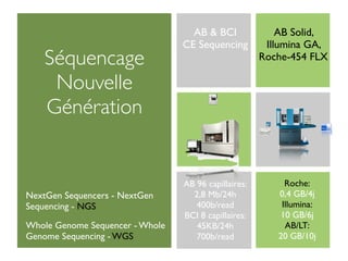
- 1. AB & BCI AB Solid, CE Sequencing Illumina GA, Séquencage Roche-454 FLX Nouvelle Génération Roche: AB 96 capillaires: 0,4 GB/4j 2,8 Mb/24h NextGen Sequencers - NextGen Illumina: 400b/read Sequencing - NGS 10 GB/6j BCI 8 capillaires: Whole Genome Sequencer - Whole AB/LT: 45KB/24h 20 GB/10j Genome Sequencing - WGS 700b/read
- 2. AB & BCI AB Solid, CE Sequencing Illumina GA, Séquencage Roche-454 FLX Nouvelle Génération Roche: AB 96 capillaires: 0,4 GB/4j 2,8 Mb/24h NextGen Sequencers - NextGen Illumina: 400b/read Sequencing - NGS 10 GB/6j BCI 8 capillaires: Whole Genome Sequencer - Whole AB/LT: 45KB/24h 20 GB/10j Genome Sequencing - WGS 700b/read
- 3. Virus: 3500 à 8 x 10⁵ bases Bactéries plus de1Mb (Escherichia coli = 4,7 Mb) Basics 1 kilobase 1kb = 1 000 bases Eucaryotes de 10 à 3 x 10⁵ Mb levure = 1,3 Mb drosophile = 165 Mb 1 mégabase 1Mb 1 000 000 bases 1 million Homo sapiens 3400 Mb 3Gb 20 000-25 000 genes Transcriptome = 2% Genome 1 gigabase 1 Gb 1000 Mb 1 milliard
- 4. Avant: le séquencage enzymatique = SANGER Sequencing ADN simple brin + ADN polymérase addition d ’un didéoxy.en petite quantité (ddNTP) 4 réactions pour les 4 bases en //, chacune avec 1 didéoxy. différent synthèse arrêtée à chaque incorporation d ’un didéoxy. statistiquement, autant de fragments avortés que de fois où la base est représentée
- 5. Avant: le séquencage enzymatique = SANGER Sequencing ADN simple brin + ADN polymérase addition d ’un didéoxy.en petite quantité (ddNTP) 4 réactions pour les 4 bases en //, chacune avec 1 didéoxy. différent synthèse arrêtée à chaque incorporation d ’un didéoxy. statistiquement, autant de fragments avortés que de fois où la base est représentée
- 6. Avant: entre gels plats et capillaires
- 7. Avant: entre gels plats et capillaires
- 8. Avant: entre gels plats et capillaires
- 10. Cout séquencage: Idée du 3+1+(0.4+4.5+0.4)x2=14.6€/1séq. ds de 700b CEQ 8 capillaires: 33.000b ds/24h (48x2x700b) cout du Cout séquencage de 33.000b ds: 688€ CEquencing Cout séquencage de 1Mb ds: 20.848€ Bioinformatique, confirmation: 5min/1000b 7hrs/33.000b
- 11. Roche GS-FLXti 0.4 Gb/run Next Generation 1m reads @ 400b Sequencers €5990/run €14.97/Mb €500k/inst. Illumina GA2 NextGen Sequencers - NextGen Sequencing 5-10 Gb/run (NGS) 60m reads @ 50b Whole Genome Sequencer - Whole Genome Sequencing (WGS) $8250(€6180)/run (5Gb) $0,33(€0,25)/Mb $460k(€344k)/inst. AB Solid 3.0 10-20 Gb/run 100m reads @ 50b €5300/run 5+5Gb The competition: €0,53/Mb Helicos Biosciences, Pacific Biosiences, George Church Lab., Nanopores sequencing, ZS-Genetics, Sequencing by TEM... €462k/inst.
- 12. The Polonator G.007 is the first quot;open sourcequot; gene sequencing instrument to hit Other Players the lab market in which the instrument's software (Web ware) and specifications are freely available to the public. At $150,000, the Polonator is the cheapest instrument on the market George Church Lab. + Danaher Motion: Polonator G.007 The HeliScope™ Single Molecule Sequencer is the first Helicos BioSciences Corp.: HeliScope SMS genetic analyzer to harness the power of direct DNA measurement, enabled by Helicos ZS-Genetics: Electron Microscopy Sequencing. By the first True Single Molecule Sequencing (tSMS)™ half of 2009, the system is expected to read complete a haploid technology. human genome in approximately 8 days, with 4X coverage, at a cost in the tens of thousands of dollars. Pacific BioSciences published technology for Single Molecule Realtime Sequencing SMRT. Instrument by 2010 Moebius Biosystems: Nexus. Over 6 Gigabases in 24hrs. Nanopore sequencing: Oxorf Nanopore, Sequenom...etc Pacific BioSciences
- 13. Roche Applied-Science GS-20, GS-FLX, GS-FLXti (454)
- 14. • GS-FLXti Data emPCR Sequencing DNA Library Preparation and Titration 4.5 h and 10.5 h 8h 10 h Genome fragmented by nebulization No cloning; no colony picking sstDNA library created with adapters A/B fragments selected using avidin-biotin purification gDNA sstDNA library Process Steps 1. DNA library preparation
- 15. • GS-FLXti Data emPCR Sequencing DNA Library Preparation and Titration 4.5 h and 10.5 h 8h 10 h Break microreactors, Anneal sstDNA to Emulsify beads and Clonal amplification enrich for DNA- an excess of DNA PCR reagents in water- occurs inside positive beads capture beads in-oil microreactors microreactors sstDNA library Clonally-amplified sstDNA attached to bead Process Steps 2. emulsion PCR
- 16. •Multiple optical fibers are fused to form an optical array. •Proprietary etching method produces wells that serve as picoliter reaction vessels. •Each well is only able to accept a single DNA bead. Load sequencing Load PicoTiterPlate Load genome into •Reactions in the reagents device on instrument PicoTiterPlate device wells are Close and Press GO! – sequence genome measured of the CCD camera. Process Steps •Titanium plate: 3. Sequencing with the PicoTiterPlate 3.4m wells device
- 17. •Multiple optical fibers are fused to form an optical array. •Proprietary etching method produces wells that serve as picoliter reaction vessels. •Each well is only able to accept a single DNA bead. Load sequencing Load PicoTiterPlate Load genome into •Reactions in the reagents device on instrument PicoTiterPlate device wells are Close and Press GO! – sequence genome measured of the CCD camera. Process Steps •Titanium plate: 3. Sequencing with the PicoTiterPlate 3.4m wells device
- 18. •Multiple optical fibers are fused to form an optical array. •Proprietary etching method produces wells that serve as picoliter reaction vessels. •Each well is only able to accept a single DNA bead. Load sequencing Load PicoTiterPlate Load genome into •Reactions in the reagents device on instrument PicoTiterPlate device wells are Close and Press GO! – sequence genome measured of the CCD camera. Process Steps •Titanium plate: 3. Sequencing with the PicoTiterPlate 3.4m wells device
- 19. •Multiple optical fibers are fused to form an optical array. •Proprietary etching method produces wells that serve as picoliter reaction vessels. •Each well is only able to accept a single DNA bead. Load sequencing Load PicoTiterPlate Load genome into •Reactions in the reagents device on instrument PicoTiterPlate device wells are Close and Press GO! – sequence genome measured of the CCD camera. Process Steps •Titanium plate: 3. Sequencing with the PicoTiterPlate 3.4m wells device
- 20. •Multiple optical fibers are fused to form an optical array. •Proprietary etching method produces wells that serve as picoliter reaction vessels. •Each well is only able to accept a single DNA bead. Load sequencing Load PicoTiterPlate Load genome into •Reactions in the reagents device on instrument PicoTiterPlate device wells are Close and Press GO! – sequence genome measured of the CCD camera. Process Steps •Titanium plate: 3. Sequencing with the PicoTiterPlate 3.4m wells device
- 21. •Multiple optical fibers are fused to form an optical array. •Proprietary etching method produces wells that serve as picoliter reaction vessels. •Each well is only able to accept a single DNA bead. Load sequencing Load PicoTiterPlate Load genome into •Reactions in the reagents device on instrument PicoTiterPlate device wells are Close and Press GO! – sequence genome measured of the CCD camera. Process Steps •Titanium plate: 3. Sequencing with the PicoTiterPlate 3.4m wells device
- 22. DNA Library Preparation and Titration emPCR Sequencing • GS-FLXti Data 4.5 h and 10.5 h 8h 10 h 3.4 m wells 3.4 m reads obtained in parallel A single clonally amplified sstDNA bead is deposited per well. Amplified sstDNA library beads Quality filtered bases DNA capture 4 bases (TACG) bead containing cycled 200 times millions of copies Chemiluminescent of a single clonal signal generation fragment Signal processing to determine base sequence and quality score Amplified sstDNA library beads Quality filtered bases Process Steps 3. Sequencing
- 23. T •Raw data is C G processed A from a series of individual T images. •Each well’s data is extracted, Signal output from a single well Metric and image viewing software quantified, (flowgram) and normalized. •Read data is converted into flowgrams. Process Steps 4. Signal-processing
- 24. •Raw data is processed from a series of individual images. Key sequence = TCAG for identifying wells and calibration •Each well’s Flow of individual bases (TCAG) is 42 times. data is TA extracted, CG quantified, and normalized. TTCTGCGAA •Read data is converted into flowgrams. Base flow Signal strength Process Steps 4. Signal-processing
- 25. • Quality filtered bases • GS-FLXti Data 400-500 bp average read length > 0.4 Gb or 1m reads with a 70 x 75 mm FLXti PicoTiterPlate device 10 hours run time • Phred-like quality score for use in available assemblers or viewers • Consensus base-called contig files - FASTA file of assembled reads mapping against known scaffold (resequencing) de novo assembly of individual bases in consensus contigs • Viewer-ready genome file - assembly file in .ace format • Assembly metric files • Run-time metrics files - summarize important information pertaining to sequencing quality for each run Process Steps 5. Data output
- 26. Software Software Mapping Mapping Image Signal Sequence Reference sequence FlowMapper Software Reference sequence Fragments (reads)
- 27. • GS-FLXti Data Sanger: Weeks 454: 4 days Sanger Technology 7 days Weeks Preparation* Total Sequencing Time - DNA Library Preparation - 180 runs (1 per 4 hours) - Cloning - 2-million-base (Mb) genome - Template Preparation - 6x coverage 454 Technology 2.5 days 1 day Preparation Total Sequencing Time - DNA Library Preparation - 1 run (10 hours) - Titration of Library Beads - 400-600 million-base (Mb) - emPCR Technology Comparison Sanger vs. 454 technology for a 2-million-base genome
- 28. NextGen Sequencers Roche GS-FLX: Workflow IT steps: Workflow 3-4 days (setup) + 1 day (run) 1. Generation of a single-stranded template DNA library (~8-16 hours) 2. Emulsion-based clonal amplification of the library (~8 hours) GS-FLX Software 3. Data generation via sequencing-by-synthesis (9 hours) 4. Image and Base calling analysis (~8 hours) ▪GS Reference Mapper 5. Data analysis using different bioinformatics tools ▪GS De Novo Assembler •Long Single Reads / Standard Shotgun (required input = 3–5μg,5μg recommended) ▪GS Amplicon Variant Analyzer ~1,000,000 single reads with an average read length of 400 bases •Paired End Reads (required input = 5μg @25 ng/μl or above, in TE; >10kb) ◦3K Long-Tag Paired End Reads. Sequence 100 bases from each end of a 3,000 base span on a single sequence read (Figure). Co-assemble GS FLX Titanium shotgun reads with 3K Third Party Software Long-Tag Paired Ends reads from Standard series runs. •Sequence Capture (required input = 3–5μg) ◦Roche NimbleGen Sequence Capture using a single microarray hybridization-based enrichment process. •Amplicon Sequencing (1-5ng or 10-50ng) ◦The DNA-sample preparation for Amplicon Sequencing with the GS FLX System consists of a simple PCR amplification reaction with special Fusion Primers. The Fusion Primer consists of a 20-25 bp target-specific sequence (3' end) and a 19 bp fixed sequence (Primer A or Primer B on the 5' end).
- 29. NextGen Sequencers Roche GS-FLX: Workflow IT steps: Workflow 3-4 days (setup) + 1 day (run) 1. Generation of a single-stranded template DNA library (~8-16 hours) 2. Emulsion-based clonal amplification of the library (~8 hours) GS-FLX Software 3. Data generation via sequencing-by-synthesis (9 hours) 4. Image and Base calling analysis (~8 hours) ▪GS Reference Mapper 5. Data analysis using different bioinformatics tools ▪GS De Novo Assembler •Long Single Reads / Standard Shotgun (required input = 3–5μg,5μg recommended) ▪GS Amplicon Variant Analyzer ~1,000,000 single reads with an average read length of 400 bases •Paired End Reads (required input = 5μg @25 ng/μl or above, in TE; >10kb) ◦3K Long-Tag Paired End Reads. Sequence 100 bases from each end of a 3,000 base span on a single sequence read (Figure). Co-assemble GS FLX Titanium shotgun reads with 3K Third Party Software Long-Tag Paired Ends reads from Standard series runs. •Sequence Capture (required input = 3–5μg) ◦Roche NimbleGen Sequence Capture using a single microarray hybridization-based enrichment process. •Amplicon Sequencing (1-5ng or 10-50ng) ◦The DNA-sample preparation for Amplicon Sequencing with the GS FLX System consists of a simple PCR amplification reaction with special Fusion Primers. The Fusion Primer consists of a 20-25 bp target-specific sequence (3' end) and a 19 bp fixed sequence (Primer A or Primer B on the 5' end).
- 30. NextGen Roche GS-FLX: Sequencers add-ons not included - Nebulizers + nitrogen tank Nebulization is required to shear fragments for DNA >70-800bp - emPCR Breaking Kit This device is required for the preparation of consistently sized reactors for emulsion PCR. - Magnetic Concentrator IVGN +€5000 - MT plate centrifuge BCI +€15.000 - Multisizer™ 3 COULTER counter +€15.000 The most versatile and accurate particle sizing and counting analyzer available today. Using The Coulter Principle, also known as ESZ (Electrical Sensing Zone Method), the Multisizer 3 COULTER COUNTER provides number, volume, mass and surface area size distributions in one measurement, with an overall sizing range of 0.4 µm to 1,200 - Agilent BioAnalyzer +€20.000 - Titanium cluster station +€29.000
- 31. Roche FLXti: Next Generation 0.5 Gb/run 1m reads @ 400b Sequencers €5990/run €14.97/Mb €585k/inst. tot The Roche Roche FLXti: Setup time: 3-4 d 0.5 Gb/run System Run time: 10 hrs images: 27 GB Primary Analysis: 15 GB PA CPU time: 80-220 hrs (6-7 hrs with cluster st) Final file size: 4 GB notes: 400-500b frag. length sequencing future dev. up to 1000b x coverage with long frag. vs x+n coverage with short reads vs cost/ Mb 10 systems in France Multiplexing capacity ≈200 publications
- 32. illumina Genome Analyzer (Solexa)
- 33. Illumina's Solexa Sequencing Technology Step 1: Sample Preparation The DNA sample of interest is sheared to appropriate size (average ~800bp) using a compressed air device known as a nebulizer. The ends of the DNA are polished, and two unique adapters are ligated to the fragments. Ligated fragments of the size range of 150-200bp are isolated via gel extraction and amplified using limited cycles of PCR. 1.5 days. Steps 2-6: Cluster Generation by Bridge Amplification In contrast to the 454 and ABI methods which use a bead-based emulsion PCR to generate quot;poloniesquot;, Illumina utilizes a unique quot;bridgedquot; amplification reaction that occurs on the surface of the flow cell. The flow cell surface is coated with single stranded oligonucleotides that correspond to the sequences of the adapters ligated during the sample preparation stage. Single-stranded, adapter-ligated fragments are bound to the surface of the flow cell exposed to reagents for polyermase-based extension. Priming occurs as the free/distal end of a ligated fragment quot;bridgesquot; to a complementary oligo on the surface. Repeated denaturation and extension results in localized amplification of single molecules in millions of unique locations across the flow cell surface. This process occurs in what is referred to as Illumina's quot;cluster stationquot;, an automated flow cell processor. 8hrs.
- 34. Illumina's Solexa Sequencing Technology Step 1: Sample Preparation The DNA sample of interest is sheared to appropriate size (average ~800bp) using a compressed air device known as a nebulizer. The ends of the DNA are polished, and two unique adapters are ligated to the fragments. Ligated fragments of the size range of 150-200bp are isolated via gel extraction and amplified using limited cycles of PCR. 1.5 days. Steps 2-6: Cluster Generation by Bridge Amplification In contrast to the 454 and ABI methods which use a bead-based emulsion PCR to generate quot;poloniesquot;, Illumina utilizes a unique quot;bridgedquot; amplification reaction that occurs on the surface of the flow cell. The flow cell surface is coated with single stranded oligonucleotides that correspond to the sequences of the adapters ligated during the sample preparation stage. Single-stranded, adapter-ligated fragments are bound to the surface of the flow cell exposed to reagents for polyermase-based extension. Priming occurs as the free/distal end of a ligated fragment quot;bridgesquot; to a complementary oligo on the surface. Repeated denaturation and extension results in localized amplification of single molecules in millions of unique locations across the flow cell surface. This process occurs in what is referred to as Illumina's quot;cluster stationquot;, an automated flow cell processor. 8hrs.
- 35. Illumina's Solexa Sequencing Technology Step 1: Sample Preparation The DNA sample of interest is sheared to appropriate size (average ~800bp) using a compressed air device known as a nebulizer. The ends of the DNA are polished, and two unique adapters are ligated to the fragments. Ligated fragments of the size range of 150-200bp are isolated via gel extraction and amplified using limited cycles of PCR. 1.5 days. Steps 2-6: Cluster Generation by Bridge Amplification In contrast to the 454 and ABI methods which use a bead-based emulsion PCR to generate quot;poloniesquot;, Illumina utilizes a unique quot;bridgedquot; amplification reaction that occurs on the surface of the flow cell. The flow cell surface is coated with single stranded oligonucleotides that correspond to the sequences of the adapters ligated during the sample preparation stage. Single-stranded, adapter-ligated fragments are bound to the surface of the flow cell exposed to reagents for polyermase-based extension. Priming occurs as the free/distal end of a ligated fragment quot;bridgesquot; to a complementary oligo on the surface. Repeated denaturation and extension results in localized amplification of single molecules in millions of unique locations across the flow cell surface. This process occurs in what is referred to as Illumina's quot;cluster stationquot;, an automated flow cell processor. 8hrs.
- 36. Illumina's Solexa Sequencing Technology Step 1: Sample Preparation The DNA sample of interest is sheared to appropriate size (average ~800bp) using a compressed air device known as a nebulizer. The ends of the DNA are polished, and two unique adapters are ligated to the fragments. Ligated fragments of the size range of 150-200bp are isolated via gel extraction and amplified using limited cycles of PCR. 1.5 days. Steps 2-6: Cluster Generation by Bridge Amplification In contrast to the 454 and ABI methods which use a bead-based emulsion PCR to generate quot;poloniesquot;, Illumina utilizes a unique quot;bridgedquot; amplification reaction that occurs on the surface of the flow cell. The flow cell surface is coated with single stranded oligonucleotides that correspond to the sequences of the adapters ligated during the sample preparation stage. Single-stranded, adapter-ligated fragments are bound to the surface of the flow cell exposed to reagents for polyermase-based extension. Priming occurs as the free/distal end of a ligated fragment quot;bridgesquot; to a complementary oligo on the surface. Repeated denaturation and extension results in localized amplification of single molecules in millions of unique locations across the flow cell surface. This process occurs in what is referred to as Illumina's quot;cluster stationquot;, an automated flow cell processor. 8hrs.
- 37. Illumina's Solexa Sequencing Technology Step 1: Sample Preparation The DNA sample of interest is sheared to appropriate size (average ~800bp) using a compressed air device known as a nebulizer. The ends of the DNA are polished, and two unique adapters are ligated to the fragments. Ligated fragments of the size range of 150-200bp are isolated via gel extraction and amplified using limited cycles of PCR. 1.5 days. Steps 2-6: Cluster Generation by Bridge Amplification In contrast to the 454 and ABI methods which use a bead-based emulsion PCR to generate quot;poloniesquot;, Illumina utilizes a unique quot;bridgedquot; amplification reaction that occurs on the surface of the flow cell. The flow cell surface is coated with single stranded oligonucleotides that correspond to the sequences of the adapters ligated during the sample preparation stage. Single-stranded, adapter-ligated fragments are bound to the surface of the flow cell exposed to reagents for polyermase-based extension. Priming occurs as the free/distal end of a ligated fragment quot;bridgesquot; to a complementary oligo on the surface. Repeated denaturation and extension results in localized amplification of single molecules in millions of unique locations across the flow cell surface. This process occurs in what is referred to as Illumina's quot;cluster stationquot;, an automated flow cell processor. 8hrs.
- 38. Illumina's Solexa Sequencing Technology Step 1: Sample Preparation The DNA sample of interest is sheared to appropriate size (average ~800bp) using a compressed air device known as a nebulizer. The ends of the DNA are polished, and two unique adapters are ligated to the fragments. Ligated fragments of the size range of 150-200bp are isolated via gel extraction and amplified using limited cycles of PCR. 1.5 days. Steps 2-6: Cluster Generation by Bridge Amplification In contrast to the 454 and ABI methods which use a bead-based emulsion PCR to generate quot;poloniesquot;, Illumina utilizes a unique quot;bridgedquot; amplification reaction that occurs on the surface of the flow cell. The flow cell surface is coated with single stranded oligonucleotides that correspond to the sequences of the adapters ligated during the sample preparation stage. Single-stranded, adapter-ligated fragments are bound to the surface of the flow cell exposed to reagents for polyermase-based extension. Priming occurs as the free/distal end of a ligated fragment quot;bridgesquot; to a complementary oligo on the surface. Repeated denaturation and extension results in localized amplification of single molecules in millions of unique locations across the flow cell surface. This process occurs in what is referred to as Illumina's quot;cluster stationquot;, an automated flow cell processor. 8hrs.
- 39. Illumina's Solexa Sequencing Technology Steps 7-12: Sequencing by Synthesis A flow cell containing millions of unique clusters is now loaded into the 1G sequencer for automated cycles of extension and imaging. The first cycle of sequencing consists first of the incorporation of a single fluorescent nucleotide, followed by high resolution imaging of the entire flow cell. These images represent the data collected for the first base. Any signal above background identifies the physical location of a cluster (or polony), and the fluorescent emission identifies which of the four bases was incorporated at that position. This cycle is repeated, one base at a time, generating a series of images each representing a single base extension at a specific cluster. Base calls are derived with an algorithm that identifies the emission color over time. At this time reports of useful Illumina reads range from 26-50 bases. The use of physical location to identify unique reads is a critical concept for all next generation sequencing systems. The density of the reads and the ability to image them without interfering noise is vital to the throughput of a given instrument. Each platform has its own unique issues that determine this number, 454 is limited by the number of wells in their PicoTiterPlate, Illumina is limited by fragment length that can effectively quot;bridgequot;, and all providers are limited by flow cell real estate. 2-6 days (18-36 cycles).
- 40. Illumina's Solexa Sequencing Technology Steps 7-12: Sequencing by Synthesis A flow cell containing millions of unique clusters is now loaded into the 1G sequencer for automated cycles of extension and imaging. The first cycle of sequencing consists first of the incorporation of a single fluorescent nucleotide, followed by high resolution imaging of the entire flow cell. These images represent the data collected for the first base. Any signal above background identifies the physical location of a cluster (or polony), and the fluorescent emission identifies which of the four bases was incorporated at that position. This cycle is repeated, one base at a time, generating a series of images each representing a single base extension at a specific cluster. Base calls are derived with an algorithm that identifies the emission color over time. At this time reports of useful Illumina reads range from 26-50 bases. The use of physical location to identify unique reads is a critical concept for all next generation sequencing systems. The density of the reads and the ability to image them without interfering noise is vital to the throughput of a given instrument. Each platform has its own unique issues that determine this number, 454 is limited by the number of wells in their PicoTiterPlate, Illumina is limited by fragment length that can effectively quot;bridgequot;, and all providers are limited by flow cell real estate. 2-6 days (18-36 cycles).
- 41. Illumina's Solexa Sequencing Technology Steps 7-12: Sequencing by Synthesis A flow cell containing millions of unique clusters is now loaded into the 1G sequencer for automated cycles of extension and imaging. The first cycle of sequencing consists first of the incorporation of a single fluorescent nucleotide, followed by high resolution imaging of the entire flow cell. These images represent the data collected for the first base. Any signal above background identifies the physical location of a cluster (or polony), and the fluorescent emission identifies which of the four bases was incorporated at that position. This cycle is repeated, one base at a time, generating a series of images each representing a single base extension at a specific cluster. Base calls are derived with an algorithm that identifies the emission color over time. At this time reports of useful Illumina reads range from 26-50 bases. The use of physical location to identify unique reads is a critical concept for all next generation sequencing systems. The density of the reads and the ability to image them without interfering noise is vital to the throughput of a given instrument. Each platform has its own unique issues that determine this number, 454 is limited by the number of wells in their PicoTiterPlate, Illumina is limited by fragment length that can effectively quot;bridgequot;, and all providers are limited by flow cell real estate. 2-6 days (18-36 cycles).
- 42. Illumina's Solexa Sequencing Technology Steps 7-12: Sequencing by Synthesis A flow cell containing millions of unique clusters is now loaded into the 1G sequencer for automated cycles of extension and imaging. The first cycle of sequencing consists first of the incorporation of a single fluorescent nucleotide, followed by high resolution imaging of the entire flow cell. These images represent the data collected for the first base. Any signal above background identifies the physical location of a cluster (or polony), and the fluorescent emission identifies which of the four bases was incorporated at that position. This cycle is repeated, one base at a time, generating a series of images each representing a single base extension at a specific cluster. Base calls are derived with an algorithm that identifies the emission color over time. At this time reports of useful Illumina reads range from 26-50 bases. The use of physical location to identify unique reads is a critical concept for all next generation sequencing systems. The density of the reads and the ability to image them without interfering noise is vital to the throughput of a given instrument. Each platform has its own unique issues that determine this number, 454 is limited by the number of wells in their PicoTiterPlate, Illumina is limited by fragment length that can effectively quot;bridgequot;, and all providers are limited by flow cell real estate. 2-6 days (18-36 cycles).
- 43. Illumina's Solexa Sequencing Technology Steps 7-12: Sequencing by Synthesis A flow cell containing millions of unique clusters is now loaded into the 1G sequencer for automated cycles of extension and imaging. The first cycle of sequencing consists first of the incorporation of a single fluorescent nucleotide, followed by high resolution imaging of the entire flow cell. These images represent the data collected for the first base. Any signal above background identifies the physical location of a cluster (or polony), and the fluorescent emission identifies which of the four bases was incorporated at that position. This cycle is repeated, one base at a time, generating a series of images each representing a single base extension at a specific cluster. Base calls are derived with an algorithm that identifies the emission color over time. At this time reports of useful Illumina reads range from 26-50 bases. The use of physical location to identify unique reads is a critical concept for all next generation sequencing systems. The density of the reads and the ability to image them without interfering noise is vital to the throughput of a given instrument. Each platform has its own unique issues that determine this number, 454 is limited by the number of wells in their PicoTiterPlate, Illumina is limited by fragment length that can effectively quot;bridgequot;, and all providers are limited by flow cell real estate. 2-6 days (18-36 cycles).
- 44. Illumina's Solexa Sequencing Technology Steps 7-12: Sequencing by Synthesis A flow cell containing millions of unique clusters is now loaded into the 1G sequencer for automated cycles of extension and imaging. The first cycle of sequencing consists first of the incorporation of a single fluorescent nucleotide, followed by high resolution imaging of the entire flow cell. These images represent the data collected for the first base. Any signal above background identifies the physical location of a cluster (or polony), and the fluorescent emission identifies which of the four bases was incorporated at that position. This cycle is repeated, one base at a time, generating a series of images each representing a single base extension at a specific cluster. Base calls are derived with an algorithm that identifies the emission color over time. At this time reports of useful Illumina reads range from 26-50 bases. The use of physical location to identify unique reads is a critical concept for all next generation sequencing systems. The density of the reads and the ability to image them without interfering noise is vital to the throughput of a given instrument. Each platform has its own unique issues that determine this number, 454 is limited by the number of wells in their PicoTiterPlate, Illumina is limited by fragment length that can effectively quot;bridgequot;, and all providers are limited by flow cell real estate. 2-6 days (18-36 cycles).
- 45. Pipeline software highlights Automated image calibration: maximizes the number of clusters used to generate sequence data Accurate cluster intensity scoring algorithms: allow efficient filtering for high-quality reads Quality-calibrated base calls: minimize the propagation of downstream sequencing errors Highly optimized genomic alignment tools: minimize the need for elaborate computer infrastructures Open source code: enables researchers to customize the software to meet their needs
- 46. Sanger: Weeks Illumina: <7 days Technology Comparison Sanger vs. Solexa technology for a 2-Gigabase genome
- 47. Sanger: Weeks Illumina: <7 days Technology Comparison Sanger vs. Solexa technology for a 2-Gigabase genome
- 48. NextGen Illumina GA2: Sequencers Workflow ▪ Tracking Samples ready for sample prep ▪ Samples ready for cluster prep Workflow 2-3 days (setup) + 2-3 days (run) ▪ Flow cells ready for sequencing 1. Non amplified DNA/RNA Sample 2. QC and possibly purify 3. Process with appropriate Sample Prep Kit 4. QC sample prep ▪ Serve analysis files to DAS2 enabled genome DAS2 server 5. Assemble 7 samples with the same number of cycles, library browsers for direct visualization of results types, and sample types without file download 6. Process grouped samples with appropriate Cluster Generation Kit ▪ Private server up and going using Authentication 7. Run cluster generation Mapping application (to handle 5-100 million 15-50bp sequences) 8. Transfer flow cell onto Genome Analyzer ▪ 9. Run sequencing 1st cycle Filter sequences by quality score ▪ 10. QC 1st cycle Count and remove identical sequences ▪ 11. Run remaining cycles Map sequences to reference genome 12. Export data Filter application ▪ Take binary map files and filter based on type of 13. Run analysis aligment and # of counts ▪ Export filtered universal binary for downstream applications Distributed Annotation System (DAS) defines a communication protocol used to exchange biological annotations
- 49. NextGen Illumina GA2: Sequencers add-ons not included - Cluster Station +$50.000 The Cluster Station is a standalone, software- controlled system for the automated generation of clonal clusters from single molecule fragments on Illumina Genome Analyzer flow cells. - Paired-End Module +$45.000 The Paired-End Module provides fully automated template preparation for the second round of sequencing in a paired-end sequencing run. - IPAR +$60.000 IPAR is a bundled hardware and software solution that provides real-time quality control and integrated online processing of primary data during sequencing runs - Agilent BioAnalyzer +€20.000 Total: €126.000
- 50. Illumina GA2: Next Generation 5-10 Gb/run (50b) $8250 (€6180)/run (5Gb) Sequencers $0,33/Mb €480/inst. tot The Illumina Illumina GA2: Setup time: 2-3 d 6-11 Gb/run System Run time: 3-6 d images: 900 GB Primary Analysis: 350 GB PA CPU time: 100 hrs Final File Size: 75 GB notes: 7/15 Gb by end of 2009 72 frag. length 9 systems in France 325 publications Multiplexing capacity
- 51. Applied BioSystems SOLID system (Agencourt BioScience)
- 52. SOLiD v2 instrument components The SOLiD™ Instrument consists of the following components: • Reagent delivery system • Electronics • Camera (4 megapixel) • Monitor stand • Independently controlled dual flow cells • Liquid waste container SOLiD v2 computer system instrument controller • Hardware: Intel® Xeon® processors • Operating system: Microsoft® Windows® XP Pro • Installed RAM: 4 GB • Hard disk storage: dual 80 GB SATA hard drives (RAID-1) head node • Hardware: Intel® Xeon® Dual Core processors (2) • Operating system: 64-bit LINUX • Installed RAM: 8 GB • Hard disk storage: dual 750 GB SATA hard drives (RAID-1) compute nodes (each) • Hardware: Intel® Xeon® Dual Core processors (2) • Operating System: 64-bit LINUX • Installed RAM: 8 GB SOLID in details • Hard disk storage: 80 GB SATA hard drives storage • Hard disk storage: 15x 750 GB SATA hard drives • Operating system: 64-bit LINUX • RAID-5 w/ hot spare
- 53. Figure 1. Library generation schematic. Sequencing on the SOLiD machine starts with library preparation. In the simplest fragment library, two different adapters are ligated to sheared genomic DNA (left panel of Fig. 1). If more rigorous structural analysis is desired, a “mate-pair” library can be generated in a similar fashion, by incorporating a circularization/ cleavage step prior to adapter ligation (right panel of Fig.1). ABI's SOLID Sequencing Technology
- 54. Figure 2. Clonal bead library generation via emulsion PCR. Once the adapters are ligated to the library, emulsion PCR is conducted using the common primers to generate “bead clones” which each contain a single nucleic acid species. ABI's SOLID Sequencing Technology
- 55. Figure 3. Depositing beads into flow cell via end modifications. Each bead is then attached to the surface of a flow cell via 3’ modifications to the DNA strands. At this point, we have a flow cell (basically a microscope slide that can be serially exposed to any liquids desired) whose surface is coated with thousands of beads each containing a single genomic DNA species, with unique adapters on either end. Each microbead can be considered a separate sequencing reaction which is monitored simultaneously via sequential digital imaging. Up to this point all next- gen sequencing technologies are very similar, this is where ABI/SOLiD diverges dramatically (see next). ABI's SOLID Sequencing Technology
- 56. Each oligo has degenerate positions at 3’ bases 1-3 (N’s), and one of 16 specific dinucleotides at positions 4-5. Positions 6 through the 5’ are also degenerate, and hold one of four fluorescent dyes. The sequencing involves: 1. Hybridization and ligation of a specific oligo whose 4th & 5th bases match that of the template 2. Detection of the specific fluor 3. Cleavage of all bases to the 5’ of base 5 4. Repeat, this time querying the 9th & 10th Figure 4. Schematic of ABI SOLiD sequencing chemistry. bases 5. After 5-7 cycles of this, perform a “reset”, in which the initial primer and all ligated portions The actual base detection is no longer done by the polymerase-driven incorporation of are melted from the labeled dideoxy terminators. Instead, SOLiD uses a mixture of labeled oligonucleotides template and and queries the input strand with ligase. Understanding the labeled oligo mixture is discarded. key to understanding SOLiD technology. 6. Next a new initial primer is used that is N-1 in length. Repeating the initial cycling (steps 1-4) now ABI's SOLID Sequencing generates an overlapping data set (bases 3/4, 8/9, etc, Technology see Fig 5).
- 57. Each oligo has degenerate positions at 3’ bases 1-3 (N’s), and one of 16 specific dinucleotides at positions 4-5. Positions 6 through the 5’ are also degenerate, and hold one of four fluorescent dyes. The sequencing involves: 1. Hybridization and ligation of a specific oligo whose 4th & 5th bases match that of the template 2. Detection of the specific fluor 3. Cleavage of all bases to the 5’ of base 5 4. Repeat, this time querying the 9th & 10th Figure 4. Schematic of ABI SOLiD sequencing chemistry. bases 5. After 5-7 cycles of this, perform a “reset”, in which the initial primer and all ligated portions The actual base detection is no longer done by the polymerase-driven incorporation of are melted from the labeled dideoxy terminators. Instead, SOLiD uses a mixture of labeled oligonucleotides template and and queries the input strand with ligase. Understanding the labeled oligo mixture is discarded. key to understanding SOLiD technology. 6. Next a new initial primer is used that is N-1 in length. Repeating the initial cycling (steps 1-4) now ABI's SOLID Sequencing generates an overlapping data set (bases 3/4, 8/9, etc, Technology see Fig 5).
- 58. For example (see Fig. 4), the dinucleotides CA, AC, TG, and GT are all encoded by the green dye. Because each base is queried twice it is possible, using the two colors, to determine which bases were at which positions. This two color query Figure 5. Sequencing coverage during SOLiD sequencing cycles system (known as “color space” in ABI- Thus, 5-7 ligation reactions followed by a 4-5 primer reset cycles are repeated speak) has some generating sequence data for ~35 contiguous bases, in which each base has interesting been queried by two different oligonucleotides. consequences with regard to the If you’re doing the math you’ve realized there are 16 possible dinucleotides identification of errors. (4^2) and only 4 dyes. So data from a single color does not tell you what base is at a given position. This is where the brilliance (and potential confusion) comes about with regard to SOLiD. There are 4 oligos for every dye, meaning there are four dinucleotides that are encoded by each dye. ABI's SOLID Sequencing Technology
- 59. For example (see Fig. 4), the dinucleotides CA, AC, TG, and GT are all encoded by the green dye. Because each base is queried twice it is possible, using the two colors, to determine which bases were at which positions. This two color query Figure 5. Sequencing coverage during SOLiD sequencing cycles system (known as “color space” in ABI- Thus, 5-7 ligation reactions followed by a 4-5 primer reset cycles are repeated speak) has some generating sequence data for ~35 contiguous bases, in which each base has interesting been queried by two different oligonucleotides. consequences with regard to the If you’re doing the math you’ve realized there are 16 possible dinucleotides identification of errors. (4^2) and only 4 dyes. So data from a single color does not tell you what base is at a given position. This is where the brilliance (and potential confusion) comes about with regard to SOLiD. There are 4 oligos for every dye, meaning there are four dinucleotides that are encoded by each dye. ABI's SOLID Sequencing Technology
- 60. NextGen AB Solid 3.0: Sequencers Workflow Workflow: 3-4 days (setup) + 4-10 days (run)
- 61. NextGen AB Solid 3.0: Sequencers Workflow Workflow: 3-4 days (setup) + 4-10 days (run)
- 62. NextGen AB Solid 3.0: Sequencers Workflow Workflow: 3-4 days (setup) + 4-10 days (run)
- 63. NextGen AB Solid 3.0: Sequencers Workflow Workflow: 3-4 days (setup) + 4-10 days (run)
- 64. NextGen AB Solid 3.0: Sequencers add-ons Covaris S2 System ULTRA-TURRAX Tube Drive from IKA The Covaris™ S2 System is required sample preparation instrument for use This device is required for the in the SOLiD™ System workflow. The preparation of consistently sized instrument is an essential part of the reactors for emulsion PCR. emulsion PCR process used to prepare the beads for emulsion PCR. The Hydroshear from Covaris System is also used to shear DNA into 60 bp fragments for fragment Genomic Solutions library preparation. The Hydroshear® from Genomic Solutions® is a reproducible and included controllable method for generating random DNA fragments of specific sizes. Use this to prepare mate- paired libraries for the SOLiD™ System. not included - Agilent BioAnalyzer +€20.000
- 65. AB Solid 3.0 Next Generation 10-20 Gb/run 100m reads @ 50b €5300/run 5+5Gb Sequencers €0,53/Mb €482k/inst. tot The SOLID AB Solid 3.0: Setup time: 3-5 d 5-12.5 Gb/run/slide System Run time: 3.5-10 d images: 2.5 TB Primary Analysis: 750 GB PA CPU time: in run time Final file size: 140 GB notes: The Scientist Top Innovation of 2008 125-400m reads in 2009 30/40Gb potential for 12x human genome @ $10.000 3 systems in France Multiplexing capacity
- 66. Roche GS-FLXti: Roche GS-FLXti: 0.5 Gb/run Setup time: 3-4 d 1m reads @ 400b 0.4Gb/run Run time: 10 hrs images: 27 GB €5990/run Primary Analysis: 15 GB PA CPU time: 220 hrs €14.97/Mb Final file size: 4 GB €585k/inst. tot Illumina GA2: Illumina GA2: 5-10 Gb/run (50b) Setup time: 2-3 d 6-11 Gb/run €6180/run (5Gb) Run time: 3-6 d images: 900 GB Primary Analysis: 350 GB €0,25/Mb PA CPU time: 100 hrs €480/inst. tot Final File Size: 75 GB AB Solid 3.0 AB Solid 3.0: 10-20 Gb/run Setup time: 3-5 d 100m reads @ 50b 5-12.5 Gb/run/slide Run time: 3.5-10 d €5300/run 5+5Gb images: 2.5 TB Primary Analysis: 750 GB PA CPU time: in run time €0,53/Mb Final file size: 140 GB €482k/inst. tot
- 67. Roche GS-FLXti General Infrastructure Laboratory 1 Controlled Room (emPCR) Amplicon Room Requirements General Laboratory 2 BioIT room Illumina GA2 - Lab space, dedicated rooms General Laboratory 1 - Hands on IT infrastructure Cluster Station room - Data Storage capacity General Laboratory 2 BioIT room -Sample and wor kflow tracability solutions General Laboratory 1 Controlled Room (emPCR) Amplicon - BioIT group support for 3rd Room General party analysis Laboratory 2 BioIT room AB Solid 3.0
- 68. NextGen Sequencing Service Providers Europe Many locations Cogenics http://www.cogenics.com/sequencing/s...ingService.cfm Many locations GATC Biotech http://www.gatc-biotech.com/en/index.php Germany dkfz http://www.dkfz.de/gpcf/ngs_sequencing.html Germany Functional Genomics Center zurich http://www.fgcz.ethz.ch/applications/gt/ngsequencing Germany Eurofins MWG Operon http://www.eurofinsdna.com/products-...equencing.html Hungary BAYGEN http://baygen.hu/ The Netherlands ServiceXS http://www.servicexs.com/servicexs+i...+ii+sequencing Spain Sistemas Genómicos http://www.sistemasgenomicos.com/ Sweden Sweden Uppsala Genome Center http://www.genpat.uu.se/node453 Switzerland Fasteris http://www.fasteris.com/ UK AGOWA - LGC http://www.lgc.co.uk/pdf/Next%20gen%...lyer%20web.p UK The Gene Pool https://www.wiki.ed.ac.uk/display/GenePool/Home UK Geneservice http://www.geneservice.co.uk/services/sequencing/ UK University of Liverpool http://www.liv.ac.uk/agf/index.html Belgium DNAVision (soon available) http://www.dnavision.be/ GATC Illumina platform based: 3500 € HT 1/8 flow cell vs 772 € Roche platform based: 10.150 € HT 1/2 picoplate vs 2995 € Cogenics 10/2008 Roche platform based: 15.000 € HT 1 full picoplate vs 5990 €
- 69. Whole genome Amplicon seq. Transcriptome seq. - Mutations / SNP - cDNA sequencing - Small RNA - de novo sequencing - comparative seq. Methylation seq. Metagenomics ChIP sequencing Les Applications
- 70. AB: Roche 1, 4, 8 regions slides 16-128 samples/slide with barcoding AB Illumina: 2, 4, 8, regions Flow Cell flow cells – 1.4mm wide channel design – 40% more usable area Roche: 2, 4, 8, 16 regions Illumina plates Multiple Sample Sequencing
- 71. Roche (192) AB (256) Illumina (96) Increase Sample Throughput via Multiplex Identifiers
- 72. AB Illumina Paired-end Sequencing Roche
- 73. Increase Selectivity via Capture all human exons on CHIP selection 7 chips