Penetrating Injuries to the Neck
•Als PPTX, PDF herunterladen•
7 gefällt mir•4,390 views

Penetrating injuries to the neck is a great summary of how to assess and manage neck wounds from lacerations to the airway to gunshot wounds. The talk covers relevant anatomy, the zones of the neck and how to investigate vascular, tracheal and oesophageal injuries. A comprehensive understanding of the relevat anatomy is essential to recognising associated injury patterns. The improvements in the accuracy of helical CTA scans has meant that the delineation of the zones of the neck has become less relevant to the further investigation and management of pemetrating neck wounds. Oesphageal injuries remain difficult to detect and require a high level of clinical suspicion to identify these.

Empfohlen
Weitere ähnliche Inhalte
Was ist angesagt?
Was ist angesagt? (20)
Andere mochten auch
Andere mochten auch (20)
Mehr von SMACC Conference
Mehr von SMACC Conference (20)
Kürzlich hochgeladen
Kürzlich hochgeladen (20)
Penetrating Injuries to the Neck
- 6. Anatomy
- 10. Zones – prognosis and managment
- 20. Questions?
- 21. Key points • Exan • Leading cause of immediate death is exsanguination • Esophageal injuries represents the most frequently missed injury and may be leading cause of delayed death • Compound difficulties in evaluation & Mx is the complicated anatomy - dense concentration of vital structures in a small space • Ongoing debate : mandatory vs selective exploration
Hinweis der Redaktion
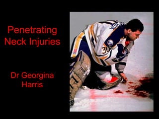
- Clint Malarchuk –1989 Goaelie for Sabre – Ice hockey team from Buffalo New York
- It is a topic where anatomy is critical…….anatomy trumps pathophysiology for once, so as a surgeon, I really lke this topic. It Makes up 10-15% of trauma pts so its important to have a good understanding of the mx While mx of the unstable pt is exciting but pretty straight forward, the management of the stable pt remains a source of debate.
- Historically, penetrating neck injuries is quite an interesting topic. Largely the domain of the military surgeons Initially mortality was huge – higher than 30%. During the first world war, there wasn’t too much that could be done. WW2 - surgical advances meant that all patients with neck injuries were explored.. As injuries to imporant sturctures began to be pciked up earlier and treated, mortality began to drop. So by the time of the korean war and vietnam way, …….
- With mandatory exploration of everyone with a PNI as well as mobile army surgical hospitals, the mortality rate was as low as 3% More recently…… mortality has started to rise again… …but largely due to wartime advances. Things such as high velocity rifles, improvised explosive devices…… Because when it comes to the damage that is caused in these inuries, it all comes down to kinetic energy.
- And with KE, velocity is much much more important than the mass or size of the projectile. With weapons , you talk about the muzzle velocities …hand guns for intstance have velocities 200-600m/s. But once you start getting speeds over 600m/s, as you do with high powered rifles, you get this effect know as cavitiation…….
- The problem with the neck is the anatomy – complex way in which a large number of essential structures are crammed into a small area. Problem with PNI is that there is a lot of vital structures, all packed into a small area. Just wanted to take a brief moment to highlight some of the more important anatomical structures…. If you know your anatomy and where exactly some of these more critical structures are, it helps in the management of any PNI Vascular structures – Jugular viens(external) in my experience is most common injury. Often easily seen on people as it is one of the most lateral structures. If you draw a line from the angle of the mandible to mid clavicle, that is its pathway, passing over the SCM obliquely Internal jugular along with the carotids lie underneath the SCM which protects it, less so in the upper neck. Carotid is obviously deeper and slightly more anterior to Jug. To outline the pathway of these vessles, you take a line from just in front of the ear, to the medial end of the clavicle. The bifurcation is around the same level as the thyroid cartilate. Vertebral arteries, deeper and more posterior Brachiocephalic/subclavial vessles become important lower neck injuries as the rise and arch over in the root of the neck.
- Laryngeal cartilages midline and susceptiable to any anterior injury Mangament of airway Eosphagus really starts around the level of your cricoid and the pharynx is everything above that.
- Nervous structures – not just cranial nerves, also branchial plexus, sympathetic chain and obviously spinal cord (injury to SC is relatively rare in PNI) Thing to remember is that these neural sturctures are often in close proximity to vascular structures so neuropathies can be a clue to possible vascualr injuries…… cranial nerves CN7,9 if higher in the neck, vagus, accessory and hypoglossal can be injured directly in the neck but also at the jugular foramen where 9,10 and 11 all come down with the internal jug. sympathetic trunk that travels within the carotid sheath – horners brachial plexus in lower neck between anterior and middle scalene so important to examine the hand spinal cord injury uncommon I’d be interested in any comments at the end about this - utiltiy of neck collars recently in the news and I would have to say that in these injuries where the incidence is so low, and the ability to be able to assess both initially and over tie is imporatnat,, that neck collars shouldn’t be used unless there are definate concerns.
- Vascular structures, neural structures, airway, digestive tract…….all intertwined in a complex manner within a small area. Overlying all of these structures, and protecting them to some degree is the platysma muscle. The essence of any PNI is that it penetrates this muscle. Attaches to fascia of pec and deltoid muscles, crosses clavicles, some fibres attaching superiorly to mandible and others interlacing with skin and superficial muscles of the face. So you can describe PNI by location, often using SCM as reference as this is boundary for anterior and posterior neck triangles…….or you can talk about the zones of the neck.
- PNI categorised by zones – carries implications for management and prognosis
- Zone one From clavicles to cricoid cartilage Includes Arch of aorta, innominate, subclavian, vertebral Lung apices Thoracic duct on the left Brachial plexus This is the zone with the highest mortality – 12% Dangerous due to proximaty to large thoracic vessels and the lungs Osseous shield which protects it on one hand but also makes surgical access difficult on the other Pt with injuries in this zone you would consider doing angiogram. Injuries are not only hard to see, but also hard to access.
- Zone 2 mid neck - Most common zone of injury (up to 75%) of injuries Cricoid cartilage to angle of mandible Structures of note in this zone are the carotids, vugular veins Cranial nerves Sympathetic chain Larynx and pharynx No osseous shield Surgical access is much better and obtaining distal and proximal control of bleeders is possible This is the zone where Elective v mandatory exploration becomes controversial Historically, around 90’s people started realising that mandatory explorations was not nesessary Large number of negative explorations Not cost effective Imaging advances such as helical CT becoming widelly available and commonlace People began advocating conservative mx but those that apposed it agued that not all injuries could be picked up with just physical examination. Literature supports both arguments…..
- Zone 3 Angle of mandible up to base of skull Vascular structures – ext/int carotids, jugular, vertebral and prevertebral plexus Oral cavity and pharynx Neural structures – consider facial nerve trunk Vascular injuries in this area are much more challenging to be repaired surgically Proximity to skull base and mandible offering osseous shield Mandatory exploration is not recommended and like zone 1, angiogram often recommended.
- So how does this help us……its all very well to be able to describe the injuries but does it really matter. As an ENT surgeon, I’m not going to talk to a critical care crowd about how to go through the management of these pts….but rather look at each zone and the implications it has on management. The only two things that I’ll say about prehospital treatment, and I’d be interested if anyone has any comments on this is ?? collars – spinal injury very unlikely and immobilisation often done at expense of easy, accurate and continued examination air embolism…..often mentioned but I’d say that if a pt has an airway injury, letting them find the best way position to help maintain their airway is best. Prehospital pearls No collars – spinal injury very unlikely and immobilisation often done at expense of an accurate examination. Bag mask ventilation can be problematic and may lead to surgical emphysema if airway injury and significant anatomical distortion Bubbling or sucking neck wounds – beware air embolism ?????Think tension pneumothorax if arresting??????
- Attacked by fighting rooster……. Initially assessed; puncture wounds to face & neck; D/C’d RTER 24 hr later w/ fever, neck swelling, & respiratory distress Neck: crepitus; inflammation; induration CXR: pneumomediastinum Endoscopic EUA: 0.5 cm perforation of lateral wall of pharynx Neck explored through lateral incision pus drained NG feeds N contrast study POD#10 D/C HD#14 on N diet
- Larynx and trachea Pharynx and eosophagus behind