Primary Maternal Care: Skills workshop Virginal examination in pregnancy
•
5 gefällt mir•1,835 views

Primary Maternal Care addresses the needs of healthcare workers in level 1 district hospitals and clinics who provide antenatal and postnatal care, but do not conduct deliveries. It is adapted from theory chapters and skills workshops from Maternal Care. This book complements the national protocol of antenatal care in South Africa. It covers: booking for antenatal care, assessing fetal growth and well being, hypertensive disorders of pregnancy, antepartum haemorrhage, preterm labour, important medical conditions

Empfohlen
Empfohlen
Weitere ähnliche Inhalte
Was ist angesagt?
Was ist angesagt? (20)
Andere mochten auch
Andere mochten auch (20)
Ähnlich wie Primary Maternal Care: Skills workshop Virginal examination in pregnancy
Ähnlich wie Primary Maternal Care: Skills workshop Virginal examination in pregnancy (20)
Mehr von Saide OER Africa
Mehr von Saide OER Africa (20)
Kürzlich hochgeladen
Kürzlich hochgeladen (20)
Primary Maternal Care: Skills workshop Virginal examination in pregnancy
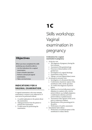
- 1. 1C Skills workshop: Vaginal examination in pregnancy A Indications for a vaginal Objectives examination in pregnancy 1. At the first visit: When you have completed this skills • The diagnosis of pregnancy during the first trimester. workshop you should be able to: • Assessment of the gestational age. • List the indications for a vaginal • Detection of abnormalities in the examination. genital tract. • Insert a bivalve speculum. • Investigation of a vaginal discharge. • Perform a bimanual vaginal • Examination of the cervix. examination. • Taking a cervical (Papanicolaou) smear. 2. At subsequent antenatal visits: • Take a cervical smear. • Investigation of a threatened abortion. • Confirmation of preterm rupture of the membranes with a sterile speculum. INDICATIONS FOR A • To confirm the diagnosis of preterm labour. VAGINAL EXAMINATION • Detection of cervical effacement and/or dilatation in a patient with a risk for A vaginal examination is the most intimate preterm labour, e.g. multiple pregnancy, examination a woman is ever subjected to. It a midtrimester abortion, previous must never be performed without: preterm labour or polyhyramnios. 1. A careful explanation to the patient about • Assessment of the ripeness of the cervix the examination. prior to induction of labour. 2. Asking permission from the patient to • Identification of the presenting part in perform the examination. the pelvis. 3. A valid reason for performing the • Performance of a pelvic assessment. examination. 3. Immediately before labour: • Performance of artificial rupture of the membranes to induce labour.
- 2. SK ILLS WORKSHOP : VAGINAL EXAMINATION IN PREGNANC Y 49 B Contraindications to a vaginal E Speculum examination examination in pregnancy 1. A speculum examination is always 1. Antepartum haemorrhage. However, there performed at the first antenatal visit. are 2 exceptions to this rule: At subsequent antenatal visits this • A cephalic presentation with the fetal examination is only done when indicated, head palpable 2/5 or less above the e.g. to investigate a vaginal discharge or in pelvic brim (i.e. engaged), thereby, the case of preterm or prelabour rupture of excluding a placenta praevia. the membranes. • Obvious signs and symptoms of 2. The Cusco or bivalve speculum is the one abruptio placentae. commonly used. 2. Preterm and prelabour rupture of the membranes without contractions (except F Insertion of a bivalve speculum with a sterile speculum to confirm or exclude rupture of the membranes). 1. The procedure must be explained to the patient. 2. The labia are parted with the fingers of the METHOD OF VAGINAL gloved left hand. 3. The patient is asked to bear down. EXAMINATION 4. The closed speculum is gently inserted posteriorly into the vagina. Great care must be taken to avoid undue contact with the C Preparation for vaginal examination anterior vaginal wall at the introitus as this 1. The bladder must be empty. causes great discomfort, or even pain, from 2. The procedure must be carefully explained pressure on the urethra. to the patient. 5. As soon as the speculum has passed 3. The patient is put in the dorsal or through the vaginal opening, the blades lithotomy position: must be slightly opened. The speculum is • The dorsal position is more comfortable now inserted deeper into the vagina. When and less embarrassing than the the cervix is reached, the speculum is fully lithotomy position and does not require opened. This method allows for inspection any equipment. This is the position of the vaginal walls during insertion and most often used. ensures that the cervix is found. • The lithotomy position provides better 6. Any vaginal discharge must be identified. access to the genital tract than the Where needed, a sample is taken with a dorsal position. Lithotomy poles and wooden spatula. stirrups are required. 7. The vagina is inspected for congenital abnormalities such as a vaginal septum, a vaginal stenosis or a double vagina and A vaginal examination must always be preceded cervix. by an abdominal examination. 8. The cervix is inspected for any laceration or tumour. A smooth, red area surroun- ding the external os that retains the normal D Examination of the vulva smooth surface, is normal during the reproductive years and is called ectopy. The vulva must be carefully inspected for any 9. If there is a history of rupture of the abnormalities, e.g. scars, warts, varicosities, membranes, the presence of liquor is noted congenital abnormalities, ulcers or discharge. and tested for. 10. A cervical (Papanicolaou) smear must be taken if a smear has not been taken recently.
- 3. 50 PRIMAR Y MATERNAL CARE 11. At the end of the examination the H Performing a bimanual examination speculum is gently withdrawn, keeping it 1. First 1 and then, where possible, 2 gloved slightly open, so that the vaginal walls can and lubricated fingers are gently inserted again be inspected all the way out. into the vagina. 2. If a vaginal septum or stenosis is present, G Taking a cervical smear the patient should be referred to a 1. A cervical (Papanicolaou) smear is taken doctor to decide whether delivery will be to detect abnormalities of the cervix, e.g. interfered with. human papilloma virus infection, cervical 3. The cervix is palpated and the following intra-epithelial neoplasia or carcinoma of are noted: the cervix. • Any dilatation. 2. Ideally the first cervical smear should be • The length of the cervix in cm, i.e. taken when the patient becomes sexually whether the cervix is effaced or not. active. In practice the first smear is usually • The surface should be smooth and taken when the patient first attends a regular. family planning or antenatal clinic. • The consistency which will become 3. If the cervical smear is normal, it should be softer during pregnancy. repeated at 30, 40 and 50 years of age. 4. Special care must be taken, when 4. The technique of taking a cervical smear is performing a bimanual examination late as follows: in pregnancy and in the presence of a high • The name, folder number and date must presenting part, not to damage a low-lying be written on the slide with a pencil placenta. If the latter is suspected, a finger beforehand. Also make sure that a spray must not be inserted into the cervical can is close at hand to fix the slide. canal. Instead, the presenting part is gently • A vaginal speculum is inserted. palpated through all the fornices. If any • The cervix must be clearly seen and is bogginess is noted between the fingers of carefully inspected. the examining hand and the presenting • A suitable spatula is inserted into part, the examination must be immediately the cervix and rotated through 360 abandoned and the patient must be degrees, making sure that the whole referred urgently for ultrasonography. circumference is gently scraped. It is 5. Where possible the presenting part is important that the smear is taken from identified. the inside of the cervical canal as well 6. A most important part of the bimanual as from the surface of the cervix. An examination is the determination of the Ayres (Aylesbury) or tongue spatula gestational age, by estimating the size of the must be used and not a brush with uterus and comparing it with the period of sharp or long points such as a spatula, amenorrhoea. This is only really accurate Cervibrush or Cytobrush. in the first trimester. Thereafter, the fundal • The material obtained is smeared onto height and the size of the fetus must be a glass slide and immediately sprayed determined by abdominal examination. with Papanicolaou’s fixative. 7. The uterine wall is palpated for any • When the slide is dry, it is sent to the irregularity, suggesting the presence of a laboratory for examination. congenital abnormality (e.g. bicornuate uterus) or myomata (fibroids). 8. Lastly, the fornices are palpated to exclude any masses, the commonest of which is an ovarian cyst or tumour.
- 4. SK ILLS WORKSHOP : VAGINAL EXAMINATION IN PREGNANC Y 51 I Explanation to the patient her how far pregnant she is, if that can be determined, and to reassure her, if everything Do not forget to explain to the patient, after appears to be normal. the examination is completed, what you have found. It is especially important to tell