The Most Attractive Hyderabad Call Girls Kothapet 𖠋 9332606886 𖠋 Will You Mis...
Neurophysiology1
1. Kisii University, School of Health Sciences, Department of
Nursing NEUROPHYSIOLOGY By Nyaboga E
Esther Nyaboga Page 1
NEUROPHYSIOLOGY
Deals with the function of the nervous system.
Nerve:
Bundle of conducting fibres enclosed in a sheath called epineurium.
Its function is to transmit impulses between any part of the body and a nerve centre.
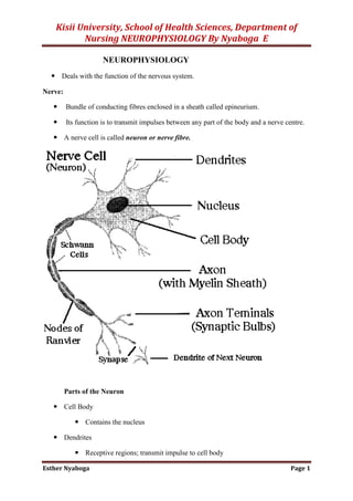
A nerve cell is called neuron or nerve fibre.
Parts of the Neuron
Cell Body
Contains the nucleus
Dendrites
Receptive regions; transmit impulse to cell body
2. Kisii University, School of Health Sciences, Department of
Nursing NEUROPHYSIOLOGY By Nyaboga E
Esther Nyaboga Page 2
Short, often highly branched
May be modified to form receptors
Axons
Transmit impulses away from cell body
Axon hillock; trigger zone
Where action potentials first develop
Presynaptic terminals (terminal boutons)
Contain neurotransmitter substance (NT)
Release of NT stimulates impulse in next neuron
Bundles of axons form nerves
In the peripheral nervous system, axons and dendrites are “wrapped” in specialized
cells called Schwann cells. During embryonic development, Schwann cells grow to
surround the neuron processes, enclosing them in several layers of Schwann cell
membrane.
These layers are the myelin sheath; myelin is a phospholipid that electrically
insulates neurons from one another.
The nuclei and cytoplasm of the Schwann cells are wrapped around the outside of the
myelin sheath and are called the neurolemma, which becomes very important if
nerves are damaged.
The Schwann cells are also believed to produce a chemical growth factor that
stimulates regeneration.
In the central nervous system, the myelin sheaths are formed by oligodendrocytes,
one of the neuroglia (glial cells), the specialized cells found only in the brain and
spinal cord.
Because no Schwann cells are present, however, there is no neurolemma, and
regeneration of neurons does not occur. This is why severing of the spinal cord, for
example, results in permanent loss of function.
SYNAPSES
The small gap or space between the axon of one neuron and the dendrites or cell body
of the next neuron is called the synapse.
3. Kisii University, School of Health Sciences, Department of
Nursing NEUROPHYSIOLOGY By Nyaboga E
Esther Nyaboga Page 3
Within the synaptic knob (terminal end) of the presynaptic axon is a chemical
neurotransmitter that is released into the synapse by the arrival of an electrical nerve
impulse. The neurotransmitter diffuses across the synapse, combines with specific
receptor sites on the cell membrane of the postsynaptic neuron, and there generates an
electrical impulse that is, in turn, carried by this neuron’s axon to the next synapse,
and so forth.
A chemical inactivator at the cell body or dendrite of the postsynaptic neuron
quickly inactivates the neurotransmitter. This prevents unwanted, continuous
impulses, unless a new impulse from the first neuron releases more neurotransmitter.
Many synapses are termed excitatory, because the neurotransmitter causes the
postsynaptic neuron to depolarize (become more negative outside as Na ions enter the
cell) and transmit an electrical impulse to another neuron, muscle cell, or gland. Some
synapses, however, are inhibitory, meaning that the neurotransmitter causes the
postsynaptic neuron to hyperpolarize (become even more positive outside as K ions
leave the cell or Cl ions enter the cell) and therefore not transmit an electrical
impulse.
Such inhibitory synapses are important, for example, for slowing the heart rate, and
for balancing the excitatory impulses transmitted to skeletal muscles. With respect to
the skeletal muscles, this inhibition prevents excessive contraction and is important
for coordination
Synaptic Functions of Neurons
As the impulses are transmitted from one neuron to the next:
Each impulse may be blocked in its transmission from one neuron to the next.
May be changed from one single impulse to repetitive impulses. Or
May be integrated with impulses from other neurons to cause highly intricate pattern
of impulses in successive neurons. All these functions can be classified as synaptic
functions of neurons.
Types of synapses:
1. Chemical synapse
Almost all synapses in the cns are chemical synapses. In these the first neuron
secretes at its nerve ending synapse a chemical substance called neurotransmitter
which acts on receptor proteins in the membrane of the next neuron to excite the
neuron, inhibit it or modify its sensitivity. Exa. of neurotransmitters are acetylcholine,
norepinephrine, epinephrine, histamine, serotonin etc.
2. Electrical synapse
4. Kisii University, School of Health Sciences, Department of
Nursing NEUROPHYSIOLOGY By Nyaboga E
Esther Nyaboga Page 4
Are characterized by direct open fluid channels that conduct electricity from one cell
to another. Most of these consist of small protein tubular structures called gap
junctions that allow free movement of ions from interior of one cell to interior of the
next.
‘one-way conduction’ at the chemical synapses
Chemical synapse transmit signals in one direction i.e. from one neuron that secretes
the transmitter substance called presynaptic neuron to the neuron on which transmitter
acts called postsynaptic neuron. This is the principle one-way conduction at chemical
synapses.
Electrical synapses often transmit signals in either direction.
The advantage of one-way conduction is that it allows signals to be directed towards
specific goals e.g. sensation, motor control, memory etc.
Physiologic Anatomy of the Synapse
Mitochondria at the prsynaptic neuron site
5. Kisii University, School of Health Sciences, Department of
Nursing NEUROPHYSIOLOGY By Nyaboga E
Esther Nyaboga Page 5
Presynaptic terminals/terminal knobs/synaptic knobs/boutons has two internal structures
important to the excitatory or inhibitory function of the synapse.
1. Transmitter vesicle: it contains the transmitter substance released to synaptic cleft
which excites or inhibits the postsynaptic neuron. It excites if the neuronal membrane
contains excitatory receptors & inhibits if the membrane contains inhibitory receptors.
2. Mitochondria: it provides ATP to supply energy for synthesis of new transmitter
substances.
Release and action of the neurotransmitter. How does it happen?
its membrane
into the cleft
in permeability characteristics of the postsynaptic neuronal membrane which leads to
excitation or inhibition of the postsynaptic neuron depending on neuronal receptor
characteristics.
Mechanism by which an action potential causes transmitter Release from the
presynaptic Terminal: Role of calcium ion.
large number of voltage-gated calcium channels
channels open and allow more calcium ions to flow into the terminal
into the synaptic cleft is directly related to the number of calcium ions that enter
The mechanism
It’s believed that when calcium ions enter the presynaptic terminal they bind with
special protein molecules on the inside surface of presynaptic membrane called
release sites. This binding causes release sites to open through the membrane
allowing a few transmitter vesicles to release their transmitter into a cleft after each
single action potential.
Action of the transmitter substance on the postsynaptic neuron: function of ‘receptor
proteins’
Postsynaptic membrane contain large number of receptor proteins. The molecules of these
receptors have two important components:
1. A binding component: protrudes outward from the membrane to synaptic cleft where it
binds the neurotransmitter coming from presynaptic terminal.
2. An ionophore component: passes all the way through the postsynaptic membrane to
the interior of the postsynaptic neuron
6. Kisii University, School of Health Sciences, Department of
Nursing NEUROPHYSIOLOGY By Nyaboga E
Esther Nyaboga Page 6
Types of ionophore :
1. An ion channel that allows passage of specified ions through the membrane
2. A secondary messanger activator that is not an ion channel, which is a molecule that
protrudes into the cell cytoplasm & activates one or more substances inside
postsynaptic neuron. These substance inturn serves as second messengers to increase
or decrease cellular function.
NERVE IMPULSE
The events of an electrical nerve impulse are the same as those of the electrical
impulse generated in muscle fibers. A neuron not carrying an impulse is in a state of
polarization, (resting state) with Na ions more abundant outside the cell, and K ions
and negative ions more abundant inside the cell. The neuron has a positive charge on
the outside of the cell membrane and a relative negative charge inside.
A stimulus (such as a neurotransmitter) makes the membrane very permeable to Na
ions, which rush into the cell. This brings about depolarization, a reversal of charges
on the membrane. The outside now has a negative charge, and the inside has a
positive charge.
As soon as depolarization takes place, the neuron membrane becomes very permeable
to K ions, which rush out of the cell. This restores the positive charge outside and the
negative charge inside, and is called repolarization.
(The term action potential refers to depolarization followed by repolarization.)
Then the sodium and potassium pumps return Na ions outside and K ions inside, and
the neuron is ready to respond to another stimulus and transmit another impulse.
Transmission of electrical impulses is very rapid. The presence of an insulating
myelin sheath increases the velocity of impulses since only the nodes of Ranvier
depolarize. This is called saltatory conduction.
At synapses, nerve impulse transmission changes from electrical to chemical and
depends on the release of neurotransmitters. Although diffusion across synapses is
slow, the synapses are so small that this does not significantly affect the velocity of
impulses.
Electrical Signals
Neurons produce electrical signals called action potentials ( = nerve impulse)
Nerve impulses transfer information from one part of body to another
e.g., receptor to CNS or CNS to effector
7. Kisii University, School of Health Sciences, Department of
Nursing NEUROPHYSIOLOGY By Nyaboga E
Esther Nyaboga Page 7
Electrical properties result from
ionic concentration differences across plasma membrane
permeability of membrane
Electrochemical Gradient of the Neuron Membrane
Electrical Gradient
Develops when there are more positive or negative charges (ions) on one side
of a membrane than on the other
Charges (ions) move toward the area of opposite charge
Positive toward negative and vice versa
Chemical Gradient
Develops when there are more ions of a substance in one area than in another
(e.g., more Na+
extracellularly than intracellularly)
Ions tend to move from an area of high concentration to an area of low
concentration; more to less (i.e., down their concentration gradient)
Electrochemical gradient
The sum of all electrical and chemical forces acting across the cell membrane
Resting Membrane Potential (RMP)
Nerve cell has an electrical potential, or voltage across its membrane of a –70 mV; (=
to 1/20th that of a flashlight battery (1.5 v)
The potential is generated by different concentrations of Na+
, K+
, Cl
, and protein
anions (A
)
But the ionic differences are the consequence of:
Differential permeability of the axon membrane to these ions
Operation of a membrane pump called the sodium-potassium pump
What Establishes the RMP?
Diffusion of Na+
and K+
down their concentration gradients
Na+
diffuses into the cell and K+
diffuses out of the cell
BUT, membrane is 75x’s more permeable to K+
than Na+
Thus, more K+
diffuses out than Na+
diffuses in
This increases the number of positive charges on the outside of the membrane
relative to the inside.
8. Kisii University, School of Health Sciences, Department of
Nursing NEUROPHYSIOLOGY By Nyaboga E
Esther Nyaboga Page 8
BUT, the Na+
-K+
pump carries 3 Na+
out for every 2 K+
in.
This is strange in that MORE K+
exited the cell than Na+
entered!
Pumping more + charges out than in also increases the number of + changes
on the outside of the membrane relative to the inside.
AND presence of anionic proteins (A-
) in the cytosol adds to the negativity of the
cytosolic side of the membrane
THEREFORE, the inside of the membrane is measured at a -70 mV (1 mv = one-
thousandth of a volt)
Changes in the Membrane Potential
Membrane potential is dynamic
Rises or falls in response to temporary changes in membrane permeability
Changes in membrane permeability result from the opening or closing of
membrane channels
Types of channels
Passive or leak channels - always open
Gated channels - open or close in response to specific stimuli;
Ligand-gated channels
Voltage-gated channels
Nongated (Leakage) channels
Many more of these for K+
and Cl-
than for Na+
.
So, at rest, more K+
and Cl-
are moving than Na+
.
How are they moving?
Protein repels Cl-
, so Cl-
moves out.
K+
are in higher concentration on inside than out, they diffuse out.
Always open and responsible for permeability when membrane is at rest.
Gated ion channels.
Gated ion channels open and close because of some sort of stimulus. When they
open, they change the permeability of the cell membrane.
Ligand-gated: open or close in response to ligand (a chemical) such as ACh
binding to receptor protein.
9. Kisii University, School of Health Sciences, Department of
Nursing NEUROPHYSIOLOGY By Nyaboga E
Esther Nyaboga Page 9
Acetylcholine (ACh) binds to acetylcholine receptor on a Na+
channel.
Channel opens, Na+
enters the cell.
Ligand-gated channels most abun- dant on dendrites and cell body;
areas where most synaptic commu-nication occurs
Voltage-gated:
open or close in response to small voltage changes across the cell membrane.
At rest, membrane is negative on the inside relative to the outside.
When cell is stimulated, that relative charge changes and voltage-gated ion channels
either open or close.
Most common voltage gated are Na+
, K+
, and Ca+2
Common on areas where action potentials develop
Axons of unipolar and multipolar neurons
Sarcolemma (including T-tubules) of skeletal muscle fibers and cardiac
muscle fibers
Local Potentials/Graded Potentials
Graded: of varying intensity; NOT all the same intensity
Changes in membrane potential that cannot spread far from site of stimulation
Can result in depolarization or hyperpolarization
Depolarization
Opening Na+
channels allows more + charges to enter thereby making interior
less negative (-70 mV -60mV); see next slide
RMP shifts toward O mV
Hyperpolarization
Opening of K+
channels allows more + charges to leave thereby making
interior more negative (-70 mV -80 mV)
RMP shifts away from O mV
Repolarization
Process of restoring membrane potential back to normal (RMP)
Degree of depolarization decreases with distance from stimulation site; called
decremental spread (see next slide)
10. Kisii University, School of Health Sciences, Department of
Nursing NEUROPHYSIOLOGY By Nyaboga E
Esther Nyaboga Page 10
Graded potentials occur on dendrites and cell bodies of neurons but also on gland
cells, sensory receptors, and muscle cell sarcolemma
Characteristics of local potentials:
1. A stimulus causes ion channels to open, resulting in increases permeability of the
membrane to Na+
, K+
or Cl-
.
2. Increased permeability of the membrane to Na+
results in depolarization. Increased
permeability to K+
or Cl-
results in hyperpolarization.
3. Local potentials are graded; that is, the size of the local potential is propotional to the
strength of the stimulus.
4. Local potentials are conducted in a decremental fashion, meaning that their magnitude
decreases as they spread over the plasma membrane. Local potentials cannot be
measures a few millimetres from the point of stimulation.
Action Potential: Resting State
Na+
and K+
channels are closed
Leakage accounts for small movements of Na+
and K+
Each Na+
channel has two voltage-regulated gates
Activation gates – closed in the resting state
Inactivation gates – open in the resting state
Action Potential: Depolarization Phase
11. Kisii University, School of Health Sciences, Department of
Nursing NEUROPHYSIOLOGY By Nyaboga E
Esther Nyaboga Page 11
Some stimulus opens Na+
gates and Na+
influx occurs
K+
gates are closed
Na+
influx causes a reversal of RMP
Interior of membrane now less negative (from -70 mV -55 mV)
Threshold – a critical level of depolarization (-55 to -50 mV)
At threshold, depolarization becomes self-generating
I.e., depolarization of one segment leads to depolarization in the next
If threshold is not reached, no action potential develops
Action Potential: Repolarization Phase
Sodium inactivation gates close
Membrane permeability to Na+
declines to resting levels
As sodium gates close, voltage-sensitive K+
gates open
K+
exits the cell and internal negativity of the resting neuron
is restored
Action Potential: Hyperpolarization
12. Kisii University, School of Health Sciences, Department of
Nursing NEUROPHYSIOLOGY By Nyaboga E
Esther Nyaboga Page 12
Potassium gates remain open, causing an excessive efflux of K+
This efflux causes hyperpolarization of the membrane (undershoot)
The neuron is insensitive to stimulus and depolarization during this time
Depolarization and Hyperpolarization
Phases of the Action Potential (figure below)
1 – RESTING STATE
RMP = -70 mV
2 – DEPOLARIZATION
Increased Na+
influx
Membrane Potential becomes less negative
If threshold is reached, depolarization continues
Peak reached at +30 mV
Total amplitude = 100 mV
3 – REPOLARIZATION
Decreased Na+
influx
Increased K+
efflux
Membrane Potential becomes more negative
4 – HYPERPOLARIZATION
13. Kisii University, School of Health Sciences, Department of
Nursing NEUROPHYSIOLOGY By Nyaboga E
Esther Nyaboga Page 13
Excess K+
efflux
Action Potential: Role of the Sodium-Potassium Pump
Repolarization
Restores the resting electrical conditions of the neuron
Does not restore the resting ionic conditions
Ionic redistribution back to resting conditions is restored by the sodium-potassium
pump
Speed of Impulse Conduction
Faster in myelinated than in non-myelinated
In myelinated axons, lipids act as insulation (the myelin sheath) forcing local currents
to jump from node to node
In myelinated neurons, speed is affected by:
Thickness of myelin sheath
Diameter of axons
14. Kisii University, School of Health Sciences, Department of
Nursing NEUROPHYSIOLOGY By Nyaboga E
Esther Nyaboga Page 14
Large-diameter conduct more rapidly than small-diameter. Large
diameter axons have greater surface area and more voltage-gated Na+
channels
Nerve Fiber Types
Type A: large-diameter (4-20 µm), heavily myelinated. Conduct at 15-120 m/s (= 300
mph).
Motor neurons supplying skeletal muscles and most sensory neurons carrying
info. about position, balance, delicate touch
Type B: medium-diameter (2-4 µm), lightly myelinated. Conduct at 3-15 m/s.
Sensory neurons carrying info. about temperature, pain, general touch,
pressure sensations
Type C: small-diameter (0.5-2 µm), unmyelinated. Conduct at 2 m/s or less.
Many sensory neurons and most ANS motor neurons to smooth muscle,
cardiac muscle, glands
Coding for Stimulus Intensity
All action potentials are alike (of the same amplitude) and are independent of stimulus
intensity.
The amplitude of the action potential is the same for a weak stimulus as it is
for a strong stimulus.
So how does one stimulus feel stronger than another?
Strong stimuli generate more action potentials than weaker stimuli.
More action potentials stimulate the release of more neurotransmitter from the
synaptic knob
The CNS determines stimulus intensity by the frequency of impulse transmission
15. Kisii University, School of Health Sciences, Department of
Nursing NEUROPHYSIOLOGY By Nyaboga E
Esther Nyaboga Page 15
Neuronal Pathways and Circuits
Organization of neurons in CNS varies in complexity
Convergent pathways: several neurons converge on a single postsynaptic
neuron. E.g., synthesis of data in brain.
Divergent pathways: the spread of information from one neuron to several
neurons. E.g., important information can be transmitted to many parts of the
brain.
Oscillating circuits: Arranged in circular fashion to allow action potentials to
cause a neuron in a farther along circuit to produce an action potential more
than once. Can be a single neuron or a group of neurons that are self
stimulating. Continue until neurons are fatigued or until inhibited by other
neurons. Respiration? Wake/sleep.
References
1. Arthur C Guyton and John E Hall (2015).textbook of Medical Physiology, 12th
edition, Elsevier Saunders.
2. Walter F Boron and Emile L Boulpaep (2015). Medical Physiology International
edition 2nd
edition, Saunders Elsevier.