Spettroscopia di neutroni e dinamica proteica
•Als PPT, PDF herunterladen•
0 gefällt mir•626 views

Seminario Prof. Joseph Zaccai 8 aprile 2010 ore 13.15 Dipartimento di Fisica

Empfohlen
Empfohlen
Weitere ähnliche Inhalte
Ähnlich wie Spettroscopia di neutroni e dinamica proteica
Ähnlich wie Spettroscopia di neutroni e dinamica proteica (20)
Mehr von nipslab
Mehr von nipslab (16)
Spettroscopia di neutroni e dinamica proteica
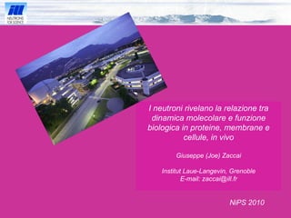
- 1. I neutroni rivelano la relazione tra dinamica molecolare e funzione biologica in proteine, membrane e cellule, in vivo Giuseppe (Joe) Zaccai Institut Laue-Langevin, Grenoble E-mail: zaccai@ill.fr
- 2. > 60 000 strutture nella PDB
- 3. Capire una struttura è capire le forze che la mantengano
- 4. Haloarcula marismortui Sale ! Archaeoglobus fulgidus Temperatura ! Malate dehydrogenase
- 5. _ H + . _ S + ∆ H+,-, 0 ∆ S +,-, 0 Privalov and Khechinashvili (1974) J. Mol. Biol., 86, 665-684 Calorimetria ∆ H+ ∆ S+
- 6. BPTI (Go et al., 1983) 3.56 ps - 1 0.21 ps - 1 Normal Modes
- 7. r i (0) r i ( t ) Neutron Incoherent Scattering Single Particle Scattering k 0 k 1 Q σ (H) >> σ (D and other nuclei)
- 8. ≈ ( 1 / Q ) 2 Å 2 ≈ 2 Å 2 on IN13 at ILL time ≈ 100 ps on the backscattering spectrometer IN13 ≈ 25 Å 2 H 2 O diffusing H on protein The elastic scan : a window in length – time
- 9. Data from Doster et al. (1989), Cordone et al. (2000) hydrated powder in trehalose glass Neutron Scattering: Mean square displacements and E ffective Force Constants in Myoglobin < u 2 > and < k’ > are experimental parameters ~ flexibility, ~ resilience respectively T’ IN 13 8 microeV 100 ps Parametri « High throughput ! » T ( K ) 0 40 80 120 160 200 240 280 0.00 0.30 0.60 0.90 1.20 k' = 0.3N/m k = 2N/m k = 3N/m < u 2 > Å 2 ( ) 320 1.50
- 10. Lavori sul Lisozima di Paciaroni e colleghi: La dinamica di una proteina dipende fortemente dall’ambiente acquoso !
- 11. Frauenfelder et al. (1988) Una chiave per capire : “ Energy landscapes or seascapes”
- 12. quasi-harmonic approximation √ < u 2 > ∆ G d √ < u 2 > < k ’ > ~ ∆G / 2d 2 Bicout and Zaccai, Biophys. J., 2001 Bicout and Zaccai, Biophys. J., 2001 ∆ G ~ k B T ’ < k > < k ’ > E a √ < u 2 > d E a d
- 13. Vita colorata del sale
- 14. 2M NaCl H 2 O 2M NaCl D 2 O 2M KCl D 2 O < k’ > = 0.1 N/m < k’ > = 0.2 N/m < k’ > = 0.5 N/m 200 mg/ml protein Fluctuations and force constants d epend on the solvent and correlate with stability Tehei et al.(2001) 2M NaCl H 2 O 2M NaCl D 2 O 2M KCl D 2 O
- 15. There are Dynamics-Function-Activity Relations Dynamics – Function-Activity
- 16. Biological Dynamics is not as simple as 'Flexibility is required for activity' - it is Function Specific !
- 17. Gabel et al., Biophys J, 2009 Human BuChE and Soman
- 18. Hb e temperatura fisiologica Hb in … solution powder Stadler et al., Biophys J, 2009 Stadler et al., Biophys J, 2008
- 19. Purple Membrane H. Salinarum Bact e riorhodopsin CP CP EC
- 20. Photocycle specific flexibility K J L N O BR 570 M 412 h M 412 CP H + CP EC EC H + A G F E D C B H +
- 21. Dry PM Wet PM Harmonic domain Dynamical transition Activation of photocyle ! ‘ transition of methyls’ H +
- 22. Labelled sample : In-vivo labelling: Fully deuterated PM with hydrogenated retinal, Trp, Met. Retinal binding pocket Extracellular moiety of BR Labelled BR: Sample preparation: EC CP
- 23. Large amplitude motions in labelled unlabelled PM 0 % r.h. 75 % r.h. 86 % r.h. 93 % r.h. < u 2 >, < k’ > Separate flexibility and rigidity
- 24. <u 2 > [Å 2 ] Lipids BR <u 2 > [Å 2 ]
- 25. Less soft core = valve H + Soft body Softer lipid environment All are hydration ... and temperature dependent Degrees of ‘Softness’
- 26. Dynamic adaptation to heat LDH of Rabbit MDH of Methanocaldococcus jannaschii Tehei, M., D. Madern, B. Franzetti & G. Zaccai, J Biol Chem (2005)
- 27. Soft Rabbit muscle LDH Hard Methanocaldococcus jannaschii MalDH
- 28. Escherichia coli mesophile 37°C Aquaspirillum arcticum psychrophile 4°C Proteus mirabilis mesophile 37°C Thermus thermophilus thermophile 65°C Aquifex pyrofilus hyperthermophile 85°C Tehei et al. (2004) Adaptation of the Proteome
- 29. E. coli : alive and dead
- 30. 1,0 1,5 2,0 2,5 270 280 290 300 310 320 0 10 20 30 40 <k'> =(0,39±0,01) N/m <u 2 > (Å 2 ) T (K) B T (°C) 1.0 1.5 2.0 2.5 270 280 290 300 310 320 0 10 20 30 40 T (K) <u 2 > (Å 2 ) <k'> =(0,67±0,11) N/m C T (°C) 1.0 1.5 2.0 2.5 270 280 290 300 310 320 0 10 20 30 40 <k'> =(0,60±0,01) N/m <u 2 > (Å 2 ) T (K) D T (°C) P. mirabilis A. arcticum A. pyrofilus T. thermophilus
- 31. The effective force constant increases to maintain stability
- 32. Same rms fluctuation at physiological temperature ! √
- 33. Una costante di forza adatta per mantere lo stesso valore della flutuazione alla temperatura fisiologica L’evoluzione ha scelto la dinamica !
- 34. Heparan Sulfate, a cell surface polysaccharide Jasnin et al., PCCP, 2010 Cartografia dinamica della cellula < u 2 > (Å 2 )
- 35. < u 2 > (Å 2 ) Proteine di membrana Lipidi poliSaccaridi 1.00 P L S <u 2 > [Å 2 ]
- 36. Michel Ferrand, Valérie Réat, Uschi Lehnert, Martin Weik, Moeava Tehei, Frank Gabel, Katy Wood, Marion Jasnin, Andreas Stadler… Bacteriorhodopsin Dieter Oesterhelt and his Lab, MPI Martinsried Proteins and Cells Dominique Madern et Bruno Franzetti, IBS, Grenoble, Margaret et Ben Zion Ginzburg, Jérusalem, Martine Moulin et Michael Haertlein, D-LAB, Marie-Thérèse Giudici-Orticoni, Marseille Red Blood Cells Gerhard Artmann and his Lab in Aachen, Georg Bueldt in Juelich Senza chi …
- 37. Grazie !