LEUCOCYTES
•Als PPT, PDF herunterladen•
107 gefällt mir•38,670 views

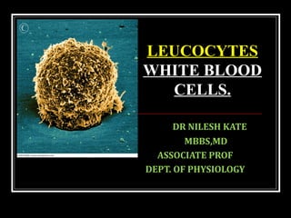
LEUCOCYTES WHITE BLOOD CELLS

Empfohlen
Weitere ähnliche Inhalte
Was ist angesagt?
Was ist angesagt? (20)
Andere mochten auch
Andere mochten auch (20)
Ähnlich wie LEUCOCYTES
Ähnlich wie LEUCOCYTES (20)
Mehr von Dr Nilesh Kate
Mehr von Dr Nilesh Kate (20)
Kürzlich hochgeladen
Kürzlich hochgeladen (20)
LEUCOCYTES
- 1. DR NILESH KATE MBBS,MD ASSOCIATE PROF DEPT. OF PHYSIOLOGY LEUCOCYTES WHITE BLOOD CELLS.
- 2. OBJECTIVES. Types of WBC and their counts Formation of WBC Morphology, life span, functions and variations in count of WBC Applied aspects. Sunday, June 19, 2016
- 3. INTRODUCTION • Crucial in the body’s defense against pathogens • These are complete cells, with a nucleus and organelles • They lack Hb so they are colorless (i.e. white) • Able to move into and out of blood vessels (Diapedesis) • Can respond to chemicals released by damaged tissues (Chemotaxis) • Some are capable of Phagocytosis
- 4. TYPES OF LEUKOCYTES • Granulocytes • Granules in their cytoplasm can be stained • Biologically active substances involved in inflammatory and allergic reactions. • Neutrophils, Eosinophil, and Basophils
- 5. TYPES OF LEUKOCYTES • Agranulocytes • Lack visible cytoplasmic granules • Lymphocytes and Monocytes
- 7. LEUKOCYTE LEVELS IN THE BLOOD • Normal levels =4,000 to 11,000 cells/mm3 • Abnormal leukocyte levels • Leucocytosis • Above 11,000 leukocytes/ml • Generally indicates an infection • Leucopenia • Abnormally low leukocyte level • Commonly caused by certain drugs
- 8. Figure 19.11 WHITE BLOOD CELLS
- 9. CONCEPT OF POOL There are 3 different areas in our body where different WBCs reside 1. Marrow pool: 90% neutrophils 2. Blood pool: 3% 3. Tissue pool: 7% Genesis of WBCs: Leucopoiesis In bone marrow PHSC (Pluripotential hemopoietic→ stem cells) differentiates committed stem cells→ → CFU-GM Granulocytes & monocytes are formed only in bone marrow lymphocytes & plasma cells are produced in various lymphogenous tissues
- 11. REGULATION OF LEUCOPOIESIS Granulopenia or Dead granulocytes and monocytes Release G-CSF M-CSF GM-CSF Interleukins IL-1, IL-3 Stimulate Bone Marrow Normal counts inhibit increased formation Granulocytes, Monocytes/Macrophages Prostaglandins: Monocytes, Lactoferrin Cytokines: glycoproteins formed by monocytes and T- lymphocytes :
- 12. NEUTROPHILS: Size:10-14 µm in diam. Nucleus: 1. Multilobed (1-6 lobes) therefore called polymorphnuclear leucocytes. 2. Young cell have single horse shoe shaped nucleus. 3. As the cells grow older nucleus becomes multilobed. Lobes are connected with one another by chromatin threads. 4. Arneth count: Cytoplasm: contains neutrally stained granules average half-life in the circulation is 6 hours
- 13. NEUTROPHILS: 4 types of granules are present 1. Primary/ Azurophilic granules: less in no. enzymes like i. myeloperoxidase, (produces HOCl for killing bacteria). ii. lysosomal granules containing acid hydrolases, which can digest bacteria, elastase, proteinase, α-1 antitrypsin iii. Antimicrobial proteins like cathepsin-G, Defensins- α, β iv. Tissue destruction during inflammation
- 14. 2. Secondary /Specific/peroxidase negative granules: more numerous. Contain i. Lactoferrin-antibacterial ii. Gelatinase, lysozyme- microbicidal, Vit B12binding protein & iii. Components of enzyme system that produce free radicals like H2O2, which kills the microbes. iv. Substances that facilitates chemotaxis. Sunday, June 19, 2016
- 15. NEUTROPHILS: 3. Tertiary granules: Gelatinase, alkaline phosphatase and cytochrome-b 4. Secretory Granules: secretory vesicles contain CD3, phospholipase and tyrosine kinase Toxic granules: During severe infections toxic coarse granules are seen. Functions: 1. Phagocytosis: 2. Reaction of inflammation: release leukotrienes, prostaglandins, thromboxanes 3. Febrile Response: They contain fever producing substance, endogenous pyrogen which is an important mediator of febrile response to bacterial pyrogens.
- 16. NEUTROPHILS: These enzymes act in a cooperative fashion with the O2 –, H2O2, and HOCl formed by the action of the NADPH oxidase and myeloperoxidase to produce a killing zone around the activated neutrophil.
- 17. PROCESS OF PHAGOCYTOSIS (CELL EATING) Phagocytes engulf and kill microorganisms Steps of Phagocytosis: 1. Margination 2. Emigration and Diapedesis 3. Chemotaxis 4. Recognition and attachment(Opsonization) 5. Engulfment and creation of phagosome 6. Fusion of phagosome with lysosome 7. Destruction and digestion 8. Residual body → Exocytosis
- 18. NEUTROPHIL RECRUITMENT Selectins/Addressins ß2 –Integrin / ICAM-1 flow rolling adhesion transmigration inflammatory mediators Tissue Injury (e.g. Bacterial infection) chemoattractant (e.g. IL-8, C5a), leukotrienes • phagocytosis • oxidant production • lysosomal granules endothelium
- 19. OPSONIZATION AND PHAGOCYTOSIS IgG and C3b opsonin proteins Fc receptors for antibody Complement receptors: (e.g. C3b) Other receptors for collectins (eg. mannose-binding protein) G protein-mediated responses, increased motor activity of the cell, exocytosis, respiratory burst
- 20. OPSONIZATION
- 22. RESPIRATORY BURST: NADPH OXIDASE NADPH oxidase activation
- 23. Superoxide anion: O2- Hydrogen peroxide: H2O2 Hydroxyl radical: OH . Hypochlorous acid: HOCl Myeloperoxidase = MPO O2 + e- 2O2- + 2H+ H2O2 + Fe2+ H2O2 O2- H2O2 + O2 OH + OH- + Fe3+ HOCl + OH-MPO REACTIVE OXYGENREACTIVE OXYGEN METABOLITESMETABOLITES
- 24. VARIATION IN NEUTROPHIL COUNT: Neutrophilia: in↑ neutrophils>10000/mm3 A. Physiological 1)After Exercise, 2)After injection of epinephrine, 3)Pregnancy, menstruation, parturition & lactation, 4)Newborn, 5)After meals, 6)Mental or emotional stress B. Pathological 1)Acute pyogenic (pus forming) bacterial infections, 2) Acute Rheumatic fever, Gout 2)Following tissue destruction, i) Burns ii) After hemorrhage, iii) myocardial infarction, iv) After surgery v) poisoning by lead, mercury,
- 25. NEUTROPENIA: IN NEUTROPHILS↓ : < 2500/mm3 In children Typhoid, paratyphoid fever Viral infection Malaria Aplasia of bone marrow Bone marrow depression due to Chloromycetin, cytotoxic drugs X-rays & radiations Chemical poisons like arsenic
- 26. Size:10-14 µm in diam. (2%) Nucleus: 1. Usually (85%) cells ‘bilobed’. 2. Lobes are connected with one another by chromatin threads thus producing spectacle appearance. 3. Remaining 15% cells have trilobed nucleus. Sunday, June 19, 2016
- 27. Cytoplasm: i. Acidophilic, appears light pink in colour after staining Granular ii. Granules 1. Coarse, stain bright brick red with acidic (eosin) dye. 2. Granules do not cover the nucleus. 3. They contain very high peroxidase content (histaminase), lysozymes, ECF-A & Major Basic Protein (MBP) Sunday, June 19, 2016
- 28. FUNCTIONS: 1. Mild Phagocytosis: less motile than neutrophils 2. Parasitic infestations: Major Basic Protein- Larvicidal Eosinophil Cationic Protein- bactericidal & major destroyer of helminths. Eosinophil Peroxidase – destruction of helminths, bacteria & tumour cells
- 29. FUNCTIONS: 3. Allergic reaction: bronchial asthma & hay fever Detoxifying inflammation inducing subs like bradykinin, histamine inhibit mast cell degranulation phagocytose & destroy Ag-Ab complexes 4. Immunity: specially abundant in the mucosa of respiratory tracts, GIT, urinary tract, where they provide mucosal immunity Sunday, June 19, 2016
- 30. VARIATION IN EOSINOPHIL COUNT Eosinophilia: in↑ eosinophils Causes are:- 1)Allergic conditions e.g. bronchial asthma, hay fever, filariasis 2) Parasitic infestation, trichinosis & schistosomiasis e.g. worms (hookworm, roundworm & tapeworm), 3) Skin disease like utricaria. Eosinopenia: in↓ eosinophils Causes are:- 1) ACTH & steroid therapy 2) Stressful conditions, & 3) Acute pyogenic infections
- 31. BASOPHILS: Size:10-14 µm in diam. Nucleus: irregular bilobed, often ‘S’ shaped & its boundary is not clear because of overcrowding with coarse granules. Cytoplasm: Is slightly basophilic & appear blue, it is full of granules. Granules: coarse, stain deep purple/blue Plenty, completely fill the cell & overload the nucleus Contain heparin, histamine & 5HT.
- 32. FUNCTIONS 1. Mild phagocytosis 2. Role in allergic reaction: Basophils release histamine, bradykinin, slow reacting substance of anaphylaxis (SRS-A) & serotonin (5HT). These substances cause local vascular & tissue reactions that cause many allergic manifestations. 3. Prevents spread of Allergic inflammatory process 4. Liberates heparin which i. Acts as anticoagulant & keeps blood in fluid state. ii. Activates the enzyme lipoprotein lipase which removes fat particles from the blood after fatty meal.
- 33. VARIATION IN BASOPHIL COUNT: Basophilia: in basophil↑ count >100/mm3 Causes are:- 1) Viral infections, e.g. influenza, small pox & chicken pox 2) Allergic diseases 3) Chronic myeloid leukemia. Basopenia: in basophil↓ count Causes are:- 1) Corticosteroid therapy, 2) Drug induced reactions & 3) Acute pyogenic infections
- 35. 2nd line of defence. Size: Largest WBC 18-20 µm. Nucleus: 1. Is large single, eccentric in position (present on one side of the cell). 2. It is notched/ indented (kidney shaped) 3. It has reticulated chromatin network. Cytoplasm: i. Is abundant, pale blue & usually clear with no granules. Granules: i. Sometimes contain fine purple dust like granules called Azur granules
- 36. FUNCTIONS 1. Role in phagocytosis: These are powerful phagocytes & capable of phagocytosis as many as 100 bacteria. They also have ability to engulf large particles such as RBCs & malarial parasites. 2. Role in tumor immunity: kill tumor cells after sensitization by lymphocytes, play a key role in the lymphocyte – mediated immunity. 3. Synthesis of Biological Substances: synthesis complement, prostaglandin E & clot promoting factors Interleukin1 ii) Hemopoietic factors iii) TNF-α, iv)Binding proteins like transferrin,v) lysosomes, Proteases vii) Acid hydrolases
- 37. VARIATION IN COUNT Monocytosis: in m↑ onocyte count Causes are:- 1) Certain bacterial infections, e.g. tuberculosis, syphilis & subacute bacterial endocarditis 2) Viral infections 3) Protozoal & rickettsial infections, e.g. malaria, kala azar Monocytopenia: in↓ monocyte count Causes are:- It is rare, may be seen in hypoplastic bone marrow.
- 38. 2 types of lymphocytes Morphologically: small & large Functionally: T & B lymphocytes Small lymphocytes: 7-10 µm Nucleus rounded, cytoplasm: just rim is seen. Older cells. Large lymphocytes: 10-14 µm Nucleus is big with indentation, definite cytoplasm is seen. Precursor of small lymphocyte.
- 39. Functional subtypes: small lymphocytes are broadly classified into 1. B lymphocyte: processed in the bone marrow, concerned with the humoral immunity. 2. T lymphocyte: processed in thymus, concerned with the cellular immunity Functions of B lymphocytes: B lymphocytes & their derivatives, plasma cells are responsible for humoral (antigen mediated) immunity. They produce antibodies (gamma globulins).This is major mechanism against the invading organisms
- 40. by direct action by making them inactive by agglutination, precipitation, neutralization or lysis and through complement system Functions of T lymphocytes: T lymphocytes are responsible for cellular (Cell mediated/ T cell) immunity. T cell immunity play imp defensive role against: viral & bacterial infections tumor cells Provide a specific immune response to infectious diseases.
- 41. VARIATION IN LYMPHOCYTE COUNT Lymphocytosis: in↑ lymphocyte count Physiological 1. healthy & young children 2. female during menstruation Pathological: 1. Chronic infections like tuberculosis, hepatitis & whooping cough 2. Lymphatic leukemia 3. Viral infections like chicken pox 4. Autoimmune disease like thyrotoxicosis Lymphocytopenia: ↓ in lymphocyte count 1. Patients on corticosteroid & immunosuppressive therapy 2. Hypoplastic bone marrow 3. Widespread irradiation 4. Acquired Immuno Deficiency syndrome (AIDS)
- 42. LIFE SPAN OF WBCS Granulocytes: after released from bone marrow, 4-8 hours circulate in blood & another 4-5 days in the tissues. Survive only for few hours in serious infection Monocytes: 72 hrs in blood. Once in tissue they swell up to much larger size to become tissue macrophage in this form they can live for month.(3)→ Lymphocytes: Life span for week or months depending on body’s need. They continually circulate in blood & move from blood to tissues & from tissues to blood and again blood to tissues.
- 43. LEUKAEMIAS Malignant diseases Increase in total WBC count->50,000/mm3 Presence of immature wbcs in peripheral blood Types of LeukaemiasTypes of Leukaemias 1. Acute myeloblastic leukaemia (AML) 2. Acute lymphoblastic leukaemia (ALL) 3. Chronic myeloid leukaemia (CML) 4. Chronic Lymphoid leukaemia (CLL)
- 44. Thank You