FDG PET/CT Characteristics of Adrenal Benignand Malignant Lesions
•
0 gefällt mir•872 views


Empfohlen
Weitere ähnliche Inhalte
Was ist angesagt?
Was ist angesagt? (20)
Andere mochten auch
Andere mochten auch (13)
Ähnlich wie FDG PET/CT Characteristics of Adrenal Benignand Malignant Lesions
Ähnlich wie FDG PET/CT Characteristics of Adrenal Benignand Malignant Lesions (20)
Kürzlich hochgeladen
Kürzlich hochgeladen (20)
FDG PET/CT Characteristics of Adrenal Benignand Malignant Lesions
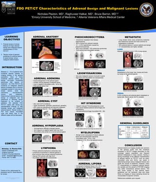
- 1. FDG PET/CT Characteristics of Adrenal Benign and Malignant Lesions Nicholas Plaxton, Raghuveer Halkar, MD 1, Bruce Barron, MD 1, MD 1,2 1Emory University School of Medicine, 2 Atlanta Veterans Affairs Medical Center LEARNING ADRENAL ANATOMY PHEOCHROMOCYTOMA METASTATIC OBJECTIVES • Uncommon neoplasm that release • Lung, breast, renal, ovary, lymphoma, leukemia catecholamine and melanoma cancers are most common to • 85% of arise from adrenal medulla metastasize to adrenals. 1. Pictorial review of clinical • 0.1 – 0.3% hypertension caused by • 50% adrenal lesions in cancer patients are benign features of major adrenal pheochromocytomas • CT characteristics are highly variable benign and malignant lesions • 90% occur sporadic, 10% endocrine syndromes characteristics in FDG LUNG • FDG activity and Hounsfield variable PET/CT. Adrenal metastasis occur 1.3% in lung cancer. 2. Tabular review of general guidelines for adrenal lesions to assist benign versus malignant determination. Liver Stomach Aorta Pancreas IFC Left adrenal INTRODUCTION Right adrenal Spleen Left kidney Bilateral adrenal metastases MIBG scan Adrenal pheochromocytoma BREAST Between 2 – 7% of patients have LEIOMYOSARCOMA CT Cross-section at level of adrenal glands Metastasize to the lungs, liver, bones and brain, incidental adrenal masses on but rarely to the adrenal glands. imaging studies in the general • Very rare malignant cancer of smooth muscle population. Most of these incidental adrenal lesions are ADRENAL ADENOMA • High SUV and Hounsfield not characteristic benign non-hyper functioning • Focal enlargement in the adrenal gland adenomas that require no • Contain varying degrees of adipose tissue treatment. On FDG PET/CT the • Low or negative Hounsfield units ( -20 to 30) incidence of malignant adrenal • FDG activity below liver lesions increases due to common metastatic spread in lung cancer, breast cancer, renal cell Bilateral adrenal metastases carcinoma, neuroendocrine RENAL tumors and melanoma. 0.03% incidence and adrenal gland metastasis is Determining if adrenal lesions are Adrenal adenoma with 0 Hounsfield, SUV below liver typical site. Renal cancer SUV is variable. benign or malignant can be Right adrenal leiomyosarcoma wrapping around the IVC with 6.2 SUV, size 6 cm paramount in directing cancer treatment to be curative or ADRENAL CYST palliative. We selected FDG • Very rare 0.01% incidence HIT SYNDROME PET/CT cases with strong key • Types (endothelial, epithelial, pseudocyst, parasitic) • Heparin Induced Thrombocytopenia is rare representative findings to help • 40% cysts are pseudocyst can become malignant • Can cause bilateral adrenal hemorrhage and illustrate benign and malignant • Hounsfield for fluid (0 – 15), no FDG activity insufficiency adrenal lesions. Tabular review of PET SUV values, Hounsfield • Hematoma/hemorrhage Hounsfield (50 – 90) units and lesion size in the different cases will be discussed. Right adrenal metastasis s/p nephrectomy Adrenal fluid filled cyst with 15 Hounsfield and no FDG activity GENERAL GUIDELINES ADRENAL HYPERPLASIA Benign Indeterminate Malignant • Homogeneous diffusely enlarged glands HIT with bilateral adrenal hemorrhage and intense SUV Size < 3 cm 3 cm < & < 6 cm > 6 cm • SUV equal to or slightly higher than liver SUV Hounsfield < 0 >0 variable • Hounsfield units similar to a normal adrenal gland MYELOLIPOMA SUV Less liver Equal liver Greater liver • Benign tumor composed of mature adipose Shape Round Irregular Smooth Heterogeneous tissue and hematopoietic elements Uniform Poorly defined • Hounsfield units ( -30 to -100) • Incidence 0.1%, 3% of all adrenal tumors CONTACT Bilateral adrenal hyperplasia 8 Hounsfield, SUV 2.0 (liver SUV 2.9) CONCLUSION Nicholas A. Plaxton M.D. LYMPHOMA Adrenal lesions on imaging studies are common in the general public and this incidence Depart of Radiology and • Primary adrenal lymphoma is extremely rare increases in PET/CT images secondary to the Imaging Sciences • Lymphoma involvement in adrenal glands is oncologic patient population and frequent Division of Nuclear Medicine more common than primary adrenal lymphoma adrenal metastases in many cancers. Having a Email: nickplaxton@emory.edu • Intense FDG activity with high SUV strong knowledge of the different characteristics Phone: 404 712 4868 • Hounsfield units are not characteristic of adrenal lesions on PET/CT such as sizes, Left adrenal myelolipoma Hounsfield units and SUVs, can assist in determining benign versus malignant lesions and ADRENAL LIPOMA guide the course of treatment. However, many CT characteristics of malignant adrenal lesions • Composed of adipose tissue ( HU -50 to -100) are variable and the advent of PET/MR imaging • Usually has no significant FDG activity may better characterize adrenal lesions. The Thanks to Eric Jablonowski for take home caveat is that adrenal lesion illustration and Dr. David Brandon guidelines are not hardened rules and close for review. follow up imaging or tissue sampling should be incorporated if suspicion remains. Poster Design & Printing by Genigraphics® - Primary adrenal lymphoma A 5 cm adrenal lipoma with - 62 Hounsfield and no FDG activity 800.790.4001 References available upon request.