NRFHRP [ Natnael Dechasa ] PPT.pdf
•
1 gefällt mir•297 views

This document discusses fetal heart rate monitoring techniques and patterns. It describes intermittent auscultation using a fetoscope or Doppler ultrasound, and continuous electronic fetal monitoring using cardiotocography. Normal fetal heart rate is between 120-160 bpm; patterns like late decelerations, variable decelerations, or a sinusoidal pattern are non-reassuring. The document outlines how to interpret and manage different fetal heart rate patterns.

Empfohlen
Weitere ähnliche Inhalte
Was ist angesagt?
Was ist angesagt? (20)
Ähnlich wie NRFHRP [ Natnael Dechasa ] PPT.pdf
Ähnlich wie NRFHRP [ Natnael Dechasa ] PPT.pdf (20)
Mehr von Dire Dawa University
Mehr von Dire Dawa University (17)
Kürzlich hochgeladen
Kürzlich hochgeladen (20)
NRFHRP [ Natnael Dechasa ] PPT.pdf
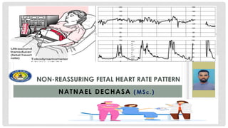
- 1. NON-REASSURING FETAL HEART RATE PATTERN NATNAEL DECHASA (MSc . )
- 2. 2 FHR monitoring in real sense is fetal brain monitoring because fetal brain responds to hypoxia by altering FHR. Methods of fetal heart rate monitoring styles . Intermittent auscultation Fetoscope. Doppler US device. Continuous electrical FHR monitoring (EFM). Cardiotocography (CTG). FHRM…
- 3. 3 Intermittent auscultation Advantages widely available, easy inexpensive. Effective if done in consistent manner at appropriate interval for the stage of labor. Limitation Sometimes difficult in obesity & polyhydramnios . Can’t detect early fetal heart beat abnormality. FHRM…
- 4. 4 Advantages of EFM over clinical monitoring. Can detect hypoxia early and can explain the mechanism of hypoxia and its specific treatment. It is an important record for medico legal purpose. FHRM…
- 5. 5 Drawbacks: Due to error of interpretation C/S rate may be high. Instruments are expensive and trained personnel are required to interpret a trace. Mother has to be confined in bed. FHRM…
- 6. 6 Indications for electronic fetal monitoring Previous history of stillbirth. Induction of Labor. Preterm labor. Non- reassuring fetal status; decreased fetal mov’t Meconium staining of amniotic fluid. FHRM…
- 7. 7 Full description of a FHR tracings. FHRM…
- 8. 8 Base line rate. Average fetal heart rate of a ten minutes recording. Draw a horizontal line by excluding acceleration and deceleration. Normal = 120 to 160 bpm Bradycardia <120 bpm Tachycardia >160 bpm FHRM…
- 9. Periodic changes… Acceleration An abrupt increase in the FHR above the baseline. Before 32 weeks of gestation, accelerations should last 10 sec and peak 10 bpm above baseline. After of 32 weeks gestation, accelerations should last 15 sec and peak 15 bpm above baseline. A prolonged acceleration is lasting 2 minutes above the base line but less than 10 minutes. An acceleration of 10 minutes or more is considered a change in baseline. 9 Periodic changes…
- 11. Periodic changes… Except those associated with variable deceleration, accelerations are physiologic response to fetal movement. Presence of acceleration –reassuring. Absence of acceleration-fetus is not moving (doesn’t necessarily mean hypoxia). 11 Periodic changes…
- 12. Deceleration; Four principal type based on timing, relationships to contractions, duration and shape. Early deceleration Gradual decrease in FHR and return to base line associated with a uterine contraction. The onset, nadir and recovery of decelerations are coincident with the beginning , peak and ending of contraction respectively. Caused by compression of fetal head by the uterine cervix (it stimulates vagal nerve). Not associated with fetal hypoxia, acidemia or low APGAR scores. 12 Periodic changes…
- 13. Late deceleration Gradual decrease and return to baseline FHR associated with uterine contraction. The onset of deceleration occur at or after the pick of uterine contraction and returning to baseline only after the contraction has ended. Causes • Excessive Ux contraction (oxytocin). • Feto-placental insufficiency . • Maternal hypotension (epidural ). 13 Periodic changes…
- 14. Variable deceleration Abrupt decrease in FHR below the base line, onset, depth and duration have no relation with contractions. 14 Periodic changes…
- 15. Causes of variable deceleration. *Cord compression/occlusion/.* Oligohydramnios Nuchal cord/cord stretching. Cord prolapse/ compression. 15 Periodic changes…
- 17. • Prolonged deceleration Decrease in FHR below the baseline ≥15bpm, lasting ≥2min but <10min from the onset to return to baseline. May be caused by any of the mechanisms mentioned so far, but are of a profound and sustained nature. 17 Periodic changes…
- 18. Sinusoid pattern Rare but significant True sinusoidal pattern – associated with fetal anemia ( iso- immunization, ruptured vasa previa, TTT). 18 Periodic changes…
- 19. Sinusoid pattern… Criteria for identifying sinusoidal FHR pattern A stable baseline FHR of 120-160bpm with regular sine wave-like oscillations. An amplitude of 5-15bpm. Oscillation of sine wave above and below the baseline and absence of accelerations. 19 Periodic changes…
- 21. Interpretation of FHR patterns The FHR pattern recorded by an electronic FHR monitor is typically interpreted as Reassuring FHRP or Non reassuring FHRP. Reassuring fetal heart rate pattern includes A baseline fetal heart rate of 120 to 160 bpm. Absence of late or variable FHR decelerations . Moderate FHR variability (6 to 25 bpm). Early decelerations may or may not be present. 21 Periodic changes…
- 22. Interpretation of FHR patterns… Nonreassuring FHRP Replaces the term fetal distress. Non reassuring FHRP includes Late decelerations (>50% of contraction). Variable deceleration. Sinusoidal tracing Prolonged decelerations./recurrent/* Bradycardia / tachycardia. 22 Periodic changes…
- 23. Management of non reassuring FHR patterns. Cause.???? Non-surgical intervention. Lateral positioning. Oxygen administration. Hydration. Discontinue Oxytocin. Tocolysis. Amnioinfusion. 23 Periodic changes…