Reverse Takotsubo Cardiomyopathy Following General Anaesthesia
•
0 gefällt mir•35 views

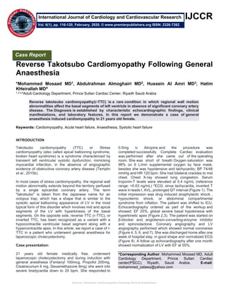
Reverse takotsubo cardiomyopathy(r-TTC) is a rare condition in which regional wall motion abnormalities affect the basal segments of left ventricle in absence of significant coronary artery disease. The Diagnosis is established by characteristic echocardiographic findings, clinical manifestations, and laboratory features. In this report we demonstrate a case of general anaesthesia induced cardiomyopathy in 21 years old female.

Empfohlen
Empfohlen
Weitere ähnliche Inhalte
Was ist angesagt?
Was ist angesagt? (20)
Ähnlich wie Reverse Takotsubo Cardiomyopathy Following General Anaesthesia
Ähnlich wie Reverse Takotsubo Cardiomyopathy Following General Anaesthesia (20)
Mehr von Premier Publishers
Mehr von Premier Publishers (20)
Kürzlich hochgeladen
Kürzlich hochgeladen (20)
Reverse Takotsubo Cardiomyopathy Following General Anaesthesia
- 1. Reverse Takotsubo Cardiomyopathy Following General Anaesthesia IJCCR II Reverse Takotsubo Cardiomyopathy Following General Anaesthesia *Mohammed Mosaad MD1, Abdulrahman Almoghairi MD2, Hussein Al Amri MD3, Hatim KHeirallah MD4 1,2,3,4Adult Cardiology Department, Prince Sultan Cardiac Center, Riyadh Saudi Arabia Reverse takotsubo cardiomyopathy(r-TTC) is a rare condition in which regional wall motion abnormalities affect the basal segments of left ventricle in absence of significant coronary artery disease. The Diagnosis is established by characteristic echocardiographic findings, clinical manifestations, and laboratory features. In this report we demonstrate a case of general anaesthesia induced cardiomyopathy in 21 years old female. Keywords: Cardiomyopathy, Acute heart failure, Anaesthesia, Systolic heart failure INTRODUCTION Takotsubo cardiomyopathy (TTC) or Stress cardiomyopathy (also called apical ballooning syndrome, broken heart syndrome) is a syndrome characterized by transient left ventricular systolic dysfunction, mimicking myocardial infarction, in the absence of angiographic evidence of obstructive coronary artery disease (Templin et al., 2015b). In most cases of stress cardiomyopathy, the regional wall motion abnormality extends beyond the territory perfused by a single epicardial coronary artery. The term "takotsubo" is taken from the Japanese name for an octopus trap, which has a shape that is similar to the systolic apical ballooning appearance of LV in the most typical form of this disorder which involves mid and apical segments of the LV with hyperkinesis of the basal segments. On the opposite side, reverse TTC (r-TTC), or inverted TTC, has been recognized as a variant with a hypocontractile ventricular basal segment along with a hypercontractile apex. In this article, we report a case of r- TTC in a patient who underwent general anesthesia for laparoscopic cholecystectomy. Case presentation: 21 years old female medically free, underwent laparoscopic cholecystectomy and during induction with general anesthesia (Fentanyl 100mcg, Propofol 200mg, Cisatracurium 6 mg, Dexamethazone 8mg) she went into severe bradycardia down to 20 bpm. She responded to 0.5mg iv Atropine and the procedure was completed successfully. Complete Cardiac evaluation was performed after she came out of the operating room. She was short of breath Oxygen saturation was 98% on 6 L/min supplemental oxygen by face mask, besides she was hypotensive and tachycardic; BP 74/48 mmHg and HR 120 bpm. She had bilateral crackles to mid chest. Chest X-ray showed lung congestion. Serum troponin-T levels were elevated at 0.4 ng/mL (reference range: <0.03 ng/mL) *ECG: sinus tachycardia, inverted T wave in leads I, AVL, prolonged QT interval (Figure 1). The initial impression was drug induced anaphylactic shock , hypovolemic shock, or abdominal compartmental syndrome from inflation. The patient was shifted to ICU. Echocardiography ordered as part of the workup and showed: EF 25%, global severe basal hypokinesia with hyperkinetic apex (Figure 2,3). The patient was started on β-blocker and angiotensin-converting-enzyme inhibitor and spironolactone. Coronary angiography and LV angiography performed which showed normal coronaries (Figure 4, 5, 6, and 7). She was discharged home after one week of hospital stay, in good shape and normalized ECG (Figure 8). A follow up echocardiography after one month showed normalization of LV with EF at 55%. *Corresponding Author: Mohammed Mosaad MD, Adult Cardiology Department, Prince Sultan Cardiac center(PSCC), Riyadh, Saudi Arabia. E-mail: mohammed_zalawy@yahoo.com Case Report Vol. 6(1), pp. 116-120, February, 2020. © www.premierpublishers.org ISSN: 2326-7262 International Journal of Cardiology and Cardiovascular Research
- 2. Reverse Takotsubo Cardiomyopathy Following General Anaesthesia Mosaad et al. 117 DISCUSSION The mechanisms involved in stress induced cardiomyopathy are postulated to include catecholamine excess, microvascular dysfunction, and coronary artery spasm. Also, dynamic mid-cavity or LV outflow tract obstruction has been documented in some patients and may contribute to apical dysfunction, but the pathogenesis is far from being precisely understood. However, the most widely accepted etiological mechanism behind both types is sympathetic nervous system over- activation. Among the various neurochemical substances associated with cardiac wall motion abnormalities, epinephrine and norepinephrine seem to be the most crucial. This catecholamine surge is believed to mediate a vascular dysfunction leading to coronary artery vasospasm, microvascular dysfunction, hyperdynamic contractility, and direct myocardial toxicity via free radicals formation (Lyon et al., 2008). Other theories stem from the possible role in protein signaling within the myocardial cells that mediates a paradoxical negative inotropic effect to protect against the intense activation of β-adrenoceptors. This effect is greatest at the apical myocardium where the β- adrenoceptor density is highest. (Lyon et al., 2008). This has been also proven by 123-meta-iodobenzylguanidine myocardial scintigraphy that implied more myocardial sympathetic innervation in the apex. (Chattopadhyay et al., 2009) This might explain the myocardial stunning affecting the apical wall in TTC. (Chattopadhyay et al., 2009) However, it does not explain the hyperkinetic apical wall motion in r- TTC, neither the hypokinesis of the basal wall. Role of coronary artery disease or dysfunction, Although the clinical presentation simulates that of an acute MI, coronary arteriography typically shows no obstructive lesions, and only a minority of patients display coronary spasm with acetylcholine provocation. The following observations support the hypothesis of coronary vascular dysfunction, which may be catecholamine-induced: • The occasional finding of multifocal coronary vasospasm on coronary angiography (Kurisu et al., 2002) • Transient myocardial perfusion abnormalities that resolve with improvement in the myopathy. The presence of abnormal Thrombolysis in Myocardial Infarction (TIMI) frame counts on angiography. The TIMI frame count is the number of cine frames required for dye to first reach standardized distal coronary landmarks. Predisposing factors Limited data are available on predisposing factors for stress cardiomyopathy. There have been reports of familial cases, raising the possibility of a genetic predisposition. Small studies of patients with stress cardiomyopathy have found genetic heterogeneity and suggest a possible polygenic basis. Patients with psychiatric and/or neurologic disorders may be predisposed to develop stress cardiomyopathy (Templin et al., 2015a). In the International Takotsubo Registry study, 55.8 percent of patients with stress cardiomyopathy had an acute, former, or chronic psychiatric (such as affective or anxiety disorder) or neurologic disorder (such as seizure or headache disorder) as compared with 25.7 percent of patients with ACS (Templin et al., 2015a). Furthermore, it deserves observation that some differences between TTC and r-TTC do exist. Ramaraj and Movahed (2010) noted that in r-TTC patients are usually younger, tend to have a lower LVEF, and sustains a quicker myocardial recovery in comparison to TTC. Moreover, since the basilar part of the ventricle is the main involved region in r-TTC, which has more myocardial tissue, cardiac biomarkers are usually more elevated in comparison to TTC. Patients have also developed rTTC while under general anesthesia for surgical and dental procedures. It is not clear if this occurrence is directly related to the use of anesthetic agents or the emotional stress associated with the procedure. (Khalil et al., 2018; Açar 2016) CONCLUSION Reverse takotsubo cardiomyopathy is a rare type of stress-induced cardiomyopathy that had been reported following neurological/psychiatric /physical stress. We presented a case of r-TTC after anaesthesia induction. Similar cases are not much reported and not well described before. ABBREVIATIONS LIST • Reverse takotsubo cardiomyopathy (r-TTC) • Takotsubo cardiomyopathy (TTC) • Electrocardiogram (ECG) • left ventricle (LV) • myocardial infarction (MI) • Acute Coronary Syndrome (ACS)
- 3. Reverse Takotsubo Cardiomyopathy Following General Anaesthesia Int. J. Cardiol. Cardiovasc. Res. 118 APPENDIX Figures Figure 1a: ECG Directly post procedure Figure 1b: ECG Directly post procedure Figure 2: Echocardiography Apical two-chamber Figure 3: Echocardiography Apical two- chamber view end-systole view end-systole
- 4. Reverse Takotsubo Cardiomyopathy Following General Anaesthesia Mosaad et al. 119 Figure 4: Right coronary angiogam Figure 5: Left coronary angiogram Figure 6: Left ventriculogram in end-systole Figure 7: Left ventriculogram in end-diastole
- 5. Reverse Takotsubo Cardiomyopathy Following General Anaesthesia Int. J. Cardiol. Cardiovasc. Res. 120 Figure 8: Normalized ECG, back to baseline FUNDING: No funding required DISCLOSURES: No conflict of interest, Author is not related to any industry. ACKNOWLEDGEMENTS I wish to express my sincere gratitude to my colleagues and my seniors for providing their guidance, comments and suggestions. I would like sincerely thank Dr. Abdulrahman Almoghairi, Dr. Hatim KHeirallah and Dr. Hussein AlAmri for their guidance and encouragement and assistance in carrying out this case report REFERENCES Açar B, Kırbaş Ö, Ünal S, et al. Reverse Takotsubo cardiomyopathy following intra-abdominal surgery. Turk Kardiyol Dern Ars 2016;44:514-6. Chattopadhyay S, John J. Tako-tsubo and reverse tako- tsubo cardiomyopathy: temporal evolution of the same disease? Eur Heart J. 2009;30:2837-2837. [PubMed] Ikram S, Saleem N, Latif RK. Acute left ventricle failure on induction of anesthesia: a case report of reverse stress cardiomyopathy presentation, diagnosis and treatment. J Anesth. 2016;30:911-914. [PubMed] Khalil A, Dabbous A, Taha S et al, Reverse Takotsubo cardiomyopathy during general anaesthesia in a 16 yr old female victim of war. J cardiothorac Vasc Anaesth 2018;32:1858-62. Kurisu S, Sato H, Kawagoe T, et al. Tako-tsubo-like left ventricular dysfunction with ST-segment elevation: a novel cardiac syndrome mimicking acute myocardial infarction. Am Heart J 2002; 143:448. Lyon AR, Rees PS, Prasad S, Poole-Wilson PA, Harding SE. Stress (takotsubo) cardiomyopathy—a novel pathophysiological hypothesis to explain catecholamine-induced acute myocardial stunning. Nat Clin Pract Cardiovasc Med. 2008;5:22-29. [PubMed] [Google Scholar] Ramaraj R, Movahed MR. Reverse or inverted takotsubo cardiomyopathy (reverse left ventricular apical ballooning syndrome) presents at a younger age compared with the mid or apical variant and is always associated with triggering stress. Congest Heart Fail. 2010;16:284-286. [PubMed] Templin C, Ghadri JR, Diekmann J, et al. Clinical Features and Outcomes of Takotsubo (Stress) Cardiomyopathy. N Engl J Med 2015a; 373:929. Templin C, Ghadri JR, Diekmann J, et al. Clinical Features and Outcomes of Takotsubo (Stress) Cardiomyopathy. N Engl J Med 2015b; 373:929. Accepted 1 February 2020 Citation: Mosaad M, Almoghairi A, Al Amri H, KHeirallah H (2020). Reverse Takotsubo Cardiomyopathy Following General Anaesthesia. International Journal of Cardiology and Cardiovascular Research: 6(1): 116-120. Copyright: © 2020 Mosaad et al. This is an open-access article distributed under the terms of the Creative Commons Attribution License, which permits unrestricted use, distribution, and reproduction in any medium, provided the original author and source are cited.