Anatomy of trachea & lungs
•Als PPTX, PDF herunterladen•
44 gefällt mir•7,639 views

Anatomy of trachea & lungs

Empfohlen
Weitere ähnliche Inhalte
Was ist angesagt?
Was ist angesagt? (20)
Ähnlich wie Anatomy of trachea & lungs
Ähnlich wie Anatomy of trachea & lungs (20)
Mehr von نصار ايوب
Mehr von نصار ايوب (16)
Kürzlich hochgeladen
Kürzlich hochgeladen (20)
Anatomy of trachea & lungs
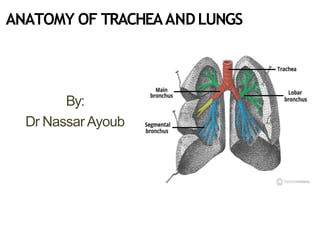
- 1. ANATOMY OF TRACHEAANDLUNGS By: Dr Nassar Ayoub
- 2. Trachea The trachea is a cartilaginous and membranous tube, extending from the cricoid cartilage of the larynx, on alevel with C6 vertebra, to the level of the angle of Louis (T4/5) vertebra, where it divides into the two bronchi, one for eachlung.
- 3. Trachea • Structure: The trachea is arigid fibroelastic structure. Incomplete rings of hyaline cartilage continuously maintain the patency ofthe lumen. • The trachea is lined internally with ciliated columnar epithelium.
- 4. Trachea • It measures about11 cm. In length. • Its diameter, from side to side, is from 2 to 2.5 cm. • It has18-22 cartilaginous rings • It is greater in themale than in thefemale.
- 5. Trachea Relations: Anteriorly: Neck: • Theisthmus of the thyroid gland (2nd, 3rd and 4th tracheal rings) • Theinferior thyroid veins • Thesternothyroid and sternohyoid muscles • Thecervical fascia Thorax: • Themanubrium sterni • Theremains of the thymus • Theleft innominate vein • Theaortic arch • Left common carotid arteries • Thedeep cardiac plexus.
- 6. Trachea Relations: Posteriorly : the esophagus Laterally: Neck: • Commoncarotid arteries • Right and left lobes ofthe thyroid gland • Inferior thyroidarteries • Therecurrent nerves
- 7. Trachea Relations: Laterally: Thorax: itlies in the superior mediastinum. Right • The pleura • Right vagus, • Theinnominate artery Left • Left recurrent nerve • Theaortic arch • Theleft common carotid • Subclavianarteries
- 8. Inferior thyroida. Recurrent laryngealn. Thoracicduct Vagusn. Sympathetic trunk Trachea
- 9. Trachea • Thetrachea divides into two main bronchi : the left and the right bronchi, at the level of the sternal angle at the anatomical point known asthe carina.
- 10. Bronchi Theright bronchus: • Wider, shorter, and more vertical in direction than theleft. • It is about 2.5 cm.Long, • It enters the right lung nearly opposite the T5 vertebra.
- 11. Bronchi TheLeft Bronchus • It is smaller incaliber but longer than the right • It is about 5 cm.long. • It enters the root of the left lung opposite the T6vertebra.
- 13. trachea Left primary bronchus Right Primary Bronchus Wider, short er, and more vertical than the left
- 14. Bronchi • Theprimary bronchi divide to form Secondary Bronchi (lobar bronchi). • There is one secondary bronchus for each lobe of the lungs. • There are 2 lobes on the left lung. • There are 3 lobes on the right lung.
- 15. Bronchi • Theleft main bronchus enters the hilum and divides into asuperior and inferior lobar bronchus. • Theright main bronchus gives off the bronchus to the upper lobe prior to entering the hilum (superior lobar bronchus) and once into the hilum divides into middle and inferior lobar bronchi.
- 16. Bronchi • Eachlobar bronchus divides within thelobe into(Tertiary Bronchi) segmental bronchi. • Eachsegmental bronchus enters a bronchopulmonary segment. • Eachbronchopulmonary segment is pyramidal in shape with its apex directed towards the hilum.
- 19. Tracheablood supply • the trachea receives its blood supply from branches of the inferior thyroid and bronchial arteries.
- 20. Lungs • Thetwo lungs are organs of respiration and lie on either sideof the mediastinum surrounded by the right and left pleuralcavities. • Right and left lungs.
- 21. Lungs • Theright lung is normally alittle larger than the left lung becausethe middle mediastinum, containin g the heart, bulges more to the left than to the right.
- 22. Lungs Eachlung hasahalf-cone shape, with a base,apex, two surfaces and three borders: • Thebase sits on the diaphragm. • Theapex projects above 1st rib and into the root of theneck.
- 23. Lungs • Thetwo surfaces-thecostal surface lies immediately adjacent to the ribs and intercostal spacesof the thoracic wall. • Themediastinal surface lies against the mediastinum anteriorly and the vertebral column posteriorly and contains the comma-shaped hilum of the lung through which structures enter and leave.
- 24. Lungs • Thethree borders-the inferior border of the lung is sharp and separates the base from the costal surface. Theanterior and posterior borders separate the costal surface fromthe medial surface. • Unlike the anterior and inferior borders, which are sharp, the posteriorborder is smooth androunded.
- 25. LungsRoot and hilum of lung: • Theroot of each lung is ashort tubular collection of structures that together attachthe lung to structures inthe mediastinum . • Thehilum, where structures enter and leave.
- 26. Lungs Structures within each root and located inthe hilum: • Apulmonary artery • Twopulmonary veins • Amain bronchus • Bronchial vessel • Nerves • Lymphatics
- 27. Lungs Structure the right lung: • It is dividedinto upper, middle and lower lobes by oblique and horizontal fissures. Theleft lung: • It hastwo lobes,upper and lower lobes. They are separated by the oblique fissure.
- 28. Lungs Read about right and left lungs relations and lungs impressions.
- 31. Bronchial Tree • Thetrachea is divided into 2 bronchi • Themain bronchus divides within the lung into lobar bronchi. • Thelobar bronchi divide into segmental bronchi which supply bronchopulmonary segments. • Eachbronchopulmonary segment, the segmental bronchi divide into bronchioles, which further subdivide and supply the respiratory surfaces.
- 32. Bronchopulmonary Segment • There are ten bronchopulmonary segments in each lung • Eachbronchopulmonary segment is shaped like an irregular cone with the apex at the origin of the segmental bronchus and the baseprojected peripherally onto the surface of thelung.
- 33. Bronchopulmonary Segment • Abronchopulmonary segment is the smallest,functionally independent region of a lung and the smallest area of lung that canbeisolated and removed without affecting adjacent regions.
- 37. Blood supply of thelung • The bronchi and parenchymal tissue of the lungs are supplied by bronchial arteries a branches of the descending thoracic aorta. • Bronchial veins, which also communicate with pulmonary veins, drain into the azygos and hemiazygos.
- 40. Nerve supply of thelungs • Apulmonary plexusis located at the root of eachlung. • Theplexus is composed of sympatheticfibres (from the sympathetic trunk) and parasympathetic fibres (from the vagus).