PRINCIPLES AND PRACTICES OF RIGHT HEART CATHETERIZATION IN CHILDREN
•Als PPTX, PDF herunterladen•
34 gefällt mir•6,524 views

This document provides an overview of right heart catheterization (RHC) in children. It begins with a brief history of RHC, describing early experiments in the 1840s-1920s. The document then covers patient preparation, venous access approaches, conducting the procedure, normal pressure values, shunt detection/quantification using oximetry, and understanding Fick's principle. The key objectives are to gain knowledge on performing tailored RH studies, the diagnostic role of RHC, and quantifying left-to-right shunts.

Empfohlen
Weitere ähnliche Inhalte
Was ist angesagt?
Was ist angesagt? (20)
Ähnlich wie PRINCIPLES AND PRACTICES OF RIGHT HEART CATHETERIZATION IN CHILDREN
Ähnlich wie PRINCIPLES AND PRACTICES OF RIGHT HEART CATHETERIZATION IN CHILDREN (20)
Mehr von Dr. Murtaza Kamal MD,DNB,DrNB Ped Cardiology
Mehr von Dr. Murtaza Kamal MD,DNB,DrNB Ped Cardiology (20)
Kürzlich hochgeladen
Kürzlich hochgeladen (20)
PRINCIPLES AND PRACTICES OF RIGHT HEART CATHETERIZATION IN CHILDREN
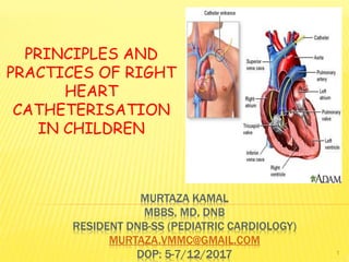
- 1. MURTAZA KAMAL MBBS, MD, DNB RESIDENT DNB-SS (PEDIATRIC CARDIOLOGY) MURTAZA.VMMC@GMAIL.COM DOP: 5-7/12/2017 PRINCIPLES AND PRACTICES OF RIGHT HEART CATHETERISATION IN CHILDREN 1
- 2. OBJECTIVES… Gain an overview of history and development of RHC Learn how to perform a RH study tailored to answer a specific clinical question Gain a better understanding of role of RHC as a diagnostic tool in specific situations 2
- 3. CLAUDE BERNARD 1844: France 1st RHC on horse Inserted glass tubes via jugular vein and carotid artery Measured temperature in both ventricles Later measured intracardiac pressures too Nossaman BD et al. H/o RHC: 100 years of experimentation and methodology development. Cardiol Rev 2010;18:94-101. 3
- 4. WERNER FORSSMANN 1929; Germany Self catheterization using urethral catheter Used lt. anticubital vein RV; (X-RAY) Against medical ethics Meyer JA. W Forssmann and catheterisation of the heart, 1929. Ann Thorac Surg 1990;49:497-9 4
- 5. FINALLY GOT RECOGNIZED… The Nobel Foundation: 1956 5
- 6. INTRODUCTION With advent of non-invasive modalities (ECHO, MRI): Cardiac catheterization has reduced dramatically Gold standard: For assessment of cardiac hemodynamics Resolves discrepancy b/w c/fs and non-invasive measurements 6
- 7. INTRODUCTION CONT… Perform through review of clinical history, physical exam, ECG, CXR, ECHO, MRI(+/-) before patient enters cath lab Why is the study being performed? If results are not going to alter the course of management: Best not to perform Have a clear idea as what is the data one wishes to seek Wild goose chase: More questions than answers 7
- 8. CONDUCT OF A CATHETERIZATION STUDY Proper study needs adherence to standard protocols Due attention to be given to pressure recordings and saturation assessments Flexibility is approach needed: Each case is different 8
- 9. PATIENT PREPARATION The parents must be informed of indication and risks of procedure Retrospective and prospective data revealed: Serious adverse event: 1.1% Mortality: 0.05% Hoeper M et al. Complications of RHC procedures in patients with PH in experienced centers. J Am Coll Cardiol 2006;48: 2546-52 9
- 10. PATIENT PREPARATION CONT… MC complications: Access site hematoma Vagal reaction Pneumothorax Arrhythmias If available: Quote individual/ departmental complications 10
- 11. PATIENT PREPARATION Rule out anemia, infections, thrombocytopenia Electrolyte/ metabolic disturbances Dehydration Digoxin toxicity Coagulopathy Safe in patients with INR <3.5 undergoing RHC via IJV or anti cubital veins Ranu H et al. A retrospective review to evaluate the safety of RHC via IJV in assessment of PH.. Clin Cardiol 2010;33: 303-6 11
- 12. OUR HOSPITAL PROTOCOL Obtain cath profile and PAC clearance before admission (1 day prior) NBM: 4 hours before Caution: OVERZELOUS FASTING PROTOCOLS MAY LEAD TO VOLUME DEPLETION: MAKING CHALLENGING VENOUS ACCESS IVFs: 1/2DNS since NMB Blood in hand Injection Cefazolin 30mg/kg i/v 1 hr before procedure 12
- 13. VENOUS ACCESS Route of access depends on: Operator experience Presence of cardiac devices and indwelling catheters Prior h/o venous cannulation and associated complications FV access commonly used in children or if LHC performed concurrently Small studies demonstrated feasibility and safety of RHC+LHC via ACV and radial artery respectively Yang CH, Guo GB, Yip HK. Bilateral cardiac catheterizations: the safety and feasibility of a superficial forearm venous and transradial arterial approach. Int Heart J 2006;47:21–7. Lo TS, Buch AN, Hall IR, et al. Percutaneous left and right heart catheterization in fully anticoagulated patients utilizing the radial artery and forearm vein: a two-center experience. J Interv Cardiol 2006;19:258–63 Gilchrist IC, Kharabsheh S, Nickolaus MJ, et al. Radial approach to right heart catheterization: early experience with a promising technique. 13
- 14. VENOUS ACCESS CONT… USG guided vs landmark based: Meta-analysis available Clear benefit of USG for IJV cannulation Higher success rate Fewer complications Faster access Hind Daniel, Calvert Neill, McWilliams Richard, et al. Ultrasonic locating devices for central venous cannulation: meta-analysis. BMJ 2003;327:361. Data very limited: USG for FV and SCV cannulation 14
- 15. VENOUS ACCESS CONT… Balloon flotation catheters (Swan- Ganz) : Balloon at distal end, facilitate passage through RH Designed to be placed without fluoroscopy, although screening helps (marked RH dilatation/ severe TR) 15
- 16. VENOUS ACCESS CONT… Catheter inserted into RA and balloon inflated Catheter then follows direction of blood flow towards PAs Advancing further should allow performer to obtain PCWP Important to avoid leaving balloon inflated for longer than necessary : Risk of pulmonary infarction/ rupture 16
- 17. CATHETERIZATION FROM FV Commonly performed using multipurpose end hole catheter using direct fluoroscopy Requires greater manipulation than balloon flotation catheters to navigate through RH: Guide wire may be required to improve steerability MP catheters can be used to cross directly into LA in patients with PFO for direct pressure 17
- 18. PROCEDURE Before starting: Confirm pressure transducers are zeroed, leveled, appropriately calibrated Establishment of “zero” value is the concept of making hydrostatic measurements with fluid filled systems relative to a reference value, usually atmospheric pressure (760mm Hg), then examining the change from that value 18
- 19. PROCEDURE Transducer should be placed at appropriate level For every 1cm above LA the catheter is referenced, the pressure measurement is underestimated by 0.74mmHg and vice versa 19
- 20. 20
- 21. THE CONCEPT OF PHLEBOSTATIC AXIS Correct reference point Midpoint b/w anterior and posterior surfaces of chest at 4th ICS Essential that level of stopcock of transducer be at this level All transducers must be at this level 21
- 22. PRINCIPALS TO BE ADHERED TO DURING CATH STUDY Data to be obtained in a steady state Essential to maintain decorum in a quiet and calm environment Appropriate sedation needed in case of agitated child Watch for over sedation: Respiratory depression, consequently changes in sats 22
- 23. PRINCIPALS TO BE ADHERED TO DURING CATH STUDY Obtain entire data in<7 mins Withdrawal pressures and saturations better than ingoing If sample can’t be obtained from a site due to ventricular premature complex, skip site until rest of run completed Complete hemodynamic data must be obtained before angiograms Obtain pressures and oxymetry samples as close in time as possible 23
- 24. PRINCIPALS TO BE ADHERED TO DURING CATH STUDY Repeated measurements : More accurate Record catheter course An end hole catheter (eg: Swan Ganz) or one with side holes close to its tip (eg. NIH) can be used Sat syringes not to be overheparinized, sample gets diluted; just quote the inner lining of the syringe Remove air bubbles: PO2 rises 24
- 25. PRINCIPALS TO BE ADHERED TO DURING CATH STUDY Glass syringes: Gold standard Plastic syringes: Porous, fall in PO2 Metabolism of WBCs: Tends to fall in PO2 Measure sats <5 mins (if delay: Transfer in ice <30 mins) 25
- 26. USE DEDICATED OXYMETRY MACHINE Should be in lab Measures directly the o2 saturation using spectrophotometry to correctly quantify oxy, deoxy, carboxy and methHb and total Hb Do not use ABG machines: O2 saturation results derived from o2 dissociation curves, using PO2 values: Which is affected by many factors ( Adult or fetal Hb, temp, ph, CO2, 2,3- DPG levels) 26
- 27. THE ACTUAL MEASUREMENTS FOR SHUNTS • Place catheter in PA (Swan Ganz) and pigtail in Ao • Measure PA and Ao pressures • Take o2 sat in PA+ Ao blood • Enter LV by retrograde crossing of Ao valve • Advance PA catheter to PCWP position • Measure simultaneously LV-PCWP pressures 27
- 28. THE ACTUAL MEASUREMENTS FOR SHUNTS CONT… • Pull back from PCWP to PA • Pull back from PA to RV for PS and record RV pressure. Take RV sample for O2 sats • Record simultaneous LV-RV pressure • Pull back from RV to Rato screen for tricuspid stenosis and record RA pressure. Take RA sample • Take SVC+IVC samples for O2 saturations • Pull back from LV to aorta for AS 28
- 29. NORMAL PRESSURE VALUES OF VARIOUS HEART CHAMBERS CHAMBER AVERAGE PRESSURE RA 6/5/3 RV 25/4 PA 25/9/15 PCWP 9 LA 10/12/8 LV 130/8 Ao 130/70/85 29
- 30. DO MAKE A NOTE OF THESE Mean RA pressure=RVEDP RVSP=Peak PA pressure PA diastolic pressure=Mean PCWP=Mean LA pressure= LVEDP LVSP=Ao pressure Presence of gradients across these chambers indicates obstruction to blood flow 30
- 31. RIGHT ATRIAL PRESSURES A: Atrial systole, just after P wave C: RV contraction/ TV closure V: Filling of RA against closed TV valve X: Atrial relaxation Y: Opening of TV in early diastole 31
- 32. RV PRESSURE A rapid upstroke during isovolumetric contraction A plateau during systolic ejection A decline to near zero during isovolumetric relaxation A slow rise to the end diastolic pressure during diastolic filling 32
- 33. PA PRESSURE PA systolic pressure= RVSP (<30mm Hg) Mean pressure< 20mm Hg PA diastolic pressure begins with dicrotic notch caused by valve closure, and the diastolic pressure is typically no more than 2-3 mm Hg higher than the wedge pressure 33
- 34. PCWP Is usually a good reflection of LA and LVEDP because of absence of valves in pulmonary circulation It has the characteristic a and v wave appearance of an atrial tracing 34
- 35. SATURATIONS Site Average Range SVC 74% 67-83% IVC 78% 65-87% RA 75% 65-87% RV 75% 67-84% PA 75% 67-84% LA 95% 92-98% LV 95% 92-98% FA 95% 92-98% 35
- 36. SHUNT DETECTION & QUANTIFICATION 36
- 37. WHEN IS IT UTILISED? When the is discrepancy b/w physical and non-invasive findings At the time of device closure Assessment of shunt operability in patients with severe PAH with borderline findings 37
- 38. SHUNT DETECTION Oximetric run used Past: Indicator dye (Indocyanine green) used Detected very small lt rt shunt missed by oxymetry No longer used Presence of unexplained arterial desaturation (FA SaO2<95%) or unexpectedly high O2 content in PA (SaO2>80%): Raises suspicion of rt lt or a lt rt shunt respectively. Follow this by a complete oximetry run 38
- 39. OXIMETRY RUN Full oximetry run involves taking serial samples at following locations: Lt+ rt. PA MPA RVOT RV mid RV tricuspid valve or apex RA low or near TV RA high SVC low (near RA junction) SVC high (near innominate vein junction) IVC high ( just at/ below diaphragm) IVC low L4-5 LV Ao (diatal to ductus insertion) 39
- 40. DETECTION OF LEFT TO RIGHT SHUNT BY OXIMETRY Antman et al, AJC 80; Barrat et al, JLCM 57, Freed et al, BHJ 79 40
- 41. CAUSES OF STEP UP AT ATRIAL LEVEL ASD PAPVC VSD with TR RSOV RA LV RA shunt Cor AV Fistula RA 41
- 42. CAUSES OF STEP UP AT VENTRICULAR LEVEL VSD RSOV RV Low ASD Cor AV Fistula RA PDA with PR AVSD 42
- 43. CAUSES OF STEP UP AT GREAT VESSEL LEVEL Patent Ductus Arteriosus Aorto-pulmonary Window Outlet VSD Coronary origin from pulmonary artery 43
- 44. LIMITATIONS Steady state may not be present: Patient agitation/ Arrhythmias Lacks sensitivity. Small shunts may be missed In conditions of high level of systemic blood flow, mixed venous o2 sats tends to be higher than normal and interchamber variablility would be reduced equalization of arterial and venous blood 44
- 45. UNDERSTANDING THE FICK’S PRINCIPAL Total uptake/release of a substance by an organ is the product of the bld flow to the organ and the AV concentration difference of the substance 45
- 46. PULMONARY BLOOD FLOW Using lung as an organ and O2 as substance: Bld flow to lung will be: Qp (L/min) =O2 consumption(VO2)/ AV O2 difference =VO2/ PV O2 content-PA O2 content 46
- 47. PBF If PV can’t be entered See systemic arterial O2 content ≥95% <95 Use this value Determine if rt lt shunt +nt –nt Use 98% value Use observed systemic arterial saturation value 47
- 48. SYSTEMIC BLOOD FLOW Using body as an organ and O2 as substance: Bld flow to body will be: Qs= o2 consumption(VO2)/ SA02-MVO2 Note: In presence of shunt lesions, the MVO2 is to be measured in the chamber immediately proximal to the shunt 48
- 49. CALCULATION OF QS IN PRESENCE OF LT->RT SHUNT 49 Grossman & Baim’s, 8th edition (FLAMM’S FORMULA)
- 50. SHUNT QUANTIFICATION Absolute terms (L/min)=Qp-Qs Relative terms (ratio)=Qp/Qs Ratio advantageous as it takes out unreliable variables like VO2 Qp/Qs=(SAO2-MVO2)/ (PVO2-PAO2) 50
- 51. QP/QS 1: No shunt <1: Rtlt shunt 1-1.5: Small lt rt shunt (in absence of PAH; would not need closure) 1.5-2: Intermediate lt rt shunts (may be closed if risk of closure low) >2: Large lt rt shunt (Needs closure) 51
- 52. CALCULATION OF BIDIRECTIONAL SHUNT Effective bld flow: Flow that would exist in absence of any lt—>rt or rt lt shunt Qeff= O2 consumption/ (PVO2-MVO2) Lt rt: Qp-Qeff Rt lt: Qs-Qeff 52
- 53. SHUNT OPERABILITY Large shunts: High PAH due to increased flow Anatomic changes takes place in pul. vasculature Reversible initially, later ir-reversible As PVRI increases> 6-8 Wood U: Poor operative outcome In these cases: If PAH irreversible; Sx tends to transform these from Eisenmenger’s syndrome to one analogous to idiopathic PAH 53
- 54. SHUNT OPERABILITY CONT… Compared to idiopathic PAH; pts. With ES have much better prognosis with 40% expected to survive till 25 yoa Assessment of operability is not an “ all or none” phenomenon Clinical and non invasive parameters too are considered 54
- 55. CLINICAL & NON INVASIVE FINDINGS TO ASSESS SHUNT OPERABILITY 55 Vijaylaxmi: Cardiac Catheterization From Pediatric to Geneatric: 1st edition
- 56. HEMODYNAMIC ASSESSMENT OF SHUNT OPERABILITY Favorable outcomes: Baseline Qp/Qs >1.5-2 PVRI <6Wood U PVR:SVR <0.3 without vasoreactive test Age <1 year (Most imp.) 56
- 57. TECHNIQUES TO ASSESS OPERABILITY Lung biopsy Exposure to vasodilator Temporary balloon occlusion of defect 57
- 58. 01. LUNG BIOPSY Gold standard Heath Edward classification Grade 4-6: Irreversible Invasive Associated with morbidity Not available at all centers Some studies have questioned reliability 58
- 60. 02. EXPOSURE TO VASODILATOR 100% O2 NO (+/- O2) Tolazoline Adenosine Epoprostenol Used to assess pulmonary reactivity in cath labs 60
- 61. PROCEDURE Pt. adequately sedated Obtain baseline rt/lt heart studies (PVRI,SVRI, Qp, Qs) 100% o2 X 10 mins Repeat rt/lt heart studies (recalculate Qp, Qs, PVRI, SVRI) If NO used: 20-80ppm by NO ventilator 61
- 62. TIPS FOR CALCULATION O2 consumption remains constant Post O2 inhalation: Dissolved O2 must be taken into account in calculating O2 content Failure to take into consideration the dissolved O2 may make an inoperable case appear operable In pts with a positive response , there is a fall in the diastolic and mean PA pressures without a fall/rise in Ao pressure/ CO 62
- 63. PRESENCE OF ALL OF THESE INDICATES FAVOURABLE OUTCOME FOLLOWING SURGERY Decrease of 20% in PVRI Decrease of 20% in PVR: SVR ratio Final PVRI <6Woods U/m2 Final ratio of PVR: SVR <0.3 63
- 64. 03. TEMPORARY BALLOON OCCLUSION Occlusion abolishes lt rt shunt Operable pts: Drop in PA pressure Inoperable pts: No drop in PA pressure; actual rise in PA pressure with/without a fall in Ao pressure Best studied in PDAs and sometimes in ASDs Technically difficult in VSDs 64
- 65. PDA BALLOON OCCLUSION 10 mins occlusion A 25% fall in PA pressures or 50% fall in ratio b/w pulmonary and Ao diastolic pressures A fall in PA pressure with a > 20 mm Hg systolic, diastolic and mean pressure difference b/w PA and FA during balloon occlusion 65
- 66. ASD BALLOON OCCLUSION 15 mins +ve response: Mean reduction in pulmonary pressure of ≥25% after balloon occlusion compared to basal levels, without a fall in systemic pressure or an increase in VEDP 66
- 67. 67 MEASUREMENT OF CARDIAC OUTPUT
- 68. JUST A GLANCE AT THE FORMULAE 68Callan P, Clark AL. Heart 2016;102:1–11. doi:10.1136/heartjnl-2015-307786
- 69. CARDIAC OUTPUT Fick method Thermo dilution method Angiographic method 69
- 70. A. FICK METHOD OF CO ESTIMATION Gold standard Fick’s principal In the absence of shunts: Qp=Qs=CO Also useful in patients with TR where thermodilution method is unreliable 2 main variables: O2 consumption (VO2) AVO2 70
- 71. 01. O2 CONSUMPTION (VO2) Earlier methods: Rarely used Douglas bag/ polarography method/ paramagnetic method Cumbersome/ specialized equipments/ experienced personnel Only means of getting accurate VO2 Children: La Farge- Miettinen tables 71
- 72. LA FARGE- MIETTINEN TABLES: BOYS 72Vijaylaxmi: Cardiac Catheterization From Pediatric to Geneatric: 1st edition
- 73. LA FARGE- MIETTINEN TABLES: GIRLS 73Vijaylaxmi: Cardiac Catheterization From Pediatric to Geneatric: 1st edition
- 74. 02. AV O2 DIFFERENCE O2 content = O2 bound to Hb+ Dissolved O2 = 1.36mlx Hbx saturation+ 0.003mlxPaO2 In pts on RA: Content of dissolved O2 low: Hence ignored (= 1.36x Hb(g/L)X 10X (AO2- MVO2) If breathing with FiO2 >50%: Take dissolved O2 too (Imp when shunt operability in severe PAH cases is assessed) 74
- 75. BEFORE STARTING THE CASE, DO HAVE THESE HANDY Hb level Ht +Wt for BSA calculation HR, age, sex: For VO2 75
- 76. LIMITATIONS OF THE FICK PRINCIPAL Use of assumed VO2 value (Errors of 10-25% can creep in) Inability to obtain steady state under certain circumstances (samples to be obtained simultaneously) Do not use this method in: Significant MR, AR 76
- 77. B. THERMODILUTION METHOD OF CO ESTIMATION Values correlate well to Fick method Involves determining the extent and rate of thermal changes in blood stream following injection of fixed vol of cold NS Temperature time curve obtained: Area gives CO 77
- 78. METHOD Distal tip of Swan Ganz catheter placed in PA, proximal port in RA 10 ml NS bolus injected rapidly in proximal port at a constant rate Resultant change in temperature in liquid measured by thermistor mounted at the distal end of catheter Result displayed on computer This is repeated 3 times 3 recordings should be within 15-20% of each other, otherwise repeat the procedure 78
- 79. LIMITATIONS OF THERMODILUTION METHOD Do not use in: Severe TR Low CO states (overestimates CO) Intracardiac shunts Marked respiratory variation Cardiac arrhythmias 79
- 80. C. ANGIOGRAPHIC METHOD OF CO ESTIMATION CO=SV X HR SV= EDV- ESV By tracing LV ED and ES images of a high quality ventriculogram, EDV and ESV can be calculated There are inherent inaccuracies of calibrating angiographic volumes: Rarely used clinically Only use: Calculation of stenotic valve areas in pts with significant AR or MR 80
- 82. RESISTANCE MEASUREMENT Ohm’s law: R=V/I Resistance= Δ Pressure/ Flow SVR= Mean Ao Pre – Mean RA pre/ Qs Wood units(mm Hg/L/min) X 80: dynes/sec/cm-5 Normal SVR: 8-20 Wood U (700-1,66 dynes/sec/cm-5) 82
- 83. RESISTANCE MEASUREMENT CONT… PVR= Mean PA pre- Mean LA (or PCWP) pre/ Qp Normal PVR: 20-130dynes/sec/cm-5(.25-1.6W U) PVRI = Mean PA- Mean PCWP/ CI = Mean PA- Mean PCWP/Qp/BSA = (Mean PA- Mean PCWP/ Qp) x BSA = PVR X BSA 83
- 85. 85 CONSTRICTIVE PERICARDITIS AND RESTRICTIVE CARDIOMYOPATHY
- 86. CONSTRICTIVE PERICARDITIS Hallmark: Diastolic pressure equalization in all heart chambers due to global inhibition of diastolic filling from a fibrous, non-compliant pericardial sac Square root sign: Dip and plateau pattern in RV pressure waveform Dip: Unimpaired early diastolic filling of ventricles, coupled with high LA+RA pressures at the moment the mitral and tricuspid valves open Plateau: Ventricles then fill rapidly and suddenly meet the constraints of a rigid pericardium: Pressure in ventricles reaching a plateau 86
- 87. SQUARE ROOT SIGN OF CP 87 Callan P, Clark AL.Heart2016;102:1–11. doi:10.1136/heartjnl-2015-307786
- 88. CP CONT… Treatment with high dose diuretics prior to cath: May result in low filling pressure, lead to incorrect exclusion of diagnosis of constriction Low filling pressures due to hypervolemia in the absence of constriction: May lead to an apparent pressure equalization and a FP result A fluid challenge can help in improving diagnostic power in both situations 88 Predictive accuracy of individual: 70-85% PPV if all 3 fulfilled> 90% Vaitkus P. Kussmaul W. Constrictive pericarditis versus restrictive cardiomyopathy: a reappraisal and update of diagnostic criteria. Am Heart J 1991;122:1431 –41.
- 89. RESTRICTIVE CARDIOMYOPATHY Pressure changes can resemble those of CP, although LV diastolic pressure is usually appreciably higher than the right Diastolic pressure may be coincidentally nearly identical in both ventricles Dip and plateau pattern: Often seen, but with the diastolic constrain in due to impaired ventricular relaxation rather than pericardial constrain 89
- 90. 90 ERRORS AT VARIOUS LEVELS
- 91. 01. ERRORS IN PRESSURE RECORDING Errors at zero level, balancing, calibration of transducers Clots or kinks in the system Loose connections/ defective transducers Use of multi hole catheter for withdrawal gradients Artifacts: Catheter whip artifact, end pressure artifact, catheter impact artifact, wedging of catheter, hybrid tracings Systolic pressure amplification in periphery Use of computer derived mean values in patients with marked respiratory variation 91
- 92. 02. ERRORS IN SAMPLING Obtaining samples in different physiologic states ( arrhythmias, acidosis, hypoventilation) Partial wedging of catheter (PA) Non representative sampling (PVs) 92
- 93. 03. ERRORS IN OXIMETRY Diluted samples (saline/ heparin) Air bubble in syringe Delay in sample sending Using ABG samples to estimate O2 sats Using non standardized equipment 93
- 94. 04. ERRORS IN CALCULATION Assumed VO2 Assumed PV saturation Failure to account for dissolved O2 during O2 study Flows corrected for BSA by dividing instead of multiplying Errors in identifying the mixing chamber correctly and using O2 sats from wrong chamber 94
- 95. 95 ANGIOGRAMS
- 96. ANGIOGRAMS Should be performed after all hemodynamic and oximetry data have been obtained In pts with elevated LVEDP/ PCWP (>25 mmHg), avoid angiograms or perform only it has been reduced to safe levels with NTG/ lasix 96
- 97. PRIOR TO PERFORMING ANGIOGRAMS, ALWAYS DO: Confirm the catheter type (Berman and not Swan) Ensure the catheter is not entrapped and no air bubble Perform a test injection to confirm that catheter has not migrated Confirm the contrast volume, flow rates and injection pressures 97
- 98. COMMONLY USED RADIOLOGICAL VIEWS 98Vijaylaxmi: Cardiac Catheterization From Pediatric to Geneatric: 1st edition
- 99. 99 COMPLICATIONS
- 100. COMPLICATIONS Access site complications: Access site hematomas Pseudoaneurysms AV fistulas IJV access: Hemo/ pneumothorax Acute/ chronic limb ischemia: Loss of pulses secondary to thrombosis Femoral vein thrombosis 100
- 101. COMPLICATIONS CONT… Arrtythmias: Ventricular/ Supraventricular- Transient Embolism: Espec in rt lt shunts Air/ blood clots Lead to stroke/ MI/ pulmonary or peripheral embolism Appropriate anticoagulation and diligence during flushing essential Avoid entry into LV in pts with LV clot/ Ao valve endocardotis 101
- 102. COMPLICATIONS CONT… Infections Bacterial endocarditis Cardiac perforation Tamponade Contrast reaction Precipitation of pulmonary edema Retained equipment ARF Rarely death 102
- 103. THANKS… Catheterization is like a puzzle: Everything must fit with everything else 103