2. Vol. 14, No. 2 Azhari et al: Atrial septal defects 149
The absence of other significant cardiac anomalies,
albeit that we included those with mitral valvar
prolapse in the absence of mitral regurgitation.
We reviewed the medical records, the 12-lead electro-cardiograms,
and the chest radiographs, as well as
the echocardiograms, of all patients. We noted the
age and weight at diagnosis and on follow-up, the
associated syndromes, the presence or absence of
symptoms and the final outcome in terms of sponta-neous
closure versus persistence of the defects and
the eventual need for intervention. We measured the
cardiothoracic ratio on the chest radiographs, taking
a ratio between 0.5 and 0.6 as evidence of mild
cardiomegaly, between 0.6 and 0.75 as moderate car-diomegaly,
and severe when greater than 0.75. The
12-lead electrocardiograms were reviewed for rhythm,
frontal QRS-axis, PR-interval and right ventricular
conduction delay. We analysed the initial and subse-quent
echocardiograms, all performed using a
5.0 MHz transducer (Vingmed CFM 800 super
Vision). The maximal size of the defect was mea-sured
from the subcostal long and short axes, and the
longest end-systolic measurement was taken. We
used the same view as taken during the initial mea-surement
for follow-up in each patient. Left-to-right
shunting was confirmed by colour Doppler echo-cardiography.
When available, we measured the right
ventricular size on M-mode parasternal long axis
recordings, comparing our findings with reference
values,13 otherwise we assessed subjectively the pres-ence
of right ventricular dilation. The pattern of
motion of the ventricular septum was evaluated using
both cross-sectional and M-mode recordings.
Patients were evaluated clinically and echocardio-graphically
at intervals ranging between 1 and 12
months, according to their clinical state. All patients
with defects not documented to have been repaired
or closed spontaneously had an echocardiogram per-formed
in January 2003 by a “blinded” cardiologist
as a final follow-up.
Classification
We divided the patients into groups, as proposed by
Radzik and colleagues,12 into those with small defects,
measuring from 3 to 5 mm, those with defects of
medium size measuring from 5 to 8 mm, and those
with large defects greater than 8mm.
Statistical analysis
Statistical analysis was performed using SPSS 10.0
for windows. Data are expressed as mean plus or
minus the standard deviation, and the range. Mean
values were compared using independent t-test, and
one-way ANOVA. A Kaplan-Meier survival function
was used to produce a graphical representation of the
closure of the defects as a function of time for the
different groups. A probability less than 0.05 was
deemed to be significant.
Results
We included a total of 121 patients for analysis. Of
these, 74 (61.2 %) were females, giving a ratio of
females to males of 1.6 to 1. There were 22 patients
(18.2%) having small defects measuring from 3 to
5 mm, 27 patients (22.3%) with defects of medium
size, measuring from 5 to 8 mm, and 72 (59.5%)
having large defects greater than 8 mm in size. Of
those with small defects, 59% were male, whereas
65.6 % of those with medium and large defects were
female.
Age and weight at diagnosis
The mean age at diagnosis was 23.0 24.3 months,
with a range from 1 day to 11 years. The mean age at
diagnosis differed significantly depending on the
initial size of the defect, being 3.3 months for patients
with small defects, compared to 32.5 months for
patients with a large defect (p 0.001 – Table 1).
The mean age at diagnosis for patients with heart
failure was 14 17.9 months, with a range from
2 months to 61 months, with 79% presenting during
the first year of life.
The body weight at the time of diagnosis was
below the 5th percentile in 50 patients (41.3%),
with 3 patients (6%) having small or medium-sized
defects being below the 5th percentile at the time of
diagnosis, as compared to 47 patients (65.3%) with
large defects (p 0.001 – Fig. 1).
Patients with medium and large defects, measur-ing
greater than 5 mm, and who experienced sponta-neous
closure, were significantly younger, at a mean
age of 9.2 months, at the time of diagnosis, with a
minority (13%) having subnormal body weight, as
compared to those with defects of the same size that
remained unchanged. The patients making up the
latter group were diagnosed at a mean age of 30.5
months, with the majority (56%) having a body
weight less than normal at the time of diagnosis
(p value 0.001 and 0.002 respectively).
Table 1. Showing the relation between the size of the defect and
the mean age at diagnosis.
Mean age at diagnosis
Size (mm) (months)
Small (3–5) 3.3
Medium (5–8) 13.5
Large (8) 32.5
3. 150 Cardiology in the Young April 2004
7
11
17
20
10
50
below 5th 5th 10th 25th 50th 75th 90th 95th
Associated anomalies
Down’s syndrome was found in 19 patients (15.7%),
but only one had Holt-Oram syndrome. In 4 patients
(3.3%) there was associated prolapse of the mitral
valve.
Clinical presentation
We found that 14 patients (11.6%) presented with
congestive heart failure, and all except one had
defects larger in size than 8 mm. The remaining 107
patients (88.4%) were detected while investigating
an incidental heart murmur, or else were referred
because of cardiomegaly or chest pain. A history of
mild exercise intolerance was obtained retrospectively
in 15 patients (12.4%). All of these also had large
defects, and were older than 4 years at diagnosis.
Those with small defects
All patients were asymptomatic, with 20 (91%),
including 8 with Down’s syndrome, detected during
the investigation of asymptomatic cardiac murmurs.
Of the other 2 patients, one was detected during
evaluation of supraventricular tachycardia due to
atrioventricular nodal re-entry, and the other was an
infant of a diabetic mother.
Those with defects of medium size
Of these patients, 23 (85.2%), including 7 with
Down’s syndrome, had been referred with an asymp-tomatic
cardiac murmur, with 3 patients being diag-nosed
incidentally during investigation of chronic
chest pain, one of whom had associated mitral valvar
prolapse, and only 1 patient presenting with conges-tive
heart failure.
Those with large defects
The mean size of the defects was 14.8 5.3mm,
with a range from 8 to 28 mm. Of the patients mak-ing
up this group, 4 had Down’s syndrome, and one
had Holt-Oram syndrome. Congestive heart failure
had been the presenting feature in 13 (18.1%), with
54 patients being detected during investigation of a
heart murmur (75%), 2 presenting with cardiomegaly
(2.8%), 2 with recurrent chest pain (2.8%), and the
final one having supraventricular tachycardia, not of
the type expected from atrial overload.
Electrocardiogram
Electrocardiographic abnormalities were documented
in 109 patients (90%). Normal sinus rhythm was
observed in all but two patients (1.7%); who pre-sented
with atrioventricular nodal reentry. The mean
frontal QRS-axis was to the right in 67 patients
(55.4%), (100° to 180°). First-degree atrio-ventricular
block was documented in 12 patients
(9.9%), and delay of right ventricular conduction
was seen in 107 patients (88.4%).
An abnormal electrocardiogram was found in 15
of 22 patients (68.2%) with small defects, and of
these, 5 (22.7%) had right-axis deviation, 13 (59%)
had right ventricular conduction delay, while all had
a normal PR-interval.
Similar abnormalities were found in 23 patients
(85.2%) with defects of medium size, with 7 (25.9%)
having right axis deviation, 4 (14.8%) first degree
heart block, and 23 (85.2%) right ventricular con-duction
delay.
The electrocardiogram was abnormal in 71 of the
patients (98.6%) with large defects, with 55
(76.4%) having right axis deviation, 8 (11.1%) first
degree heart block, and 71 (98.6%) right ventricular
conduction delay.
Chest radiographs
Cardiomegaly was found in 53 patients (44%), with
mild changes seen in 2 patients (9%) with small
defects, and mild-to-moderate changes in 7 (26%)
with defects of medium size. In 44 (61%) of those
with larger defects, there was mild-to-severe car-diomegaly.
The presence of cardiomegaly on chest
radiograph was always associated with echocardio-graphic
evidence of right-heart dilation.
Echocardiographic findings
Right heart dilation was found in 83 patients
(68.6%). This was associated with flattened or para-doxical
motion of the ventricular septum in 75
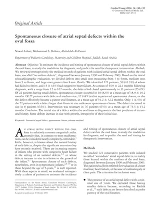
Body weight (percentile)
Number of patients
60
50
40
30
20
10
0 3 3
Figure 1.
The centiles for weight at the time of diagnosis of our 121 patients.
Note that the distribution is skewed toward the lower centiles.
4. Vol. 14, No. 2 Azhari et al: Atrial septal defects 151
patients (62%). Signs of right heart volume overload
were found in 2 patients (9%) with small defects, 10
(37%) with medium defects, and 71 (98.6%) with
large defects. We found signs of volume overload in
all patients with a defect measuring more than 6mm
who were diagnosed after, or followed-up until, the
age of 5 years. Absence of dilation of the right heart
on echocardiography was always associated with
normal cardiac size on chest radiographs, while the
presence of mild dilation was associated with an
increased cardiothoracic ratio in two-fifths of cases.
Moderate or severe dilation was always associ-ated
with radiographic evidence of cardiomegaly
(p 0.001).
Pulmonary arterial hypertension
Pulmonary arterial pressures greater than 30mmHg
were found in 8 patients (6.6%); 6 being male and
2 female, with the mean size of the defect being
15.5 6.7 mm, with a range from 9 to 28 mm. The
mean age at diagnosis was 17.6 20.9 months,
with a range from 2 to 61 months, with 5 patients
(62.5%) being below the age of 1 year at the
time of diagnosis. The body weight at diagnosis was
below the 5th centile in 6 patients (75%). Only one
had Down’s syndrome, and all had signs of
congestive heart failure. Cardiomegaly and signs of
right heart volume overload were also present in all
patients.
Follow-up and outcome
Small defects: At a mean period of follow-up of
21.3 13.4 months, with a range from 4 to 56
months, the defect had closed spontaneously in 18
patients (81.8%). In 2 patients (9.1%), the defects
remained at their original size, while in another 2
(9.1%), the defects had increased in size from 3 and
4 to 8 and 10 mm, respectively. The defects were
closed surgically in the last 2 patients, at ages of 60
and 61 months, respectively.
Defects of medium size
At a mean period of follow-up of 45.5 21.1 months,
with a range from 12 to 102 months, the defects had
closed spontaneously in 8 patients (29.6%), while in
4 (14.8%), the defects had decreased in size, effec-tively
becoming patent oval foramens. In 1 patient
(3.7%), the defect decreased in size from 7 to 4mm.
This was the only patient who presented with symp-toms
and signs of heart failure and he is still under-going
follow-up. In 11 patients (40.7%), the defects
remained at their original size. Of these, 4 were
referred for closure, while 7 are still undergoing
follow-up. In 3 patients (11.1%), the defects had
increased their sizes from 6, 6, and 7 to 10, 12, and
16 mm, respectively.
Intervention was considered in 7 patients (26%),
and was achieved surgically in 4, with the others
having transcatheter closure. Radiological evidence
of cardiomegaly, as well as echocardiographic evidence
of progressive right ventricular volume overload,
was seen in 6 of these patients, including the three in
whom the size of the defect had increased. Elective
intervention was offered to the seventh patient,
whose defect measured 7 mm, despite the absence of
symptoms, normal weight gain, and only mild vol-ume
overload, because there had been no change in the
size of the defect at the age of 5 years after follow-up
of 55 months.
Large defects
At a mean follow-up of 55.1 17.5 months, with a
range from 20 to 99 months, only 1 patient had
experienced spontaneous closure (1.4%), the defect
in this patient measuring 8 mm. In another patient,
the defect had decreased in size from 9 to 4 mm. In
67 patients (93%), the defects did not change in size,
but in 3 (4.2%), the defects increased from 11, 14,
and 16 to 20, 20, and 22 mm, respectively.
Intervention was considered in a total of 67
patients (93%), including the 8 with elevated pul-monary
arterial pressures. All did well except one,
who died during attempted surgical closure. This
was a 4-month old infant weighing 2.9 kg, with a
defect measuring 12 mm, intractable cardiac failure,
and elevated pulmonary arterial pressures. We are
still following 4 patients (5.6%), one because of
a progressive decrease in the size of the defect, and
3 who will probably be candidates for elective inter-vention,
but who currently are asymptomatic and
thriving, with only signs of mild volume overload.
The outcomes for the patients with defects of differ-ent
size are shown in Table 2.
Indications for intervention
Intervention was considered in a total of 76 patients
(63%), including 11 patients (16.4%) with indica-tions
for early intervention. These were the uncon-trolled
symptoms of cardiac failure associated with
failure to grow and signs of volume overload despite
aggressive medical treatment. In the other cases,
intervention was considered mainly because of the
presence of right heart volume overload, an increase
in the size of the defect, or as an elective procedure
prior to the age for schooling in those with large
defects that showed no evidence of a spontaneous
decrease in size.
5. 152 Cardiology in the Young April 2004
Table 2. Summarizes the outcome of 121 patients with defects of different sizes.
Number of patients Number of Number of Number of
Number of with spontaneous patients with patients with patients with
Size (mm) patients closure/POF (%) decreased size (%) increased size (%) unchanged size (%)
Small (3–5) 22 18 (82) – 2 (9) 2 (9)
Medium (5–8) 27 12 (44) 1 (3.7) 3 (11) 11 (40.7)
Large (8) 72 1 (1.4) 1 (1.4) 3 (4.2) 67 (93)
Total 121 31 (25.6) 2 (1.6) 8 (6.6) 80 (66)
Abbreviation: POF: patent oval foramen
Age at intervention
The mean age for those having early intervention
was 16.6 6.2 months, with a range from 4 to 24
months, otherwise the mean age at intervention was
75.5 15.2 months.
Spontaneous closure
Spontaneous closure occurred in a total of 31
patients, giving an overall incidence of 25.6%. The
mean age at spontaneous closure for patients with
small defects was 18.9 months, with a range from 6
to 55 months, with 94% of the closures documented
before the age of 2 years. The mean age at sponta-neous
closure for patients with defects of medium
size was 51.2 months, with a range from 15 to 137
months, with 92% of closures documented before
the age of 6 years (Table 3). The timing of sponta-neous
closure differed significantly according to the
sizes of the defects (p 0.005). The probability of
spontaneous closure as a function of time in relation
to initial size of the defect is shown in Figure 2.
Since only one patient with a large defect experi-enced
spontaneous closure, this patient was not
included in the analysis. The presence or absence of
cardiac failure did not influence the incidence of
spontaneous closure, (p 0.33), nor were there dif-ferences
in the incidence and timing of spontaneous
closure (p 0.05) between the genders for those
with defects of the same size. Spontaneous closure
occurred in 10 (52.6%) of 19 patients with Down’s
syndrome, with no statistical difference in their inci-dence
and timing of spontaneous closure (p 0.05)
compared to chromosomally normal patients with
defects of the same size.
Growth-related increase in the size of defects
An increase in the size of the defect was documented
in 8 patients (6.6%); 2 initially having small defect,
3 having defects of medium size, and 3 with large
defects. The sizes increased at a rate of 1.9 mm per
year, with a range of 0.5 to 3 mm per year. All these
patients were candidates for intervention.
Table 3. The mean age at spontaneous closure, and the period of
follow-up for patients with defects of different sizes.
Mean age of spontaneous Follow-up
closure (months) period (months)
Size (mm) Mean SD Mean SD
Small (3–5) 18.9 10.2 21.3 13.4
Medium (5–8) 51.2 32.2 45.5 21.1
Large (8) – 55.1 17.6
Total 34.0 29.0 44.9 22.1
0 20 40 60 80 100 120 140
Proportion of ASDs open
1.00
0.75
0.50
0.25
0.00
Discussion
We have collected data retrospectively from a large
series of patients with so-called isolated secundum
atrial septal defects, in other words those with the
defects confined within the oval fossa. The patients
formed a group with a wide age range, with defects
of variable sizes, and with follow-up of long dura-tion.
The mean period of follow-up, however, was
shorter for those with small defects, since the major-ity
of these underwent spontaneous closure before
the age of 2 years (Table 3).
Small defects
Medium defects
Months
Figure 2.
Kaplan-Meier curve showing the proportion of defects remaining
open as a function of time according to their initial size for patients
with small and medium defects.
6. Vol. 14, No. 2 Azhari et al: Atrial septal defects 153
We found a significant relation between the initial
size of the defect and the age at diagnosis; larger
defects, compared to the smaller ones, were found in
older patients. It has been postulated that an atrial
septal defect is usually small in infancy, growing large
enough to produce symptoms only later in life.1
Because of this, it is argued that, at an older age, when
most of the smaller defects have closed spontaneously,
others that had escaped initial clinical detection
would have increased the sizes of their defects, with
increased left-to-right shunting and obvious abnor-malities
in the physical examination, thus permitting
their detection as larger defects. In our study, an
increase in the size of the defect was confirmed in only
8 (6.6%) patients. McMahon et al.,6 in contrast, found
that the defects increased in size in two-thirds of their
patients, and they endorsed the concept that small
defects, initially believed to be hemodynamically
insignificant, could grow into major defects. The con-tinuous
shunting through the defects, the intrinsi-cally
compliant nature of the atrial septum,6 and the
stretching in the opposite direction to the short axis of
the elliptoid shape of the defect,7 are proposed as
mechanisms to explain the growth of defects.
While patients with defects in the oval fossa are
typically held to be asymptomatic in childhood,
infants are often symptomatic.3–5,14–19 Our data sup-ports
this belief, with almost nine-tenths of our
patients being asymptomatic, while four-fifths of those
presenting in cardiac failure did so at ages lower than
1 year.
We noted a body weight less than the fifth centile
at the time of diagnosis in two-fifths of our patients,
with a significant statistical relation to the size of the
defect at diagnosis, suggesting that the more hemo-dynamically
significant the defect, the greater is the
risk of having a subnormal body weight. Even after
excluding patients with congestive cardiac failure,
the size of the defect was found to be an independent
risk factor for failure to thrive (p 0.001).
The incidence and severity of the abnormalities
seen in electrocardiograms, chest radiographs and
echocardiograms correlated positively with the
increased size of the defects (p 0.001). Almost all
patients (98.6%) with large defects had signs of
right ventricular conduction delay and right heart
volume overload on their electrocardiograms and
echocardiograms, respectively. The detection of right
sided dilation by chest radiography correlated well
with that found on echocardiogram, with the appar-ent
ability of the echocardiogram to detect mild
dilation that was not visualized on chest radiography.
First degree atrioventricular block has been
reported to occur in between one-tenth and one-third
of patients with defects in the oval fossa.1 We
found this feature in one-tenth of our patients.
Spontaneous closure of atrial septal defects con-firmed
by echocardiography has been reported to
range from one-sixth20 to nine-tenths.21 The wide
range of reported incidence is probably due to the wide
variation in sizes of the defects among the different
studies. Our study showed an overall incidence of
spontaneous closure in one-quarter. This is expected,
since three-fifths of our cohort had large defects,
with an average size of almost 15 mm. Those with
small defects, measuring less than 5 mm, had a high
chance, over four-fifths, of spontaneous closure, com-pared
to a very low chance, only just over 1%, for
those with defects larger than 8 mm. Those with
defects of medium size had an unpredictable course,
with two-fifths of patients experiencing spontaneous
closure at a mean age near 4-years, with this event
occurring before the age of 6 years in nine-tenths of
this cohort. We had the chance to document sponta-neous
closure in patients with defects measuring 7
and 8 mm at ages of 137 and 101 months, respec-tively,
because the parents refused intervention. The
defects decreased gradually in size until complete
closure was confirmed echocardiographically by mul-tiple
studies in both patients.
Radzik and colleagues12 limited their study to
those diagnosed before the age of 3 months, with the
majority (77%) of their group having small defects.
We studied a group with broader age range, and
three-fifths of our patients had large defects. Despite
these differences in age, sex, and the size of the
defects, our study endorsed that of Radzik and col-leagues12
in showing that the classical female pre-dominance
is observed only for defects of medium or
large size, specifically those measuring greater than
5 mm, and that the incidence and timing of sponta-neous
closure is highly correlated with the size of the
defect at diagnosis.
Other factors that influenced the incidence of
spontaneous closure in our patients were the weight
and age of the patient at diagnosis. A normal body
weight at diagnosis was associated with a higher rate
of spontaneous closure (p 0.002). Moreover,
younger patients at diagnosis had a higher rate of
spontaneous closure (p 0.001). This finding has
previously been reported by Cockerham et al.,22 and
by Mody,10 both of whom reported an increased like-lihood
of spontaneous closure in children diagnosed
before the age of 2 years.
We documented Down’s syndrome in 16% of our
patients, albeit that having the syndrome did not pre-dict
the incidence and timing of spontaneous closure.
If untreated, atrial septal defects may cause a variety
of complications. These include the eventual devel-opment
of pulmonary hypertension, atrial arrhyth-mias,
and paradoxical embolisation, with infarction
of organs.23 Those with persistent defects permitting
7. 154 Cardiology in the Young April 2004
a ratio of pulmonary to systemic flows of more than
1.5 should undergo therapeutic closure before school
age, or whenever the diagnosis is made if later.24
There is no obvious advantage in delaying the repair
beyond this age, for the long-standing volume over-load
of the right heart causes irreversible changes that
contribute to the subsequent complications.25 Since
cardiac catheterization is rarely indicated, echocar-diographic
evidence of right ventricular volume over-load
is usually taken as an evidence of a significant
shunt, and hence an indication for intervention. This
was the main reason behind intervention in our series.
Earlier intervention is indicated if there is marked
cardiomegaly, failure to thrive, or congestive cardiac
failure.1
In the light of our experience, we now propose the
following strategy for the management of atrial sep-tal
defect in our institution, permitting us to inform
the parents about our expectation and plan once we
know the size of the defect.
Patients with small defects measuring from 3 to
5 mm are followed periodically using echocardio-graphy
to document either the greater chance of
spontaneous closure or the minimal risk of an
increase in the size of the defect.
Patients with defects of medium size, between 5
and 8 mm, are followed by echocardiography on a
yearly basis until the age of 5 years. Those show-ing
no evidence of a spontaneous decrease in the
size of the defect are planned electively for inter-vention
before the age of 6 years. A longer period
of conservative follow-up may be allowed for
those showing a progressive decrease in the size of
the defect.
Patients with defects of 8 mm or larger are
planned electively for intervention prior to com-mencing
school at the age of 4 years, or earlier if
indicated, to avoid the inevitable risk of volume
overload that is associated with their very low
chance of spontaneous closure.
Early intervention is considered in a patient with
a defect of any size should evidence develop of
uncontrolled cardiac failure, failure to thrive,
progressive increase in the size of the defect, or
significant right heart volume overload.
Based on our results, and experience, we now employ
a prognostic scoring system devised by Azhari and
Shihata, using a number of selected clinical and
diagnostic criterions (Table 4). This will help to
decide upon the management and predict the need,
and timing, of intervention in patients with isolated
atrial septal defects in the oval fossa. Patients will be
scored at each follow-up evaluation. When there is
an increase or a decrease in the size of the defect, the
Table 4. The proposed prognostic score devised by Azhari and
Shihata for patients with atrial septal defects within the oval fossa.
Criterion 0 1 2
Age (years) 2 2–4 4
Size (mm) 3–5 5–8 8
Failure to thrive Absent Present –
Cardiac failure Absent Controlled Uncontrolled
Cardiomegaly/ Absent Mild Moderate/Severe
RV-overload
Increasing Absent Present –
size of defect*
Pulmonary Absent – Present
hypertension
Abbreviation: RV: right ventricle.
• A score of less than 3 is an indication for conservative follow-up
• A score of 4 is an indication for elective intervention
• A score of 5 or greater is an indication for early intervention
*With any documented decrease in size, 1-point is deducted from the
total score
new size will be scored, and one point will be added
or deducted from the total score for the documented
increase or decrease in size. Patients with score of 3
or less will be followed conservatively, while those
with a score of 4 are planned for elective intervention
prior to starting school at the age of 4 years. Those
with score of 5 or more are candidates for intervention
at an earlier age, with the higher the score the more
urgent the need for intervention.
Acknowledgement
We are grateful to Dr. Ziad R. Bulbul, Head of the
Section of Pediatric Cardiology at King Faisal
Specialist Hospital and Research Center, Riyadh,
and Dr. Raja Al-Radadi, for their valuable contribu-tions
to this manuscript.
References
1. Beerman LB, Zuberbuhler JR. Atrial septal defect. In:
Anderson RH, Macartney FJ, Shinebourne EA, Tynan M (eds).
Paediatric Cardiology. Churchill Livingstone, Edinburgh, 1987,
pp 541.
2. Hoffman JIE, Rudolph AM, Danilowicz D. Left to right atrial
shunts in infants. Am J Cardiol 1972; 30: 868–875.
3. Hunt CE, Lucas RV. Symptomatic atrial septal defect in infancy.
Circulation 1973; 47: 1042–1048.
4. Dimich I, Steinfeld L, Park SC. Symptomatic atrial septal defect
in infants. Am Heart J 1973; 85: 601–604.
5. Toews WH, Nora JJ, Wolfe RR. Presentation of atrial septal
defect in infancy. JAMA 1975; 234: 1250–1251.
6. McMahon CJ, Feltes TF, Fraley JK, Bricker JT, Grifka RG,
Tortoriello TA, Blake R, Bezold LI. Natural history of growth of
secundum atrial septal defects and implications for transcatheter
closure. Heart 2002; 87: 256–259.
7. Helgason H, Jonsdottir G. Spontaneous closure of atrial septal
defects. Pediatr Cardiol 1999; 20: 195–199.
8. Vol. 14, No. 2 Azhari et al: Atrial septal defects 155
8. Timmis GC, Gordon S, Reed JO. Spontaneous closure of an atrial
septal defect. JAMA 1966; 196: 137–139.
9. Hartmann AF Jr, Elliott LP. Spontaneous physiologic closure of
an atrial septal defect after infancy. Am J Cardiol 1967; 19:
290–292.
10. Mody MR. Serial hemodynamic observations in secundum atrial
septal defect with special reference to spontaneous closure. Am J
Cardiol 1973; 32: 978–981.
11. Brassard M, Fouron JC, van Doesburg N, Mercier LA, De Guise P.
Outcome of children with atrial septal defect considered too small
for surgical closure. Am J Cardiol 1999; 83: 1552–1555.
12. Radzik D, Davignon A, van Doesburg N, Fournier A, Marchand T,
Ducharme G. Predictive factors for spontaneous closure of atrial
septal defects diagnosed in the first 3 months of life. J Am Coll
Cardiol 1993; 22: 851–853.
13. Roge CL, Silverman NH, Hart PA, Ray RM. Cardiac structure
growth pattern determined by echocardiography. Circulation
1978; 57: 285–290.
14. Mahoney LT, Truesdell SC, Krzmarzick TR, Lauer RM. Atrial
septal defects that present in infancy. AJDC 1986; 140: 1115–1118.
15. Hastreiter AR, Wennemark JR, Miller RA. Secundum atrial sep-tal
defects with congestive heart failure during infancy and early
childhood. Am Heart J 1962; 64: 467–472.
16. Nakamura FF, Hauck AJ, Nadas AS. Atrial septal defect in
infants. Pediatrics 1964; 34: 101–106.
17. Ainger LE, Page JW. Ostium secundum atrial septal defects and
congestive heart failure in infancy. Am J Cardiol 1965; 15: 380–385.
18. Weinberg M, Miller RA, Hastreiter AR. Congestive heart failure
in children with atrial septal defect. J Thorac Cardiovasc Surg
1966; 51: 81–87.
19. Kavanagh-Gray D. Atrial septal defect in infancy. Can Med Assoc J
1963; 89: 491–499.
20. Ghisla RP, Hannon DW, Meyer RA, Kaplan S. Spontaneous
closure of isolated secundum atrial septal defects in infants: an
echocardiographic study. Am Heart J 1985; 109: 1327–1333.
21. Fukazawa M, Fukushige J, Ueda K. Atrial septal defects in
neonates with reference to spontaneous closure. Am Heart J 1988;
116: 123–127.
22. Cockerham JT, Martin TC, Gutierrez FR, Hartmann AF,
Goldring D, Strauss AW. Spontaneous closure of secundum atrial
septal defect in infants and young children. Am J Cardiol 1983;
52: 1267–1271.
23. Craig RJ, Selzer A. Natural history and prognosis of atrial septal
defect. Circulation 1968; 37: 805–815.
24. Kirklin JW, Barratt-Boyes BG. Cardiac Surgery, 2nd edn.
Churchill Livingstone, Edinburgh, 1993, pp 609–644.
25. Porter CJ, Feldt RH, Edward WD, Seward JB, Schaff HV. Atrial
Septal Defect. In: Moss AJ, Adams FH (eds). Heart Disease in
Infants, Children, and Adolescents Including the Fetus and Young
Adults, 5th edn. Williams and Wilkins, Baltimore, 1995, pp 687.