SlideShare ist ein Scribd-Unternehmen logo
1 von 58
Downloaden Sie, um offline zu lesen
Triple Rule Out CT
Lt. Myo Thiha
Objective
To assess the validity and efficacy of Triple-rule-out (TRO) computed
tomographic (CT) angiography in diagnosis of different vascular
causes of chest pain.
Triple Rule Out CT
TRO-CTA is a specialized computed tomography (CT) imaging exam
tailored to evaluate for pathology within the coronary arteries,
pulmonary arteries, and the aorta in a single CT study, hence the
name ‘triple-rule out’.
Why we take TRO-CT?
Acute chest pain is the second most common presentation after abdominal pain
in the emergency department.
represents a major diagnostic challenge in emergency care as it has a broad
differential diagnosis varying from benign causes to life-threatening conditions.
TRO studies are most appropriate and cost-effective when there is a suspicion
for acute coronary syndrome along with other diagnoses such as pulmonary
embolism, acute aortic syndrome, or nonvascular disease in the thorax.
Most common causes of Chest Pain
Acute Coronary Syndrome
Pulmonary Embolism
Acute Aortic Syndrome
Causes of Chest pain
Heart related causes
Heart-attack
Angina
Aortic Dissection
Digestive Causes
Chest pain can be caused by disorders of the digestive system,
including:
Heartburn
Swallowing disorders
Gallbladder or pancreas problems
Muscle and bone causes
Some types of chest pain are associated with injuries and other
problems affecting the structures that make up the chest wall,
including:
Costochondritis
Injured rib
Lung-related causes
lung disorders can cause chest pain, including:
Pulmonary embolism
Pleurisy
Pulmonary hypertension
In many clinical situations, a definite diagnosis of ED chest pain is not
possible solely based on clinical symptoms and laboratory findings.
In addition, most diagnostic modalities (i.e., ECG, cardiac enzymes,
exercise treadmill testing, radionuclide perfusion imaging and stress
echocardiography) other than MDCT are focused on the diagnosis or
exclusion of ACS and do not exclude other life-threatening causes of
acute chest pain.
For this reason, Triple Role Out (TRO) protocol with ECG-gating
technology has been proposed to encompass the entire thorax,
allowing simultaneous evaluation of coronary arteries, thoracic aorta
and pulmonary arteries for improving diagnosis of acute chest pain in
a single study.
Criteria for TRO_CT study
Clinical presentation – low to moderate risk of ACS
Clinical presentation – non ACS diagnosed considered
Normal ECG or non-specific changes
Patient able to tolerate CT and hold breath
Cardiac rhythm acceptable ECG gated scan
Adequate renal function
Exclusion criteria
 Contra-indications to iodinated contrast material including known
allergy and renal insufficiency (serum creatinine more than 1.4
mg/dl).
Marked heart failure.
Clinically unfit patients (unable to stop breathing during the
examination).
 Extensive calcium score above 1000.
Why don’t we take coronary angiography above 1000 calcium score?
Because of blooming artifacts, which can cause erroneous
enlargement of calcification, make less accuracy to determine the
coronary arteries lumen that results in false positive diagnosis.
Fig: (A) shows enlargement in the lumen of RCA because of the blooming artifacts
(B) shows normal in state of RCA
Anatomy of Aorta, Pulmonary and Coronary
Aorta
the largest blood vessel in the body.
responsible for transporting oxygen rich blood from your heart to the
rest of the body.
begins at the left ventricle of the heart, extending upward into the
chest to form an arch.
downward into the abdomen, where it branches into the iliac arteries
just above the pelvis.
Aortic Root
the portion of the aorta that is attached to the heart.
 major part of the aortic root is the aortic valve
allows blood to flow from the heart
to the rest of the body
when it is open and prevents blood
from flowing backwards into the heart when it is closed.
Ascending Aorta
begins at the sinotubular junction of the aortic root and extends up
and out from the heart until it connects with the aortic arch.
Aortic Arch
the portion of the aorta that is in the shape of an arch and connects the
ascending aorta with the descending aorta.
 The major arteries : the brachiocephalic artery,
the left carotid artery and the left subclavian artery.
Descending Thoracic Aorta
begins at the end of the aortic arch and continues down into the
abdomen. Two parts:
1. T. aorta(provides blood to the muscles of the chest wall and the
spinal cord.)
2. Ab.Aorta (five arteries that branch from the abdominal aorta: the
celiac artery, the superior mesenteric artery, the inferior
mesenteric artery, the renal arteries and the iliac arteries)
celiac artery superior mesenteric artery
inferior mesenteric artery renal arteries iliac arteries
Stomach
Liver
Pancreas
supplies blood to the small
intestine
supplies blood to the large
intestine
blood to the kidneys as well
as the muscles of the
abdominal wall and the lower
spinal cord
blood to the legs and the
organs in the pelvis.
Figure: shows five arteries from the abdominal aorta and their blood
supply
Thoracic aorta disease
1. aortic aneurysms and dissections,
2. atherosclerotic disease,
3. infections and
4. traumatic injuries.
Note: Ruptured thoracic aortic aneurysms and aortic dissections represent
life-threatening emergencies that require immediate medical attention.
Thoracic aortic aneurysms affect approximately 15,000 people in the United
States each year
Symptoms
1. Chest pain
2. Back pain
3. Hoarseness
4. Wheezing
5. Difficulty swallowing
6. Cough
The most common types of aortic aneurysms are thoracic and
abdominal. In addition, the following can signal a more serious
condition:
1. Sudden and severe chest pain
2. Fainting
3. Leg pain or numbness
4. Shortness of breath
5. Weakness
Coronary Artery
heart is mostly supplied by the two coronary arteries which arise
from the ascending aorta immediately above the aortic valve.
coronary arteries and their branches run on the surface of the heart.
RCA
arise from the anterior aortic sinus of the ascending aorta
immediately above the aortic valve.
first runs forwards between the pulmonary trunk and the right
auricle.
Then, it descends almost vertically enter the rt. Atrioventricular
groove, the rt. anterior coronary sulcus. At the inferior border of the
heart, it turns posteriorly and runs into the atrioventricular groove,
after the posterior interventricular groove finally anastomosing with
the LCA.
LCA
arise from the left posterior aortic sinus of the ascending aorta, immediately
above the aortic valve and enters the left between the pulmonary trunk and the
left auricle. then divides into the anterior interventricular artery also know as left
anterior descending artery which runs downwards in the anterior interventricular
groove to the apex of the heart.
Clinical Correlation
Angina Pectoris – since coronary arteries are narrowed, the blood
supply to the cardiac muscles is reduced.
As a result, on exertion, the patient feels moderately severe pain in
region of the left pericardium last as long as 20 mins.
Pain is often referred to the left shoulder and medial side of the arm
and forearm.
Angina pectoris pain occurs on exertion and relieved by rest.
Myocardial Infarction
sudden block of the larger branches of either coronary artery usually
leads to myocardial ischaemia followed by the myocardial necrosis
(myocardial infarction).
Part of the heart suffering from MI, stops functioning and often
causes death.
This condition is termed as the heart attack or coronary attack.
Clinical features of MI
Sensation of pressure, sinking and pain in the chest that lasts longer
than 30 minutes.
Nausea (or) vomiting, sweating, shortness of breath, and tachycardia.
Pain radiates to the medial side of the arm, forearm, and hand.
Sometimes, it may be referred to jaw or neck.
Sites of coronary artery occlusion
The three most common site of CAO are
1. Anterior interventricular artery or LAD (40-50%),
2. RCA (30-40%)
3. Circumflex branch of LCA (15-20%)
NOTE: MI mostly occurs at rest whereas angina occurs on exertion.
Blood supply of the major coronary arteries
The 2 main coronary arteries are the left main and right coronary arteries.
Left main coronary artery (LMCA).
The left main coronary artery supplies blood to the left side of the heart
muscle (the left ventricle and left atrium). The left main coronary divides into
branches:
1. The left anterior descending artery branches off the left coronary artery and
supplies blood to the front of the left side of the heart.
2. The circumflex artery branches off the left coronary artery and encircles the
heart muscle. This artery supplies blood to the outer side and back of the heart.
Right coronary artery (RCA)
 The right coronary artery supplies blood to the right ventricle, the right
atrium, and the SA (sinoatrial) and AV (atrioventricular) nodes, which
regulate the heart rhythm.
The right coronary artery divides into smaller branches, including the right
posterior descending artery and the acute marginal artery.
Together with the left anterior descending artery, the right coronary
artery helps supply blood to the middle or septum of the heart.
Fig: Coronary arteries and their blood supply
Fig: Coronary Arteries and their branches
Types of Circulation
Pulmonary Circulation
the portion of the cardiovascular system that carries oxygen-poor
(deoxygenated) blood from the heart to the lungs and returns
oxygenated blood back to the heart.
deoxygenated blood from the body leaves the right ventricle through
the pulmonary arteries, which carry the blood to each lung.
34
Cont.;
pulmonary arteries are the only arteries that carry deoxygenated
blood.
In the lungs, red blood cells release carbon dioxide and pick up
oxygen during respiration.
The oxygenated blood then leaves the lungs through the pulmonary
veins, which return it to the left side of the heart and complete the
pulmonary cycle.
Cont.;
The oxygenated blood is then distributed to the body through the
systemic circulation before returning again to the pulmonary
circulation.
Fig: The pulmonary circulation carries blood between the heart and lungs.
Systemic Circulation
the portion of the cardiovascular system that carries oxygenated
blood from the heart to the body and returns deoxygenated blood
back to the heart.
Oxygenated blood from the lungs leaves the left ventricle through the
aorta.
From here it is distributed to the body's organs and tissues, which
absorb the oxygen through a complex network of arteries, arterioles,
and capillaries.
Cont.;
The deoxygenated blood is then collected by venules and flows into
veins before reaching the inferior and superior venae cavae, which
return it to the right heart, completing the systemic cycle.
The blood is then re-oxygenated through the pulmonary circulation
before returning again to the systemic circulation.
Fig: The systemic circulation. The systemic circulation brings oxygenated blood to the body cells and tissues and
transports cellular wastes away from the cells and tissues. It is also responsible for temperature regulation and
transport of hormones and other substances around the body.
Coronary Circulation
the heart needs its own blood supply, which it gets through the
coronary circulation.
the heart muscle tissue is so thick that it needs blood vessels to
deliver oxygen and nutrients deep within it.
The vessels that deliver oxygen-rich blood to the heart muscle are
called coronary arteries.
Cont.;
branch directly from the aorta, just above the heart.
The vessels that remove the deoxygenated blood from the heart
muscle are known as cardiac veins.
Fig: coronary circulation
Patient preparation for TRO
An 18–20-gauge intravenous catheter is placed into a large vein in the
antecubital fossa.
The patient is lying in a supine position with arm in front of him.
ECG leads are positioned above and below the level of the scan to prevent
streak artifact.
The ideal heart rate for ECG-gated studies is a slow regular rhythm, usually
a sinus bradycardia at 50–60 beats per minute.
Oral-blockers may be given at least 1 h before the scan for control of heart
rate.
Contrast material and scanning protocol
In order to image both the coronary and pulmonary arteries, a biphasic
injection technique was used: 70 mL of undiluted (ultravist 370) was
injected at 5 mL/s, followed by 25 mL of the same contrast material diluted
with 25 mL of saline, also injected at 5 mL/s.
For injection Protocol we used a bolus tracking technique where we
started contrast medium injection when the HU in the left atrium reached
100 HU then in the second phase we depended on the observation, for
assessment of the opacification of the pulmonary artery.
The first phase of the injection opacifies the coronary arteries during
image acquisition, while the second phase of the injection, provides
simultaneous homogeneous enhancement of the pulmonary arteries.
Data acquisition starts from the level of the medial end of the
clavicles to the lower border of the heart in cranio–caudal direction.
Fig: Typical Z axis coverage in dedicated coronary CT angiography versus triple rule-out study. A: the field of view in a
dedicated coronary CT angiography is demonstrated. B: note the increased Z axis length in the triple rule-out study
compared with dedicated coronary CT angiography.
Data evaluation
For coronary assessment every case was evaluated in the axial plane
and with slab maximum intensity projection images that were rotated
to visualize each vessel in multiple planes.
 Vessels with complex plaque were also evaluated with curved
multiplanar reconstruction by using vessel tracking software with
automatic centerline determination.
Different parts of the thoracic aorta regarding their diameter,
contrast filling, presence of filling defects, dissections, wall
irregularities, calcification, mural thrombus.
The main pulmonary artery, right and left pulmonary arteries, their
segmental and subsegmental branches, regarding their diameter,
contrast filling, presence of filling defects, wall irregularities,
calcification, mural thrombus.
Other extra-vascular structures such as cardiac size, pleural,
pericardial and parenchymal abnormalities have to be assessed.
Female patient 37 year old presenting with acute chest pain, dyspnea and hemoptysis. CT axial (A and B),
sagittal (C) and (D) images of pulmonary angiography showing left main and segmental pulmonary embolism
and left sided pleural effusion.
Triple rule-out CT angiography images (sagittal and axial views) of two patients with aortic dissection,
with standard (a, b) and low dose (c, d). The diagnostic and image quality were excellent in both patients
in the left descending artery (white arrow), the aorta(white star) and the pulmonary artery (arrowhead).
The first patient had an history of surgical replacement of the ascending aorta.
Pros
TRO CT can reduce
(a) time for patient triage,
(b) number of required diagnostic tests,
(c) costs, and
(d) radiation exposure to the patient.
Limitations
Beta-blockers that are required for coronary CTA may not be safe in
patients with pulmonary embolism.
Obesity and calcifications limit interpretation, rapid heart rate,
arrhythmias, renal dysfunction and contrast allergies.
Conclusion
Since there are a lot of chest pain cases in emergency department , it is difficult
to know the real cause of chest pain.
triple rule out is a relatively new technique, examination the coronary arteries,
pulmonary arteries and aorta in just a single study, which gives us the advantage
of screening emergency patients presenting with chest pain in a rapid and safe
way for detection of their vascular diseases.
In addition, it can reduce the radiation dose to the patient because it investigates
the coronary arteries, pulmonary arteries and aorta in just a single study and we
don’t need to separated studies to check those of them.
That’s why, it is most effective investigation to know the origin of abnormalities
even though it has still challenges.
References
 Pitts SR, Niska RW, Xu J, Burt CW. National hospital ambulatory
medical care survey: 2006 emergency department summary. Natl
Health Stat Rep 2008;7:1–8.
 Stillman AE, Oudkerk M, Ackerman M. Use of multidetector
computed tomography for the assessment of acute chest pain:
aconsensus statement of the North American Society of Cardiac
Imaging and the European Society of Cardiac Radiology. Int J
Cardiovascular Imaging 2007;23:415–27.
Thomas J, Rideau AM, Paulson EK, Bisset 3rd GS. Emergency
department imaging: current practice. J Am Coll Radiol
2008;5(7):811–816e2.
 Kevin M, Ethan J. Evaluation of a ‘‘Triple Rule-Out’’ coronary CT
angiography protocol: use of 64-section CT in low-tomoderate risk
emergency department patients suspected of having acute coronary
syndrome. Radiology 2008;248(2).
Rubinshtein R, Halon D, Gaspar T, et al. Usefulness of 64-slice cardiac
computed tomographic angiography for diagnosing acute coronary
syndromes and predicting clinical outcome in emergency department
patients with chest pain of uncertain origin. Circulation
2007;115:1762–8.
Kevin M, Ethan J. Evaluation of a ‘‘Triple Rule-Out’’ coronary CT
angiography protocol: use of 64-section CT in low-tomoderate risk
emergency department patients suspected of having acute coronary
syndrome. Radiology 2008;248(2)
THANK YOU

Weitere ähnliche Inhalte

Ähnlich wie tro-200922025135 2.pdf

Aorta, SVC, PULMONARY TRUNK.pptx
Aorta, SVC, PULMONARY TRUNK.pptxAorta, SVC, PULMONARY TRUNK.pptx
Aorta, SVC, PULMONARY TRUNK.pptxMovieDownload83
 
Tricuspid valve disease by T.N. Shanta
Tricuspid valve disease by T.N. ShantaTricuspid valve disease by T.N. Shanta
Tricuspid valve disease by T.N. ShantaTania Nusrat Shanta
 
Valvular heart diseases imaging
Valvular heart diseases imagingValvular heart diseases imaging
Valvular heart diseases imagingDev Lakhera
 
Cynotic heart disease plane radiography
Cynotic heart disease plane radiography Cynotic heart disease plane radiography
Cynotic heart disease plane radiography Dr-Girish Gunari
 
Aorta&its branches . pptx.pptx
Aorta&its branches . pptx.pptxAorta&its branches . pptx.pptx
Aorta&its branches . pptx.pptxKannurLion
 
The structure and Function of the Heart
The structure and Function of the Heart The structure and Function of the Heart
The structure and Function of the Heart TessMiller519
 
Heart Slides
Heart SlidesHeart Slides
Heart SlidesSatnam
 
A presentation on structural and functional properties of heart
A presentation on structural and functional properties of heartA presentation on structural and functional properties of heart
A presentation on structural and functional properties of heartMahade Hashan
 
cardiovascular system
cardiovascular systemcardiovascular system
cardiovascular systemJADHAVDEEPALI
 
Atherosclerosis
AtherosclerosisAtherosclerosis
Atherosclerosisshas595
 
Cardiovascula system
Cardiovascula systemCardiovascula system
Cardiovascula systemLesly Malonda
 
MYOCARDIAL INFARACTION.pptx
MYOCARDIAL INFARACTION.pptxMYOCARDIAL INFARACTION.pptx
MYOCARDIAL INFARACTION.pptxDINESH SINGH
 
Atherosclerosis
AtherosclerosisAtherosclerosis
Atherosclerosishiratufail
 
The_Heart__anatomy and physiology AHscience
The_Heart__anatomy and physiology AHscienceThe_Heart__anatomy and physiology AHscience
The_Heart__anatomy and physiology AHsciencesudheendrapv
 

Ähnlich wie tro-200922025135 2.pdf (20)

Cardiovascular System Pathology 2014 edited by @jennings argwing
Cardiovascular System Pathology 2014 edited by @jennings argwingCardiovascular System Pathology 2014 edited by @jennings argwing
Cardiovascular System Pathology 2014 edited by @jennings argwing
 
Aorta, SVC, PULMONARY TRUNK.pptx
Aorta, SVC, PULMONARY TRUNK.pptxAorta, SVC, PULMONARY TRUNK.pptx
Aorta, SVC, PULMONARY TRUNK.pptx
 
heart anatomy.pptx
heart anatomy.pptxheart anatomy.pptx
heart anatomy.pptx
 
Coronary CT
Coronary CTCoronary CT
Coronary CT
 
Tricuspid valve disease by T.N. Shanta
Tricuspid valve disease by T.N. ShantaTricuspid valve disease by T.N. Shanta
Tricuspid valve disease by T.N. Shanta
 
Valvular heart diseases imaging
Valvular heart diseases imagingValvular heart diseases imaging
Valvular heart diseases imaging
 
Cynotic heart disease plane radiography
Cynotic heart disease plane radiography Cynotic heart disease plane radiography
Cynotic heart disease plane radiography
 
Aorta&its branches . pptx.pptx
Aorta&its branches . pptx.pptxAorta&its branches . pptx.pptx
Aorta&its branches . pptx.pptx
 
The structure and Function of the Heart
The structure and Function of the Heart The structure and Function of the Heart
The structure and Function of the Heart
 
Heart Slides
Heart SlidesHeart Slides
Heart Slides
 
A presentation on structural and functional properties of heart
A presentation on structural and functional properties of heartA presentation on structural and functional properties of heart
A presentation on structural and functional properties of heart
 
CVS Anaesthesiology.pptx
CVS Anaesthesiology.pptxCVS Anaesthesiology.pptx
CVS Anaesthesiology.pptx
 
cardiovascular system
cardiovascular systemcardiovascular system
cardiovascular system
 
Atherosclerosis
AtherosclerosisAtherosclerosis
Atherosclerosis
 
Cardiovascula system
Cardiovascula systemCardiovascula system
Cardiovascula system
 
MYOCARDIAL INFARACTION.pptx
MYOCARDIAL INFARACTION.pptxMYOCARDIAL INFARACTION.pptx
MYOCARDIAL INFARACTION.pptx
 
Atherosclerosis
AtherosclerosisAtherosclerosis
Atherosclerosis
 
The_Heart__anatomy and physiology AHscience
The_Heart__anatomy and physiology AHscienceThe_Heart__anatomy and physiology AHscience
The_Heart__anatomy and physiology AHscience
 
Atherosclerosis
AtherosclerosisAtherosclerosis
Atherosclerosis
 
Cardiac cycle (Human Heart)
Cardiac cycle (Human Heart)Cardiac cycle (Human Heart)
Cardiac cycle (Human Heart)
 

Mehr von monicaaneesha

obstructivejaundice-190919140942.pdf
obstructivejaundice-190919140942.pdfobstructivejaundice-190919140942.pdf
obstructivejaundice-190919140942.pdfmonicaaneesha
 
cardiomyopathy-161006095911.pdf
cardiomyopathy-161006095911.pdfcardiomyopathy-161006095911.pdf
cardiomyopathy-161006095911.pdfmonicaaneesha
 
interventionalradiologypart1-170807180227.pdf
interventionalradiologypart1-170807180227.pdfinterventionalradiologypart1-170807180227.pdf
interventionalradiologypart1-170807180227.pdfmonicaaneesha
 
bronchialarteryembolization-dr-150106080847-conversion-gate01.pdf
bronchialarteryembolization-dr-150106080847-conversion-gate01.pdfbronchialarteryembolization-dr-150106080847-conversion-gate01.pdf
bronchialarteryembolization-dr-150106080847-conversion-gate01.pdfmonicaaneesha
 
eus-200527113847.pdf
eus-200527113847.pdfeus-200527113847.pdf
eus-200527113847.pdfmonicaaneesha
 
digitalsubtractionangiography-140522112440-phpapp01.pdf
digitalsubtractionangiography-140522112440-phpapp01.pdfdigitalsubtractionangiography-140522112440-phpapp01.pdf
digitalsubtractionangiography-140522112440-phpapp01.pdfmonicaaneesha
 
interventionradiologyhepatobiliarysystem-140107210458-phpapp02.pdf
interventionradiologyhepatobiliarysystem-140107210458-phpapp02.pdfinterventionradiologyhepatobiliarysystem-140107210458-phpapp02.pdf
interventionradiologyhepatobiliarysystem-140107210458-phpapp02.pdfmonicaaneesha
 
pacs-140504092954-phpapp01.pdf
pacs-140504092954-phpapp01.pdfpacs-140504092954-phpapp01.pdf
pacs-140504092954-phpapp01.pdfmonicaaneesha
 
pit-190330014429.pdf
pit-190330014429.pdfpit-190330014429.pdf
pit-190330014429.pdfmonicaaneesha
 

Mehr von monicaaneesha (9)

obstructivejaundice-190919140942.pdf
obstructivejaundice-190919140942.pdfobstructivejaundice-190919140942.pdf
obstructivejaundice-190919140942.pdf
 
cardiomyopathy-161006095911.pdf
cardiomyopathy-161006095911.pdfcardiomyopathy-161006095911.pdf
cardiomyopathy-161006095911.pdf
 
interventionalradiologypart1-170807180227.pdf
interventionalradiologypart1-170807180227.pdfinterventionalradiologypart1-170807180227.pdf
interventionalradiologypart1-170807180227.pdf
 
bronchialarteryembolization-dr-150106080847-conversion-gate01.pdf
bronchialarteryembolization-dr-150106080847-conversion-gate01.pdfbronchialarteryembolization-dr-150106080847-conversion-gate01.pdf
bronchialarteryembolization-dr-150106080847-conversion-gate01.pdf
 
eus-200527113847.pdf
eus-200527113847.pdfeus-200527113847.pdf
eus-200527113847.pdf
 
digitalsubtractionangiography-140522112440-phpapp01.pdf
digitalsubtractionangiography-140522112440-phpapp01.pdfdigitalsubtractionangiography-140522112440-phpapp01.pdf
digitalsubtractionangiography-140522112440-phpapp01.pdf
 
interventionradiologyhepatobiliarysystem-140107210458-phpapp02.pdf
interventionradiologyhepatobiliarysystem-140107210458-phpapp02.pdfinterventionradiologyhepatobiliarysystem-140107210458-phpapp02.pdf
interventionradiologyhepatobiliarysystem-140107210458-phpapp02.pdf
 
pacs-140504092954-phpapp01.pdf
pacs-140504092954-phpapp01.pdfpacs-140504092954-phpapp01.pdf
pacs-140504092954-phpapp01.pdf
 
pit-190330014429.pdf
pit-190330014429.pdfpit-190330014429.pdf
pit-190330014429.pdf
 

Kürzlich hochgeladen

Beyond the EU: DORA and NIS 2 Directive's Global Impact
Beyond the EU: DORA and NIS 2 Directive's Global ImpactBeyond the EU: DORA and NIS 2 Directive's Global Impact
Beyond the EU: DORA and NIS 2 Directive's Global ImpactPECB
 
fourth grading exam for kindergarten in writing
fourth grading exam for kindergarten in writingfourth grading exam for kindergarten in writing
fourth grading exam for kindergarten in writingTeacherCyreneCayanan
 
social pharmacy d-pharm 1st year by Pragati K. Mahajan
social pharmacy d-pharm 1st year by Pragati K. Mahajansocial pharmacy d-pharm 1st year by Pragati K. Mahajan
social pharmacy d-pharm 1st year by Pragati K. Mahajanpragatimahajan3
 
Presentation by Andreas Schleicher Tackling the School Absenteeism Crisis 30 ...
Presentation by Andreas Schleicher Tackling the School Absenteeism Crisis 30 ...Presentation by Andreas Schleicher Tackling the School Absenteeism Crisis 30 ...
Presentation by Andreas Schleicher Tackling the School Absenteeism Crisis 30 ...EduSkills OECD
 
SOCIAL AND HISTORICAL CONTEXT - LFTVD.pptx
SOCIAL AND HISTORICAL CONTEXT - LFTVD.pptxSOCIAL AND HISTORICAL CONTEXT - LFTVD.pptx
SOCIAL AND HISTORICAL CONTEXT - LFTVD.pptxiammrhaywood
 
9548086042 for call girls in Indira Nagar with room service
9548086042  for call girls in Indira Nagar  with room service9548086042  for call girls in Indira Nagar  with room service
9548086042 for call girls in Indira Nagar with room servicediscovermytutordmt
 
Software Engineering Methodologies (overview)
Software Engineering Methodologies (overview)Software Engineering Methodologies (overview)
Software Engineering Methodologies (overview)eniolaolutunde
 
1029-Danh muc Sach Giao Khoa khoi 6.pdf
1029-Danh muc Sach Giao Khoa khoi  6.pdf1029-Danh muc Sach Giao Khoa khoi  6.pdf
1029-Danh muc Sach Giao Khoa khoi 6.pdfQucHHunhnh
 
Explore beautiful and ugly buildings. Mathematics helps us create beautiful d...
Explore beautiful and ugly buildings. Mathematics helps us create beautiful d...Explore beautiful and ugly buildings. Mathematics helps us create beautiful d...
Explore beautiful and ugly buildings. Mathematics helps us create beautiful d...christianmathematics
 
Sanyam Choudhary Chemistry practical.pdf
Sanyam Choudhary Chemistry practical.pdfSanyam Choudhary Chemistry practical.pdf
Sanyam Choudhary Chemistry practical.pdfsanyamsingh5019
 
Q4-W6-Restating Informational Text Grade 3
Q4-W6-Restating Informational Text Grade 3Q4-W6-Restating Informational Text Grade 3
Q4-W6-Restating Informational Text Grade 3JemimahLaneBuaron
 
Student login on Anyboli platform.helpin
Student login on Anyboli platform.helpinStudent login on Anyboli platform.helpin
Student login on Anyboli platform.helpinRaunakKeshri1
 
Paris 2024 Olympic Geographies - an activity
Paris 2024 Olympic Geographies - an activityParis 2024 Olympic Geographies - an activity
Paris 2024 Olympic Geographies - an activityGeoBlogs
 
Z Score,T Score, Percential Rank and Box Plot Graph
Z Score,T Score, Percential Rank and Box Plot GraphZ Score,T Score, Percential Rank and Box Plot Graph
Z Score,T Score, Percential Rank and Box Plot GraphThiyagu K
 
Call Girls in Dwarka Mor Delhi Contact Us 9654467111
Call Girls in Dwarka Mor Delhi Contact Us 9654467111Call Girls in Dwarka Mor Delhi Contact Us 9654467111
Call Girls in Dwarka Mor Delhi Contact Us 9654467111Sapana Sha
 
Grant Readiness 101 TechSoup and Remy Consulting
Grant Readiness 101 TechSoup and Remy ConsultingGrant Readiness 101 TechSoup and Remy Consulting
Grant Readiness 101 TechSoup and Remy ConsultingTechSoup
 
Activity 01 - Artificial Culture (1).pdf
Activity 01 - Artificial Culture (1).pdfActivity 01 - Artificial Culture (1).pdf
Activity 01 - Artificial Culture (1).pdfciinovamais
 
microwave assisted reaction. General introduction
microwave assisted reaction. General introductionmicrowave assisted reaction. General introduction
microwave assisted reaction. General introductionMaksud Ahmed
 
BAG TECHNIQUE Bag technique-a tool making use of public health bag through wh...
BAG TECHNIQUE Bag technique-a tool making use of public health bag through wh...BAG TECHNIQUE Bag technique-a tool making use of public health bag through wh...
BAG TECHNIQUE Bag technique-a tool making use of public health bag through wh...Sapna Thakur
 

Kürzlich hochgeladen (20)

Beyond the EU: DORA and NIS 2 Directive's Global Impact
Beyond the EU: DORA and NIS 2 Directive's Global ImpactBeyond the EU: DORA and NIS 2 Directive's Global Impact
Beyond the EU: DORA and NIS 2 Directive's Global Impact
 
fourth grading exam for kindergarten in writing
fourth grading exam for kindergarten in writingfourth grading exam for kindergarten in writing
fourth grading exam for kindergarten in writing
 
social pharmacy d-pharm 1st year by Pragati K. Mahajan
social pharmacy d-pharm 1st year by Pragati K. Mahajansocial pharmacy d-pharm 1st year by Pragati K. Mahajan
social pharmacy d-pharm 1st year by Pragati K. Mahajan
 
Presentation by Andreas Schleicher Tackling the School Absenteeism Crisis 30 ...
Presentation by Andreas Schleicher Tackling the School Absenteeism Crisis 30 ...Presentation by Andreas Schleicher Tackling the School Absenteeism Crisis 30 ...
Presentation by Andreas Schleicher Tackling the School Absenteeism Crisis 30 ...
 
SOCIAL AND HISTORICAL CONTEXT - LFTVD.pptx
SOCIAL AND HISTORICAL CONTEXT - LFTVD.pptxSOCIAL AND HISTORICAL CONTEXT - LFTVD.pptx
SOCIAL AND HISTORICAL CONTEXT - LFTVD.pptx
 
9548086042 for call girls in Indira Nagar with room service
9548086042  for call girls in Indira Nagar  with room service9548086042  for call girls in Indira Nagar  with room service
9548086042 for call girls in Indira Nagar with room service
 
Software Engineering Methodologies (overview)
Software Engineering Methodologies (overview)Software Engineering Methodologies (overview)
Software Engineering Methodologies (overview)
 
1029-Danh muc Sach Giao Khoa khoi 6.pdf
1029-Danh muc Sach Giao Khoa khoi  6.pdf1029-Danh muc Sach Giao Khoa khoi  6.pdf
1029-Danh muc Sach Giao Khoa khoi 6.pdf
 
Explore beautiful and ugly buildings. Mathematics helps us create beautiful d...
Explore beautiful and ugly buildings. Mathematics helps us create beautiful d...Explore beautiful and ugly buildings. Mathematics helps us create beautiful d...
Explore beautiful and ugly buildings. Mathematics helps us create beautiful d...
 
Sanyam Choudhary Chemistry practical.pdf
Sanyam Choudhary Chemistry practical.pdfSanyam Choudhary Chemistry practical.pdf
Sanyam Choudhary Chemistry practical.pdf
 
INDIA QUIZ 2024 RLAC DELHI UNIVERSITY.pptx
INDIA QUIZ 2024 RLAC DELHI UNIVERSITY.pptxINDIA QUIZ 2024 RLAC DELHI UNIVERSITY.pptx
INDIA QUIZ 2024 RLAC DELHI UNIVERSITY.pptx
 
Q4-W6-Restating Informational Text Grade 3
Q4-W6-Restating Informational Text Grade 3Q4-W6-Restating Informational Text Grade 3
Q4-W6-Restating Informational Text Grade 3
 
Student login on Anyboli platform.helpin
Student login on Anyboli platform.helpinStudent login on Anyboli platform.helpin
Student login on Anyboli platform.helpin
 
Paris 2024 Olympic Geographies - an activity
Paris 2024 Olympic Geographies - an activityParis 2024 Olympic Geographies - an activity
Paris 2024 Olympic Geographies - an activity
 
Z Score,T Score, Percential Rank and Box Plot Graph
Z Score,T Score, Percential Rank and Box Plot GraphZ Score,T Score, Percential Rank and Box Plot Graph
Z Score,T Score, Percential Rank and Box Plot Graph
 
Call Girls in Dwarka Mor Delhi Contact Us 9654467111
Call Girls in Dwarka Mor Delhi Contact Us 9654467111Call Girls in Dwarka Mor Delhi Contact Us 9654467111
Call Girls in Dwarka Mor Delhi Contact Us 9654467111
 
Grant Readiness 101 TechSoup and Remy Consulting
Grant Readiness 101 TechSoup and Remy ConsultingGrant Readiness 101 TechSoup and Remy Consulting
Grant Readiness 101 TechSoup and Remy Consulting
 
Activity 01 - Artificial Culture (1).pdf
Activity 01 - Artificial Culture (1).pdfActivity 01 - Artificial Culture (1).pdf
Activity 01 - Artificial Culture (1).pdf
 
microwave assisted reaction. General introduction
microwave assisted reaction. General introductionmicrowave assisted reaction. General introduction
microwave assisted reaction. General introduction
 
BAG TECHNIQUE Bag technique-a tool making use of public health bag through wh...
BAG TECHNIQUE Bag technique-a tool making use of public health bag through wh...BAG TECHNIQUE Bag technique-a tool making use of public health bag through wh...
BAG TECHNIQUE Bag technique-a tool making use of public health bag through wh...
 

tro-200922025135 2.pdf

  • 1. Triple Rule Out CT Lt. Myo Thiha
  • 2. Objective To assess the validity and efficacy of Triple-rule-out (TRO) computed tomographic (CT) angiography in diagnosis of different vascular causes of chest pain.
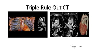
  • 3. Triple Rule Out CT TRO-CTA is a specialized computed tomography (CT) imaging exam tailored to evaluate for pathology within the coronary arteries, pulmonary arteries, and the aorta in a single CT study, hence the name ‘triple-rule out’.
  • 4. Why we take TRO-CT? Acute chest pain is the second most common presentation after abdominal pain in the emergency department. represents a major diagnostic challenge in emergency care as it has a broad differential diagnosis varying from benign causes to life-threatening conditions. TRO studies are most appropriate and cost-effective when there is a suspicion for acute coronary syndrome along with other diagnoses such as pulmonary embolism, acute aortic syndrome, or nonvascular disease in the thorax.
  • 5. Most common causes of Chest Pain Acute Coronary Syndrome Pulmonary Embolism Acute Aortic Syndrome
  • 6. Causes of Chest pain Heart related causes Heart-attack Angina Aortic Dissection Digestive Causes Chest pain can be caused by disorders of the digestive system, including: Heartburn Swallowing disorders Gallbladder or pancreas problems
  • 7. Muscle and bone causes Some types of chest pain are associated with injuries and other problems affecting the structures that make up the chest wall, including: Costochondritis Injured rib Lung-related causes lung disorders can cause chest pain, including: Pulmonary embolism Pleurisy Pulmonary hypertension
  • 8. In many clinical situations, a definite diagnosis of ED chest pain is not possible solely based on clinical symptoms and laboratory findings. In addition, most diagnostic modalities (i.e., ECG, cardiac enzymes, exercise treadmill testing, radionuclide perfusion imaging and stress echocardiography) other than MDCT are focused on the diagnosis or exclusion of ACS and do not exclude other life-threatening causes of acute chest pain.
  • 9. For this reason, Triple Role Out (TRO) protocol with ECG-gating technology has been proposed to encompass the entire thorax, allowing simultaneous evaluation of coronary arteries, thoracic aorta and pulmonary arteries for improving diagnosis of acute chest pain in a single study.
  • 10. Criteria for TRO_CT study Clinical presentation – low to moderate risk of ACS Clinical presentation – non ACS diagnosed considered Normal ECG or non-specific changes Patient able to tolerate CT and hold breath Cardiac rhythm acceptable ECG gated scan Adequate renal function
  • 11. Exclusion criteria  Contra-indications to iodinated contrast material including known allergy and renal insufficiency (serum creatinine more than 1.4 mg/dl). Marked heart failure. Clinically unfit patients (unable to stop breathing during the examination).  Extensive calcium score above 1000.
  • 12. Why don’t we take coronary angiography above 1000 calcium score? Because of blooming artifacts, which can cause erroneous enlargement of calcification, make less accuracy to determine the coronary arteries lumen that results in false positive diagnosis. Fig: (A) shows enlargement in the lumen of RCA because of the blooming artifacts (B) shows normal in state of RCA
  • 13. Anatomy of Aorta, Pulmonary and Coronary Aorta the largest blood vessel in the body. responsible for transporting oxygen rich blood from your heart to the rest of the body. begins at the left ventricle of the heart, extending upward into the chest to form an arch. downward into the abdomen, where it branches into the iliac arteries just above the pelvis.
  • 14. Aortic Root the portion of the aorta that is attached to the heart.  major part of the aortic root is the aortic valve allows blood to flow from the heart to the rest of the body when it is open and prevents blood from flowing backwards into the heart when it is closed.
  • 15. Ascending Aorta begins at the sinotubular junction of the aortic root and extends up and out from the heart until it connects with the aortic arch.
  • 16. Aortic Arch the portion of the aorta that is in the shape of an arch and connects the ascending aorta with the descending aorta.  The major arteries : the brachiocephalic artery, the left carotid artery and the left subclavian artery.
  • 17. Descending Thoracic Aorta begins at the end of the aortic arch and continues down into the abdomen. Two parts: 1. T. aorta(provides blood to the muscles of the chest wall and the spinal cord.) 2. Ab.Aorta (five arteries that branch from the abdominal aorta: the celiac artery, the superior mesenteric artery, the inferior mesenteric artery, the renal arteries and the iliac arteries)
  • 18. celiac artery superior mesenteric artery inferior mesenteric artery renal arteries iliac arteries Stomach Liver Pancreas supplies blood to the small intestine supplies blood to the large intestine blood to the kidneys as well as the muscles of the abdominal wall and the lower spinal cord blood to the legs and the organs in the pelvis. Figure: shows five arteries from the abdominal aorta and their blood supply
  • 19. Thoracic aorta disease 1. aortic aneurysms and dissections, 2. atherosclerotic disease, 3. infections and 4. traumatic injuries. Note: Ruptured thoracic aortic aneurysms and aortic dissections represent life-threatening emergencies that require immediate medical attention. Thoracic aortic aneurysms affect approximately 15,000 people in the United States each year
  • 20. Symptoms 1. Chest pain 2. Back pain 3. Hoarseness 4. Wheezing 5. Difficulty swallowing 6. Cough
  • 21. The most common types of aortic aneurysms are thoracic and abdominal. In addition, the following can signal a more serious condition: 1. Sudden and severe chest pain 2. Fainting 3. Leg pain or numbness 4. Shortness of breath 5. Weakness
  • 22. Coronary Artery heart is mostly supplied by the two coronary arteries which arise from the ascending aorta immediately above the aortic valve. coronary arteries and their branches run on the surface of the heart. RCA arise from the anterior aortic sinus of the ascending aorta immediately above the aortic valve. first runs forwards between the pulmonary trunk and the right auricle.
  • 23. Then, it descends almost vertically enter the rt. Atrioventricular groove, the rt. anterior coronary sulcus. At the inferior border of the heart, it turns posteriorly and runs into the atrioventricular groove, after the posterior interventricular groove finally anastomosing with the LCA.
  • 24. LCA arise from the left posterior aortic sinus of the ascending aorta, immediately above the aortic valve and enters the left between the pulmonary trunk and the left auricle. then divides into the anterior interventricular artery also know as left anterior descending artery which runs downwards in the anterior interventricular groove to the apex of the heart.
  • 25.
  • 26. Clinical Correlation Angina Pectoris – since coronary arteries are narrowed, the blood supply to the cardiac muscles is reduced. As a result, on exertion, the patient feels moderately severe pain in region of the left pericardium last as long as 20 mins. Pain is often referred to the left shoulder and medial side of the arm and forearm. Angina pectoris pain occurs on exertion and relieved by rest.
  • 27. Myocardial Infarction sudden block of the larger branches of either coronary artery usually leads to myocardial ischaemia followed by the myocardial necrosis (myocardial infarction). Part of the heart suffering from MI, stops functioning and often causes death. This condition is termed as the heart attack or coronary attack.
  • 28. Clinical features of MI Sensation of pressure, sinking and pain in the chest that lasts longer than 30 minutes. Nausea (or) vomiting, sweating, shortness of breath, and tachycardia. Pain radiates to the medial side of the arm, forearm, and hand. Sometimes, it may be referred to jaw or neck.
  • 29. Sites of coronary artery occlusion The three most common site of CAO are 1. Anterior interventricular artery or LAD (40-50%), 2. RCA (30-40%) 3. Circumflex branch of LCA (15-20%) NOTE: MI mostly occurs at rest whereas angina occurs on exertion.
  • 30. Blood supply of the major coronary arteries The 2 main coronary arteries are the left main and right coronary arteries. Left main coronary artery (LMCA). The left main coronary artery supplies blood to the left side of the heart muscle (the left ventricle and left atrium). The left main coronary divides into branches: 1. The left anterior descending artery branches off the left coronary artery and supplies blood to the front of the left side of the heart. 2. The circumflex artery branches off the left coronary artery and encircles the heart muscle. This artery supplies blood to the outer side and back of the heart.
  • 31. Right coronary artery (RCA)  The right coronary artery supplies blood to the right ventricle, the right atrium, and the SA (sinoatrial) and AV (atrioventricular) nodes, which regulate the heart rhythm. The right coronary artery divides into smaller branches, including the right posterior descending artery and the acute marginal artery. Together with the left anterior descending artery, the right coronary artery helps supply blood to the middle or septum of the heart.
  • 32. Fig: Coronary arteries and their blood supply
  • 33. Fig: Coronary Arteries and their branches
  • 34. Types of Circulation Pulmonary Circulation the portion of the cardiovascular system that carries oxygen-poor (deoxygenated) blood from the heart to the lungs and returns oxygenated blood back to the heart. deoxygenated blood from the body leaves the right ventricle through the pulmonary arteries, which carry the blood to each lung. 34
  • 35. Cont.; pulmonary arteries are the only arteries that carry deoxygenated blood. In the lungs, red blood cells release carbon dioxide and pick up oxygen during respiration. The oxygenated blood then leaves the lungs through the pulmonary veins, which return it to the left side of the heart and complete the pulmonary cycle.
  • 36. Cont.; The oxygenated blood is then distributed to the body through the systemic circulation before returning again to the pulmonary circulation.
  • 37. Fig: The pulmonary circulation carries blood between the heart and lungs.
  • 38. Systemic Circulation the portion of the cardiovascular system that carries oxygenated blood from the heart to the body and returns deoxygenated blood back to the heart. Oxygenated blood from the lungs leaves the left ventricle through the aorta. From here it is distributed to the body's organs and tissues, which absorb the oxygen through a complex network of arteries, arterioles, and capillaries.
  • 39. Cont.; The deoxygenated blood is then collected by venules and flows into veins before reaching the inferior and superior venae cavae, which return it to the right heart, completing the systemic cycle. The blood is then re-oxygenated through the pulmonary circulation before returning again to the systemic circulation.
  • 40. Fig: The systemic circulation. The systemic circulation brings oxygenated blood to the body cells and tissues and transports cellular wastes away from the cells and tissues. It is also responsible for temperature regulation and transport of hormones and other substances around the body.
  • 41. Coronary Circulation the heart needs its own blood supply, which it gets through the coronary circulation. the heart muscle tissue is so thick that it needs blood vessels to deliver oxygen and nutrients deep within it. The vessels that deliver oxygen-rich blood to the heart muscle are called coronary arteries.
  • 42. Cont.; branch directly from the aorta, just above the heart. The vessels that remove the deoxygenated blood from the heart muscle are known as cardiac veins.
  • 44. Patient preparation for TRO An 18–20-gauge intravenous catheter is placed into a large vein in the antecubital fossa. The patient is lying in a supine position with arm in front of him. ECG leads are positioned above and below the level of the scan to prevent streak artifact. The ideal heart rate for ECG-gated studies is a slow regular rhythm, usually a sinus bradycardia at 50–60 beats per minute. Oral-blockers may be given at least 1 h before the scan for control of heart rate.
  • 45. Contrast material and scanning protocol In order to image both the coronary and pulmonary arteries, a biphasic injection technique was used: 70 mL of undiluted (ultravist 370) was injected at 5 mL/s, followed by 25 mL of the same contrast material diluted with 25 mL of saline, also injected at 5 mL/s. For injection Protocol we used a bolus tracking technique where we started contrast medium injection when the HU in the left atrium reached 100 HU then in the second phase we depended on the observation, for assessment of the opacification of the pulmonary artery.
  • 46. The first phase of the injection opacifies the coronary arteries during image acquisition, while the second phase of the injection, provides simultaneous homogeneous enhancement of the pulmonary arteries. Data acquisition starts from the level of the medial end of the clavicles to the lower border of the heart in cranio–caudal direction.
  • 47. Fig: Typical Z axis coverage in dedicated coronary CT angiography versus triple rule-out study. A: the field of view in a dedicated coronary CT angiography is demonstrated. B: note the increased Z axis length in the triple rule-out study compared with dedicated coronary CT angiography.
  • 48. Data evaluation For coronary assessment every case was evaluated in the axial plane and with slab maximum intensity projection images that were rotated to visualize each vessel in multiple planes.  Vessels with complex plaque were also evaluated with curved multiplanar reconstruction by using vessel tracking software with automatic centerline determination.
  • 49. Different parts of the thoracic aorta regarding their diameter, contrast filling, presence of filling defects, dissections, wall irregularities, calcification, mural thrombus. The main pulmonary artery, right and left pulmonary arteries, their segmental and subsegmental branches, regarding their diameter, contrast filling, presence of filling defects, wall irregularities, calcification, mural thrombus.
  • 50. Other extra-vascular structures such as cardiac size, pleural, pericardial and parenchymal abnormalities have to be assessed.
  • 51. Female patient 37 year old presenting with acute chest pain, dyspnea and hemoptysis. CT axial (A and B), sagittal (C) and (D) images of pulmonary angiography showing left main and segmental pulmonary embolism and left sided pleural effusion.
  • 52. Triple rule-out CT angiography images (sagittal and axial views) of two patients with aortic dissection, with standard (a, b) and low dose (c, d). The diagnostic and image quality were excellent in both patients in the left descending artery (white arrow), the aorta(white star) and the pulmonary artery (arrowhead). The first patient had an history of surgical replacement of the ascending aorta.
  • 53. Pros TRO CT can reduce (a) time for patient triage, (b) number of required diagnostic tests, (c) costs, and (d) radiation exposure to the patient.
  • 54. Limitations Beta-blockers that are required for coronary CTA may not be safe in patients with pulmonary embolism. Obesity and calcifications limit interpretation, rapid heart rate, arrhythmias, renal dysfunction and contrast allergies.
  • 55. Conclusion Since there are a lot of chest pain cases in emergency department , it is difficult to know the real cause of chest pain. triple rule out is a relatively new technique, examination the coronary arteries, pulmonary arteries and aorta in just a single study, which gives us the advantage of screening emergency patients presenting with chest pain in a rapid and safe way for detection of their vascular diseases. In addition, it can reduce the radiation dose to the patient because it investigates the coronary arteries, pulmonary arteries and aorta in just a single study and we don’t need to separated studies to check those of them. That’s why, it is most effective investigation to know the origin of abnormalities even though it has still challenges.
  • 56. References  Pitts SR, Niska RW, Xu J, Burt CW. National hospital ambulatory medical care survey: 2006 emergency department summary. Natl Health Stat Rep 2008;7:1–8.  Stillman AE, Oudkerk M, Ackerman M. Use of multidetector computed tomography for the assessment of acute chest pain: aconsensus statement of the North American Society of Cardiac Imaging and the European Society of Cardiac Radiology. Int J Cardiovascular Imaging 2007;23:415–27. Thomas J, Rideau AM, Paulson EK, Bisset 3rd GS. Emergency department imaging: current practice. J Am Coll Radiol 2008;5(7):811–816e2.
  • 57.  Kevin M, Ethan J. Evaluation of a ‘‘Triple Rule-Out’’ coronary CT angiography protocol: use of 64-section CT in low-tomoderate risk emergency department patients suspected of having acute coronary syndrome. Radiology 2008;248(2). Rubinshtein R, Halon D, Gaspar T, et al. Usefulness of 64-slice cardiac computed tomographic angiography for diagnosing acute coronary syndromes and predicting clinical outcome in emergency department patients with chest pain of uncertain origin. Circulation 2007;115:1762–8. Kevin M, Ethan J. Evaluation of a ‘‘Triple Rule-Out’’ coronary CT angiography protocol: use of 64-section CT in low-tomoderate risk emergency department patients suspected of having acute coronary syndrome. Radiology 2008;248(2)