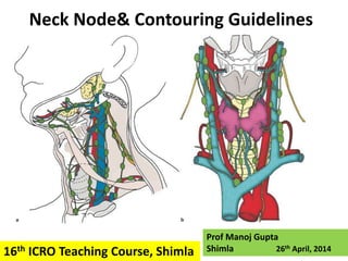
Neck node & Contouring Guidelines
•
323 gefällt mir•29,347 views

Normal Neck Node anatomical boundaries on CT scan and contouring guidelines for in N0 neck for Head and Neck IMRT

Empfohlen
Empfohlen
Weitere ähnliche Inhalte
Was ist angesagt?
Was ist angesagt? (20)
Ähnlich wie Neck node & Contouring Guidelines
Ähnlich wie Neck node & Contouring Guidelines (20)
Kürzlich hochgeladen
Kürzlich hochgeladen (20)
Neck node & Contouring Guidelines
- 1. Neck Node& Contouring Guidelines Prof Manoj Gupta Shimla 26th April, 201416th ICRO Teaching Course, Shimla
- 2. Location & Pattern of Involvement. Why is it needed ?? • Cancer treatment does not mean only cure but also to provide good quality of life. • Therapeutic gain can be accomplished either by increasing the control rate or by decreasing the side effect of treatment. • That is what modern radiotherapy like IMRT has done so far. • Not all the neck nodes are involved in all sites. • Gone are the days when with 2-D radiation every thing between the radiation portal are treated irrespective of need.
- 3. Location & Pattern of Involvement. Why is it needed ?? • Depending upon the primary site and stage of the disease, Surgeons practice selective neck dissection. • If location and pattern of spread are known, then Radiation Oncologist can also plan selective neck irradiation thus avoiding many normal structures like parotid and swallowing muscles leading to better quality of life. • With high precision radiotherapy, the goal of providing better quality of life is further achieved.
- 4. Classification of neck nodes - levels • Robbins Classification:- Surgeons usually follow this system. • For Radiation Oncologists 2 systems of classifications are commonly used - Brussels system -Rotterdam system • Due to discrepancies in different systems , a consensus guidelines was derived (RTOG Consensus guidelines).
- 5. Changes in Robbins classification • Based on surgical boundaries like muscles, nerves and vessels. • But these structures may not be identifiable on CT. • Cranial limit for level II was defined by surgeons at insertion of post belley of digastric muscle at mastoid. • But this point may not be identifiable on CT. • So cranial limit was modified to bony land mark like cervical vertibrae.
- 6. Changes in Robbins classification • Similarly, Robbins defined the caudal limit of level III as the point at which the omohyoid muscle crossed the internal jugular vein (IJV); again not clearly identifiable on CT. • Again easily identifiable landmark is choosen like lower border of cricoid cartilage.
- 7. Changes in Robbins classification • Robbins used the spinal accessory nerve (SAN) to sub-divide level II into IIa (anterior to a vertical plane defined by the nerve) and IIb (posterior to that plane). • SAN cannot be identified on CT scans, • So, posterior edge of the IJV for the subdivision between levels IIa and IIb
- 8. Classification of Neck Nodes and Contouring Guidelines
- 9. Classification of the Neck Nodes
- 10. Myelohyoid Post. Belly of digastric Sterno-cleido mastoid Omohyoid Trpezius Ant. Belly of digastric Hyoid bone Thyroid Cartilage Cricoid Cartilage Jugular vein Submandibular gland
- 12. Lateral Medial
- 15. Lymph Node Regions • Level I Submental Nodes (Ia) Submental triangle: –Bounded by two anterior belly of digastric
- 16. Primary for Ia • Floor of the mouth. • Anterior oral tongue. • Anterior mandibular alveolar ridge. • lower lip.
- 17. Lateral-> Medial edge of ant belly of two digastric muscles Anterior-> Platysma muscle and the symphysis menti, Level Ia Posterior -> body of the hyoid bone,
- 18. Medial-> region continues into the contralateral level Ia Level Ia
- 19. Cranial-> Geniohyoid muscle or a plane tangent to the basilar edge of the mandible.
- 20. Caudal-> hyoid bone Level Ia
- 22. Lymph Node Regions • Level Ib: Submandibular Triangle –Formed by the anterior and posterior belly of the digastric muscle and the body of the mandible
- 23. Primary for Ib • cancers of the oral cavity, • anterior nasal cavity, • soft tissue structures of the mid-face and • the submandibular gland.
- 24. Anteriorly -> Platysma muscle Level Ib
- 25. Posterior-> Posterior edge of the submandibular gland, Level Ib
- 26. Medial-> lateral edge of the ant belly of digastric muscle Level Ib
- 27. Lateral-> Inner side of the mandible, Platysma and skin. Level Ib
- 28. Cranial-> Mylohyoid muscle and cranial edge of the submandibular gland. Level Ib
- 29. Caudal-> Plane crossing the central part of Hyoid bone Level Ib
- 31. Most of the jugular nodes(lev. II-IV) present ant., post., and lateral to the IJV. So medial boundary is medial edge of the vessel bundle. Jugular nodes
- 32. Level II • Cranial limit for level II was defined by surgeons at insertion of post belly of digastric muscle at mastoid. • But this point may not be identifiable on CT. • Surgeons were asked to put the clips at the upper level of dissection for level II nodes in node negative neck.
- 33. How consensus was made for cranial border for level II Nodes? • Clips cluster around caudal border of transverse process of vertebra C1. • So cranial border of level II is taken at caudal edge of transverse process of C1. Parotid projection so if cranial limit is taken at base of skull then more parotid will be irradiated.
- 34. Usually the cranial limit of level II is caudal border of transverse process of C1 vertebrae. But few nodes also present superior to this up to base of skull. This region cranial to cranial limit of Level II is called Retro Styloid region. Level II Retrostyloid Region
- 35. When to treat Retro Styloid Region • Ca Nasopharynx. Bilateral • In +ve level II node Ipsilateral
- 36. Caudal-> Carotid Bifurcation (Surgical Boundry) Caudal edge of body of the hyoid bone. Level II
- 37. • anteriorly by the – the anterior edge of the carotid artery – posterior edge of the submandibular gland, – the posterior belly of the digastric muscle, Level II Anterior Relation Cranial Caudal
- 38. Anterior-> Anterior edge of the carotid artery Level II
- 39. Anterior->Posterior edge of the submandibular gland, Level II
- 40. Anterior->posterior belly of the digastric muscle, Level II
- 41. Posterior-> Posterior edge of the sternocleidomastoid (SCM) muscle, Level II
- 42. Medial-> Medial edge of the carotid artery and the paraspinal muscles (levator scapulae and splenius capitis) Level II
- 43. Lateral-> Medial edge of the SCM Level II
- 45. Primary for II • Nasal cavity. • Oral cavity. • Oropharynx. • Hypopharynx. • Larynx. • Major salivary glands. • Nasopharynx,
- 46. IIa IIb • Level II is further subdivided into two compartments. – IIa – IIb • Surgeons demarcate between the two by spinal accessory nerve (SAN). • From a radiological point of view, the posterior edge of the IJV is taken as the boundary between levels IIa and IIb. Sub division of Level II
- 47. IIa IIb
- 48. Level III • contains the middle jugular lymph nodes located around the middle third of the IJV. • It is the caudal extension of level II • Primary. Oral cavity. Oropharynx. Hypopharynx. Larynx. Nasopharynx,
- 49. Cranial-> caudal edge of body of the hyoid bone. Level III
- 50. Caudal-> Caudal edge of the cricoid cartilage. Level III
- 51. Anterior-> Posterolateral edge of the sternohyoid muscle and the anterior edge of the SCM muscle, Level III
- 52. Posterior-> Posterior edge of the SCM muscle Level III
- 53. Lateral-> Medial edge of the SCM muscle Level III
- 54. Medial-> Medial edge of the internal carotid artery and the paraspinal muscles (scalenius). Level III
- 56. includes the lower jugular lymph nodes located around the inferior third of the IJV. According to Robbins, it extends from the caudal limit of level III to the clavicle. But since surgeons never dissect upto clavicle so consensus is that the caudal limit is 2cm cranial to the cranial edge of sterno-clavicular joint. 2 cm Level IV
- 57. Caudal->2cm cranial to the cranial edge of sterno- clavicular joint.
- 58. Cranial-> Caudal edge of the cricoid cartilage. Level IV
- 59. Anterior Posterior Lateral. Level IV Anterior edge , posterior edge and medial edge of the SCM muscle, respectively
- 60. Medial-> Medial edge of the internal carotid artery and the paraspinal muscles (scalenius) Level IV
- 62. Primary for level IV • Hypopharynx. • Larynx • Oropharynx. • Skip metastasis from ant tongue. • Cervical esophagus
- 64. The uppermost part of level V contains superficial occipital lymph node(s), which are not involved in head and neck ca except skin cancer. So cranial limit is a horizontal plane crossing the cranial edge of the body of the hyoid bone Level V
- 65. Cranial -> Horizontal plane crossing the cranial edge of the body of the hyoid bone Level V
- 66. • For the caudal limit of level V, it appears from critical examination of neck dissection procedure, that surgeons never dissect up to clavicle but go only up to to the transverse cervical vessels. • Hence, caudal limit of level V is kept at CT slices encompassing the cervical transverse vessels Level V Caudal
- 67. Caudal -> CT slices at the level of transverse Cervical vessels Level V
- 68. Lateral-> Platysma muscle and the skin, Level V
- 69. Medial -> Paraspinal muscles (splenius capitis, levator scapulae and scaleni (posterior, medial and anterior) muscles) Level V
- 70. Anterior-> Posterior edge of the SCM muscle Level V
- 71. Posterior -> Antero-lateral border of the trapezius muscles .Practically, a virtual line joining the antero-lateral border of both trapezius muscles can be use to set the posterior limit of level V Level V
- 73. Primary for Level V • Nasopharynx. • Oropharynx. • Subglottic larynx. • Apex of the pyriform sinus. • Cervical esophagus. • Thyroid gland.
- 74. Va Vb Level V is divided into Va and Vb by omohyoid muscle where it crosses the internal jugular vein. But this crossing point can not be appreciated on CT film. Level V
- 75. Hyoid Thyroid Cricoid For practical purpose, use of the plane between levels III and IV extended posteriorly is recommended, which means lower border of cricoid can be taken as dividing line between Va and Vb Level V Level III Level IV Level Va Level Vb
- 76. Located in anterior neck compartment They are pre- and para tracheal nodes including the pre- cricoid (Delphian) node and lymph nodes along the recurrent laryngeal nerves. Level VI
- 77. Cranial -> Caudal edge of the body of the thyroid cartilage, Level VI
- 78. Caudal -> Cranial edge of the sternum manubrium Level VI
- 79. Anterior-> Platysma and the skin Level VI
- 80. Posterior -> Separation between the trachea and the esophagus. Level VI
- 81. . Lateral -> Medial edge of the thyroid gland and the antero-medial edge of the SCM muscle Level VI
- 83. Primary for Level VI • cancers of the thyroid gland, • the Trans glottic and subglottic larynx, • the apex of the piriform sinus and • the cervical esophagus.
- 84. Typically, retropharyngeal nodes are divided into Medial Group Lateral Group. The medial group is an inconsistent group which consist of one to two lymph nodes The lateral group lies medial to the carotid artery. The most superior lymph node of this group is also called the lymph node of Rouvie`re. lymph node of Rouvie`re. RP Nodes
- 85. Cranial-> Base of the skull Mastoid Process RP nodes
- 86. Caudal -> Cranial edge of the body of the hyoid bone RP nodes
- 87. Anterior-> Levator Veli palatini muscle. RP nodes
- 88. Posterior-> Pre-vertebral Muscles. . RP nodes
- 89. Lateral-> Medial edge of the carotid vessel. RP nodes
- 94. Node positive Neck • With N+ve, one adjacent extra nodal level is also at high risk of occult metastasis, for eg. the level IV for oral cavity tumors and the level I and V for oropharyngeal, hypopharyngeal, and to a lesser extent laryngeal tumors.
- 95. Retrostyloid Region S/C fossa Till Sternum Point No 1 In level II node positive include retro styloid region and in level IV and Vb nodes enlargement include s/c fossa up to sternum on the side of the positive node.
- 97. S/C Fossa
- 98. Point No 2 In fact, this recommendation will only apply to patients with a single involved lymph node (pN1) for whom post-operative radiotherapy is considered (e.g. because of a capsular rupture) and for whom selective treatment may be advocated.
- 99. When an involved lymph node abuts a muscle (e.g. sterno-cleido- mastoid or para-spinal) it is recommended to include this muscle at the vicinity of the node in the CTV for the entire invaded level and at least with a 1 cm margins in cranio-caudal direction. Point No 3 1 cm 1 cm
- 100. Extra Capsular Extension(ECE) Incidence of ECE is 20 to 40% in metastatic node <1cm size while goes up to 75% if size is >3 cm. The incidence of local recurrence increases, and the survival rate decreases by greater than 50% when metastatic nodal disease expands beyond the capsule i. e. if ECE present.
- 101. Magnitude of ECE The majority of the ECE extend <5mm from the capsule of node. None extend >10mm
- 102. Margins of 1 cm from the nodal GTV to the CTV would be sufficient to fully cover any subclinical nodal extension for lymph nodes smaller than 3 cm in head-and-neck cancer patients receiving IMRT. For larger nodes or matted nodes more generous margins should be given. Point No 4
- 103. Post Operative Neck
- 104. Point No 1
- 106. Point No 3
- 107. Point No 4
- 108. Pattern of Spread •Prophylactic neck node irradiation is required if the incidence of occult metastasis is >5%.
- 109. • Typically, nasopharyngeal and hypopharyngeal tumors have the highest propensity of nodal involvement which occurs in 80 and 70%, respectively. • Interestingly, the node distribution follows the same pattern in the contralateral neck as in the ipsilateral neck. • Contra lateral level V is usually not involeved
- 110. Incidence and distribution of regional metastasis for Levels I–V for clinically N0 neck • Tumor site Levels involved (%) I II III IV V • Oral cavity 20 17 9 3 0.5 • Oropharynx 2 25 19 8 2 • Hypopharynx 0 13 13 0 0 • Larynx 5 19 20 9 2.5 • In non-nasopharyngeal cancers of head and neck, level V is not included in N0 neck as incidence of involvement is <5%. • Similarly, in oro-pharynx, hypo-pharynx and larynx, level I is not included as again incidence of occult metastasis is <5%.
- 111. Non Nasopharyngeal N0 Neck • Oral Cavity Ca • Oro pharynx • Hypo pharynx • larynx Level I, II, III and in ca tongue level IV Level II, II, IV
- 112. Incidence and distribution of regional metastasis for Levels I–V for clinically N+ve neck • Tumor site Levels involved (%) • I II III IV V • Oral cavity 48 39 31 15 4 • Oropharynx 15 71 42 27 9 • Hypopharynx 10 75 72 45 11 • Larynx 6 61 54 30 6 • Nasopharynx* 13 95 60 21 44 • In non-nasopharyngeal cancers of head and neck, level V is included in N+ve neck except in ca oral cavity where incidence is <5%. • Similarly level I should be included in neck positive disease except in larynx.
- 113. Primary site for RP Nodes inclusion N0 Neck Nasopharynx and Pharyngeal Wall N+ Neck All sites except larynx In non nasopharyngeal cancers usually lateral RP nodes are involved
- 115. Thanks Questions???