1. MUSCULAR AND SKELETAL
SYSTEM OF POULTRY
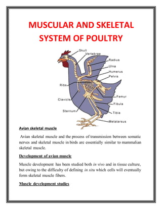
Avian skeletal muscle
Avian skeletal muscle and the process of transmission between somatic
nerves and skeletal muscle in birds are essentially similar to mammalian
skeletal muscle.
Development of avian muscle
Muscle development has been studied both in vivo and in tissue culture,
but owing to the difficulty of defining in situ which cells will eventually
form skeletal muscle fibers.
Muscle development studies
2. Muscle development has been studied both in vivo and in tissue culture,
but owing to the difficulty of defining in situ which cells will eventually
form skeletal muscle.
Myogenesis
Derives from tissue culture studies from single developing muscle cell
myogenesis, both in vivo and in vitro, consists of the fusion of spindle-
shaped, uninucleated myoblast to form multinucleated myo tubes that
will eventually grow into mature muscle fibers
Myoblasts
The change in numbers of muscle precursors cells (myoblasts) has been
studied in chick limb muscle.Myoblasts amenable to cloning appear on
about Day 3 in ova.
Ultramicroscopic structural studies of myosin and actin
Numerous close junctions with an intercellular distance of 2.5–10 nm
and some focal tight junctions with no discernible gap can be detected
between pairs of myogenic cells. It is likely that the fusion process is
initiated by the formation of close contact between cells, which is
followed by the appearance of vesicles and tubules between the adjacent
cytoplasms. At the final stage, remnants of broken cell membranes
disappear and a common cytoplasm is
3. FIGURE 1:
Schematic representation of the band pattern of a striated muscle
myofibril related to the arrangement of the actin (thin) and myosin
(thick) filaments. Two sarcomeres are shown (the area between two
adjacent Z-lines is a sarcomere). The myofibrils within the muscle fiber
are aligned in a parallel pattern that
Stretches right across the muscle fiber as a whole. In the contracted state
(bottom) the thick and thin filaments
Slide over one another but neither change in length. This causes a
narrowing of the H and I bands, but no
Change in the width of the A band which reflects the constant length of
the myosin filaments. (Reproduced from Bowman, 1964, with
permission.).
4. formed. Both thick myosin (15–16 nm in diameter) and thin actin (5–6
nm in diameter) filaments are synthesized by clusters of cytoplasmic
ribosomes (polysomes).
Sarcoplaslmic reticulum
During the earliest myotube stage in embryonic chick muscle, both in
vivo and in vitro, the sarcoplasmic reticulum
develops in isolated portions from rough-surfaced endoplasmic
reticulum. Subsequently, the isolated portions of sarcoplasmic reticulum
join together to create a network around the contractile filaments
The transverse tubular system
The transverse tubular system develops more slowly than the
sarcoplasmic reticulum the surface membrane of the myotubes
invaginates to form the T-tubules. Initially the T-tubules consist of
shallow vesicles connected to the sarcolemma, but with time they project
deeper and deeper into the myotube until contact is made with the
sarcoplasmic reticulum.
Fibers length description
Fibers lengthen by the addition of new sarcomeres and broaden with the
increase in myofibrils per fiber. An example is the chicken latissimus
dorsi muscles where mean fiber diameter increases tenfold from 4 to 6
um in 18-day in old embryos to 40–60 um in 8-month-old chicken.
There are also structural changes in Z-disks during growth in chickens.
Mechanical strength increases in the weeks after hatching, and the disks
in leg muscle become stronger than those in breast muscle.
Normal contractile activity
Normal contractile activity is essential for post- hatching muscle
growth. Immobilization of chicken pos- terior latissimus dorsi (PLD)
5. muscles for periods up to 11 months results in a dramatic reduction of
fiber size.
Muscle fiber types
As in amphibians and reptiles, as well as in mammals, some of the avian
muscle fibers adapt for rapid, intermit tent contraction whereas others
adapt for more continuous contraction.
Fictional difference in muscle fiber types
The functional differences require differences in the structure and
biochemistry of fibers. Muscles are usually described as slow or fast
contracting. However, this represents an oversimplification of the
situation .The color of the muscles (red or white) does not adequately
describe the variety of fiber types that exist either.
I IIA IIB IIIA IIIB
Histochemical criteria
ATPase(pH 9.4) No staining Strong Strong Medium Strong
ATPase(pH 4.6) Strong No or weak Weak Weak Medium
ATPase(pH 4.3) Strong No staining No staining Weak Medium
NADH-TR Medium Weak or medium No staining Medium Mediumor strong
Phosphorylase None or weak Strong Srong Weak Medium
Fiber innervation Multiple Focal Foca Multiple Multiple
Histological characteristics
Fiber shape Polygonal Polygonal Polygonal Rounded Rounded
Fascicle shape Polygonal Polygonal Polygonal Rounded Rounded
Mitochondrial density Very high High Low Very high Very high
Fiber lipid droplets No Yes No No No
Relative fiber size Small/medium Medium Medium Large Medium
6. Myonucleidistribution Peripheral Usually peripheral usually central Peripheral Peripheral
Fiber typecomposition (%)
Pectora 0 <1 >99 0 0
PLD <3 5–20 80–95 0 0
ALD 0 0 0 65–80 20–35
Sartorius (red) 30–45 35–50 15–25 0 0
Sartorius (white) 0 10–20 80–90 0 0
Plantaris 0 0 0 65–75 25–35
.aAdapted from Barnard et al. (1982) with permission.
bNADH-tetrazolium reductase.
Ultrastructure of Avian white and red fibers
Generally, white fibers have a very definite fibrillar appearance
(Fibrillen struktur) similar to that of mammalian muscle, whereas red
fibers have a more granular and indefinite appearance (Felderstruktur).
Description about fibrillenstrukur
In Fibrillenstruktur fibers the myofibrils are polygonal in cross section
and uniform in diameter and have a regular arrangement, being
separated from each other by a granular sarcoplasm. The cross
granstriations
are evident; the dark A (anisotropic)- and light I (isotropic)-bands. Each
I-band is bisected by a smooth
Z-line running directly across the fibril. The H-zone, where only thick
filaments are found,
Can be observed in the midsection of the A-band.
Development avian fibers
During development, fibers of posterior latissimus dorsi muscles of the
chicken become faster-contracting than ALD fibers at about the same
time as the density of triads becomes higher. The sarcoplasmic reticulum
also begins to differentiate around this stage, but the final fiber-type-
specific distribution of T-tubules occurs after hatching. Other studies
have correlated the development of the functional Ca2+ channels of the
sarcoplasmic reticulum with that of specific forms of foot proteins.
Isoforms of sarcoplasmic reticulum
They have similar conductances but different activation and
inactivation properties (Percival). The a-isoform appears to be essential
form normal excitation–contraction coupling myoblasts are the
7. predominant cell type, but at this early stage there is no evidence of
specialization of the nerve ending or of localization of acetyl
cholinesterase, which is used as a marker of functional transmission.
Developmental changes expression of isoforms of troponin
There are also developmental changes in expression of isoforms of
troponin T that correlate with differences in Ca2+sensitivity and
contractility in fibers from ALD PLD, and pectoralis major muscles
(Reiser et al., 1992).
The fast-contracting PLD and pectoralis major fibers become more
sensitive to Ca21 during maturation, which
corresponds to changes in iso forms of troponin T, but not of troponin C
or I or troponomyosin. There are overall
changes in troponin C expression during development.
Innervations
The final maturation and long-term survival of skeletal muscle is
highly de pendent on innervation by the motor neurons.
8. Neuromuscular juntion
The development of the neuromuscular junction is required before
individual muscle fibers can fulfill their adult role.
The first development of primitive neuromuscular junctions occurs
between Days 7 and 10 neuromuscular junctions occurs between Days 7
and 10 and by Days 15–16 mature neuromuscular junctions can be
found; these are associated with fully developed muscle fibers. From
then on the size of the neuromuscular junction increases with muscle
growth, but the basic morphology remains essentially unchanged.
Development of anterior latissmus dorsi
In embryonic chick ALD the morphological channels de velopment of
neuromuscular junctions has been correlated with the onset of
transmitter release measured using electrophysiological techniques.
Development of posterior latissmus dorsi
The development of posterior lattismus dorsi has been studied by many
scientist in case of chick and they also determined the sequence of
9. innervatiion in chick muscle by Bourgeios and Toutant (19882)
Additionally, Adachi (1983) has observed that the neuromuscular
junctions of different muscles mature at different times, with proximal
muscles preceding distal ones.
Difference between fibrstruker fibers and felder striker fibers
(innervation)
White Fibrillenstruktur fibers are focally innervated by one or only a
few nerve terminals, as in mammalian muscle, whereas the Felder
struktur-containing red fibers are multiply innervated by many nerve
terminals (Ginsborg and Mackay, (1961).
ELECTRICAL PROPERTIES OF MUSCLE FIBERS
The resting membrane potential of mature avian exmuscle fibers is
similar to that of other skeletal muscles (i.e., around -70 to -90 mV). In
general, adult muscle (i.e., around -70 to -90 mV). In general, adult
muscle fiber membranes are much more permeable to K1 than to Na+,
and this differential permeability develops during growth. The
membrane-passive electrical properties of muscle fiber determine its
response to an electrical stimulus A high fiber input resistance will result
in a large voltage response to a given current pulse; long space and time
constants will allow the response to spread over a large area of the
membrane.
In-vivo and in-vitro explanation of ALD MUSCLE
It has been found that
in vivo the ALD muscle of the chick responds to single-shock nerve
stimulating with only local endplate potentials; no action potential is
produced. Propagated muscle action potentials can only be
elicited in vivo by closely spaced twin pulses or by single shocks after a
period of high-frequency nerve stimulation.
However, in vitro, either nerve stimulation or direct muscle stimulation
can elicit propagated muscle contraction potentials in the ALD muscle
Electrical properties of ALD AND PLAD FIBERS
In terms of the development of the differences between the fiber types,
at 14 days in ovo, the electrical properties of ALD and PLD fibers are
found to be similar. The properties of the PLD change within the first 2
10. weeks of hatching; some of the changes are associated with the
membrane becoming permeable to Cl- ions.
Contractile properties of muscles
Avian skeletal muscle contains actin and myosin filaments arranged in
the classical interdigitated pattern. It is also known to contain the
regulatory .It is also known to contain the regulatory contractile proteins
troponin, tropomyosin, and a-actinin (Allen et al., 1979; Devlin and
Emerson 1978, 1979). It is therefore assumed that the process of
excitation–contraction coupling in avian muscle is essentially the same
as that in mammalian muscle.
Difference between multiply innervate and singly innervated muscle
The contraction times of multiply innervated muscles with a
Felderstruktur are 5 to 10 times slower than those of singly innervated
muscles with a Fibrillenstruktur.
Contractile property of ALD and PLD muscles of chicken
Contractile property development has been studied by Gordon in
chicken ALD and PLD muscles. After 14–16 days incubation the
contraction speeds of both embryo muscles were similar (time to half-
maximal tension response to 40 Hz stimulation was p400–500 msec)..
Neuromuscular transmission
The neurotransmitter at avian skeletal muscle neuro acetylchomuscuar
Junctions are acetylcholine. Evidence for this includes the facts that
choline acetyltransferase, the en-zyme that synthesizes acetylcholine, is
present in chickenALDand PLDmuscles and its activity increases
innerduring
development. Drugs such as hemicholinium, which inhibits choline
uptake; vesamicol, which blocks synaptic vesicular transport of
acetylcholine; and b-bungarotoxin, which blocks acetylcholine release,
block neuromuscular transmission in chicken. Thus, it is likely that
acetylcholine is synthesized from its precursors by choline acetyl
transferase in the cytoplasm of the nerve terminal. It is now known that
acetylcholine is loaded into synaptic vesicles, their storage structures, by
a two-stage concentrative mechanism.
Transportation of proton into vesicle
Active transport
11. In this, protons enter the vesicle by an active transport
mechanism involving a V-type AT-Pase. Intravesicular protons are then
exchanged for acetylcholine via the acetylcholine transporter itself. The
vesicles are thought to be anchored to the intraterminal cytoskeleton,
including actin strands, by a family of synaptic vesicle-associated
proteins, the synapsins.
12.
13. Uses of Avian muscle in neuromuscular pharmacology
It has long been known that avian and amphibian muscles respond quite
differently from mammalian muscle to the addition of acetylcholine and
other nicooftinic agonist drugs such as nicotine and decamethonium
The difference between the responses of avian and an mammalian
muscle to endplate depolarizing drugs is related to the previously
described innervations and excitation–contraction coupling
mechanisms of multi-ply and focally innervated muscles
In multiply innervated muscles, the local endplate depo-larizations
directly excite the contractile mechanism without the necessity for
action potential generation
The contracture response of avian multiply inner-vated muscle has
been used to study the actions of nicotinic agonist drugs in the same
way as in other multiply innervated muscles, such as the frog rectus
abdominis and the leech dorsal muscle.
Isolated muscles that have been used for this purpose are the anterior
latissimus dorsi the semispinalis