Anatomy of thoracic gage (bones)
•Als PPTX, PDF herunterladen•
11 gefällt mir•1,606 views

bony thoracic wall

Empfohlen
Weitere ähnliche Inhalte
Was ist angesagt?
Was ist angesagt? (20)
Ähnlich wie Anatomy of thoracic gage (bones)
Ähnlich wie Anatomy of thoracic gage (bones) (20)
Mehr von Alio Hersi
Kürzlich hochgeladen
Kürzlich hochgeladen (20)
Anatomy of thoracic gage (bones)
- 1. ANATOMY OF THORACIC GAGE (BONES) ALIO MOHAMED HERSI
- 2. REVIEW
- 3. REGIONS OF THE SPINE • Cervical • Thoracic: •Lumbar: •Sacrococcygeal:.
- 6. THE ATLAS (C1) Transverse Process Transverse Foramen Anterior Tubercle Articular Facet for Dens Lateral Mass LaminaPosterior Tubercle Superior Articular Facet Superior View
- 7. THE AXIS (C2) Odontoid Process (Dens) Body Transverse Process Inferior Articular Facet Superior Articular Facet Anterior View Posterior View Lateral Mass Spinous Process
- 8. LOWER CERVICAL VERTEBRAE C3 - C7 Transverse ProcessBody Sulcus for Spinal Nerve Lateral Mass Lamina Pedicle Superior Articular Facet Vertebral Foramen Bifid Spinous Process Transverse Foramen Axial View
- 9. • Body - L1 to L5 progressive increase in mass, cylindrical • Pedicles - longer and wider than thoracic; oval shaped • Spinous processes - horizontal, square shaped • Transverse processes - smaller than in thoracic region • Intervertebral foramen – large, triangular shape LUMBAR VERTEBRAE, L1-L5
- 10. THE SACRUM Sacral Horns Sacral Ala Pedicles Dorsal Foramina Sacral Hiatus Coccyx Posterior View Inverted triangle shape
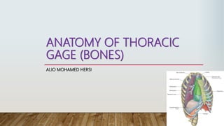
- 11. INTRODUCTION The trunk of the body is divided by the diaphragm into an upper part, called the thorax, and a lower part called the abdomen.
- 12. THORACIC WALL • Structure: skin fascia muscle bone blood vessels & nerves • Functions: 1. protection of thoracic viscera 2. provides the mechanical function of breathing 3. Conduit for the structure that pass through the thorax
- 13. THORACIC CAGE • It forms a conical enclosure for the lungs and heart and provides attachment for the pectoral girdle and upper limb. • It has a broad base and a narrower superior apex; it is rhythmically expanded by the respiratory muscles to create a vacuum that draws air into the lungs. • The inferior border of the thoracic cage is formed by a downward arc of the ribs called the costal margin. • The ribs protect the thoracic organs and spleen, most of the liver, and to some extent the kidneys.
- 14. STRUCTURE OF THE THORACIC WALL • Outside skin and by muscles attaching the shoulder girdle to the trunk. • Inside lined with parietal pleura. • Framework Posteriorly :the thoracic part of the vertebral column Anteriorly : the sternum and costal cartilages Laterally : the ribs and intercostal spaces Superiorly : the suprapleural membrane Inferiorly : the diaphragm
- 15. The Bony Thorax
- 16. COMPONENTS OF THORACIC CAGE: • Sternum • Manubrium, Body (Gladiolus), Xiphoid process • Ribs • 7 True Ribs • 5 False Ribs (including 2 floating ribs) • Clavicle Pectoral • Scapula girdle • 12 Thoracic Vertebrae (T1 - T12)
- 17. THORACIC VERTEBRAE • Body - progressive increase in mass from T1 to T12 • Pedicles - small diameter • Laminae - vertical, with “roof tile” arrangement • Spinous processes - long, overlapping, projected downward • Intervertebral foramen - larger
- 18. THORACIC VERTEBRAE, T1-T12 • Body - heart shaped when viewed superiorly. • Vertebral foramen - round • Pedicles - small in diameter • Spinous processes - long and projected downwards
- 19. • Articular processes Superior Articular Process Inferior Articular Process THORACIC VERTEBRAE, T1-T12 2 facet to articulate with the ribs
- 22. RIBS • Ribs are curved, flat bones that form most of the thoracic cage • They are remarkably light in weight yet highly resilient. • Each rib has a spongy interior containing bone marrow (hematopoietic tissue), which forms blood cells. • There are 12 pairs of ribs, all of which are attached posteriorly to the thoracic vertebrae .
- 23. • True ribs :The upper seven pairs are attached anteriorly to the sternum by their costal cartilages • False ribs :The 8th, 9th, and 10th pairs of ribs are attached anteriorly to each other and to the 7th rib by means of their costal cartilages and small synovial joints. • Floating ribs :The 11th and 12th pairs have no anterior attachment.
- 25. • Long, twisted, flat bone having a rounded, smooth superior border and a sharp, thin inferior border .The inferior border overhangs and forms the costal groove , which accommodates the intercostal vessels and nerve. • The anterior end of each rib is attached to the corresponding costal cartilage • A rib has a head, neck, tubercle, shaft, and angle . Typical rib
- 26. •The head has two facets for articulation with the numerically corresponding vertebral body and that of the vertebra immediately above. • The neck is a constricted portion situated between the head and the tubercle. • The tubercle is a prominence on the outer surface of the rib at the junction of the neck with the shaft. It has a facet for articulation with the transverse process of the numerically corresponding vertebra. • The shaft is thin and flattened and twisted on its long axis. Its inferior border has the costal groove. The angle is where the shaft of the rib bends sharply forward.
- 27. Fifth right rib, as seen from the posterior aspect .
- 28. ATYPICAL RIBS 1st rib Flat, scalene tubercle & grooves for subclavian v. 2nd rib rough tuberosity for serratus anterior m. 10th rib one facet on the head 11th & 12th one facet on the head & no neck or tubercle
- 29. TYPICAL RIB ARTICULATION • Dorsal (P) Attachment Thoracic Vertebrae • Head of Rib 2 Demifacets • Superior demifacet • Inferior demifacet of vertebra above it • Intervertebral disc • Tubercle of Rib Transverse Costal Facet • e.g. Rib No. 4 articulates with Superior Demifacet and Transverse Costal Facet of T4 & Inferior demifacet of T3 • Ventral (A) Attachment to Sternum • Via costal cartilage
- 30. APPLIED NOTES • Cervical Rib : A rib arising from the anterior tubercle of the transverse process of the seventh cervical vertebra occurs in about 0.5% of humans May be connected to the first rib by a fibrous band, or may articulate with the first rib. Pressure the lower trunk of the brachial plexus the subclavian artery • Rib Excision
- 31. COSTAL CARTILAGES • Costal cartilages are bars of cartilage connecting the upper seven ribs to the lateral edge of the sternum and the 8th, 9th, and 10th ribs to the cartilage immediately above. The cartilages of the 11th and 12th ribs end in the abdominal musculature. • The costal cartilages contribute significantly to the elasticity and mobility of the thoracic walls. • In old age, the costal cartilages tend to lose some of their flexibility as the result of superficial calcification.
- 32. STERNUM BONE • Flat bone, with 3 parts: 1. Manubrium sterni 2. Body/Gladiolus 3. Xiphoid process
- 33. PARTS OF STERNUM: 1. Manubrium sterni • Jugular/suprasternal notch -Articulates with Clavicles and Ribs 1 and 2 -Lies opposite to T3 and T4 vertebrae • Manubriosternal joint inferiorly – called Sternal Angle/Angle of Louis – opposite articulation with 2nd rib – at the level of intervertbral disc between T4 and T5 vertebrae, Easily palpated • Facet for 1st costal cartilage and 2nd costal cartilage.
- 34. PARTS OF STERNUM: 2. Body/Gladiolus • Articulates with Ribs 2-7 costal cartilage. • Xiphisternal joint inferiorly- opposite to T9 vertebra 3. Xiphoid process • Cartilaginous - calcifies through time • Allows attachment of muscles • Tip of xiphoid – at level of T10 • Facet for articulation with 7th costal cartilage.
- 35. Sternum.
- 36. APPLIED NOTES • Since the sternum possesses red hematopoietic marrow throughout life, it is a common site for marrow biopsy. • • The sternum may also be split (median sternotomy) at operation to allow the surgeon to gain easy access to the heart, great vessels, and thymus.
- 37. JOINTS OF THORACIC CAGE Posteriorly: 1. Intervertebral joints (cartilaginous) 2. Costovertebral joints(synovial plane): The first rib and the three lowest ribs have a single synovial joint with their corresponding vertebral body. the second to the ninth ribs, a synovial joint with the corresponding vertebral body and that of the vertebra above it. 3. Costotransverse joints (synovial plane): with the transverse process of the corresponding vertebra . 1 2 3
- 39. Anteriorly: 1. Costochondral joints (cartilaginous): No movement is possible. 2. Sternocostal joints(synovial plane, except 1st CC): The first costal (cartilaginous) No movement 2nd -7th synovial 3. Manubriosternal joint ( cartilaginous): Small angular movement. 4. Xiphisternal joint (cartilaginous): fuses at middle age.
- 40. MOVEMENTS • Cartilaginous joints are immobile (thus 1st rib and all costochondral joints do not move during respiration) • Synovial joints are slightly mobile (due to movements in both the joints between head, tubercle and vertebrae, necks of Ribs rotate along their axis, helping in raising and lowering of ribs during respiration)