Extra-Ossoues TaloTarsal Stabilization Leads to Normalized Navicular Position.
•
0 gefällt mir•281 views

Partial talotarsal dislocation may lead to a drop in the height of the navicular bone. This leads to a loss of arch height, more commonly known as fallen arches. This could be considered a cosmetic concern but more importantly it is a structural deformity that leads to increased strain on the support structures of the medial column of the foot. Learn more at www.GraMedica.com.
Empfohlen

Empfohlen
Weitere ähnliche Inhalte
Mehr von GraMedica
Mehr von GraMedica (20)
Kürzlich hochgeladen
Kürzlich hochgeladen (20)
Extra-Ossoues TaloTarsal Stabilization Leads to Normalized Navicular Position.
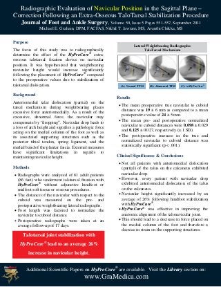
- 1. Radiographic Evaluation of Navicular Position in the Sagittal Plane – Correction Following an Extra-Osseous TaloTarsal Stabilization Procedure Journal of Foot and Ankle Surgery, Volume 50, Issue 5 Pages 551-557, September 2011 Michael E. Graham, DPM, FACFAS, Nikhil T. Jawrani, MS, Avanthi Chikka, MS Purpose Lateral Weightbearing Radiographs: The focus of this study was to radiographically TaloTarsal Mechanism determine the effect of the HyProCure® extra- osseous talotarsal fixation device on navicular position. It was hypothesized that weightbearing navicular height would increase significantly following the placement of HyProCure® compared to the preoperative values due to stabilization of talotarsal dislocation. (A): Normal TTM (B): Abnormal TTM (C): w/HyProCure® Background Results Anteriomedial talar dislocation (partial) on the The mean preoperative true navicular to cuboid tarsal mechanism during weightbearing places distance was 19 ± 6 mm as compared to a mean excessive force anteriomedially. As a result of the postoperative value of 24 ± 5 mm. excessive, abnormal force, the navicular may compensate by “dropping”. Navicular drop leads to The mean pre- and postoperative normalized a loss of arch height and signifies a pathologic force navicular to cuboid distances were 0.098 ± 0.029 acting on the medial column of the foot as well as and 0.125 ± 0.027, respectively (± 1 SD). its associated supporting structures such as the The postoperative increase in the true and posterior tibial tendon, spring ligament, and the normalized navicular to cuboid distance was medial band of the plantar fascia. External measures statistically significant (p < .001). have significant limitations in regards to maintaining navicular height. Clinical Significance & Conclusions Not all patients with anteriomedial dislocation Methods (partial) of the talus on the calcaneus exhibited Radiographs were analyzed of 61 adult patients navicular drop. (86 feet) who underwent talotarsal fixation with However, every patient with navicular drop HyProCure® without adjunctive hindfoot or exhibited anteriomedial dislocation of the talus midfoot soft tissue or osseous procedures. on the calcaneus. The distance of the navicular with respect to the Navicular height significantly increased by an cuboid was measured on the pre- and average of 26% following hindfoot stabilization postoperative weightbearing lateral radiographs. with HyProCure®. Foot length was factored to normalize the HyProCure® was effective in improving the navicular to cuboid distance. anatomic alignment of the talonavicular joint. Postoperative radiographs were taken at an This should lead to a decrease in force placed on average follow-up of 17 days. the medial column of the foot and therefore a decreae in strain on the supporting structures. Talotarsal joint stabilization with HyProCure® lead to an average 26% increase in navicular height. Additional Scientific Papers on HyProCure® are available. Visit the Library section on: www.GraMedica.com