
Empfohlen
Weitere ähnliche Inhalte
Was ist angesagt?
Was ist angesagt? (20)
Ähnlich wie The Arteries
Ähnlich wie The Arteries (20)
Mehr von meducationdotnet
Mehr von meducationdotnet (20)
The Arteries
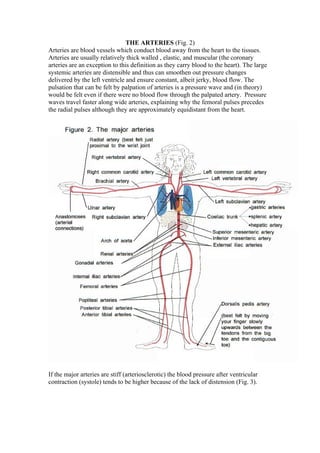
- 1. THE ARTERIES (Fig. 2) Arteries are blood vessels which conduct blood away from the heart to the tissues. Arteries are usually relatively thick walled , elastic, and muscular (the coronary arteries are an exception to this definition as they carry blood to the heart). The large systemic arteries are distensible and thus can smoothen out pressure changes delivered by the left ventricle and ensure constant, albeit jerky, blood flow. The pulsation that can be felt by palpation of arteries is a pressure wave and (in theory) would be felt even if there were no blood flow through the palpated artery. Pressure waves travel faster along wide arteries, explaining why the femoral pulses precedes the radial pulses although they are approximately equidistant from the heart. If the major arteries are stiff (arteriosclerotic) the blood pressure after ventricular contraction (systole) tends to be higher because of the lack of distension (Fig. 3).
- 2. Peripheral resistance Peripheral resistance mostly depends on the radius of the arterioles (=minute branches of the arteries). Sympathetic vasoconstrictor tone is derived from the vasomotor centre in the brain medulla. Increase in vasomotor tone leads to arterial vasoconstriction and causes a rise in arterial blood pressure (if cardiac output remains constant). If there is rapid onset of arterial blockage the areas supplied become painful, pale, cold, anaesthetic (lost sensation) and gangrenous if the obstruction is not rapidly relieved (Fig. 4). Fig 4.
- 3. Arterial disease may be associated with smoking, diabetes mellitus, hypertension, some inherited conditions including high blood fat levels and inflammatory diseases of arteries (arteritis) Atheroma (derived from a Greek word meaning porridge) furs up arteries, usually at sites of vessel wall stress, particularly in the abdominal aorta and its medium sized branches. Atheromatous plaques form which comprise a fat rich necrotic central area, surrounded by muscle cells and fibrous tissue on which platelet thrombi (clots) can develop. Ischaemic (=locally and temporarily suffering from a lack of blood) tissues are often painful because of accumulation of anoxic metabolites. Death of heart muscle (known variously as myocardial infarction, heart attack, coronary thrombosis) occurs when there is severe local restriction in blood flow through the coronary arteries. Principles underlying some arterial problems Aortic dissection occurs when blood forces its way between the layers of the arterial wall (Fig. 5). Coarctation is a focal narrowing of the aorta which tends to occur between the left subclavian artery and the ductus arteriosus (Fig. 6). Coarctation may be associated with aortic valve abnormalities and/or weakness of the aortic wall. The pulse amplitude beyond the narrowing may be diminished, the blood pressure may be lower in the lower limbs, and the femoral pulse may be delayed when compared with the radial pulse (normally the pressure wave travels faster along the wider aortic route to
- 4. the femoral arteries) and is usually felt first. There may be a murmur, caused by abnormal blood flow, distal to the coarctation,. Raynaud’s disease is a specific type of vasospasm that occurs in relation to cold or emotion. The extremities (usually the hands) become white (lack of blood caused by vasospasm), then cyanosed (because of deoxygenation of what blood there is) and then a bright red flush develops once vasospasm remits. Thrombosis (=a clot developing in situ) may occur in veins or, more rarely on atheromatous arteries. An embolism (=a wedge) is the transfer of an intravascular mass (usually a blood clot) from its point of origin to a distant site where it causes obstruction. A pulmonary embolism is impaction of a blood clot (formed as a thrombus, usually in the leg veins) in the lungs. Although the clot originates and travels to the right side of the heart in
- 5. systemic veins, it then travels to its site of impaction in the lungs in the pulmonary arteries.