1. Mod.1-4 – Invasive markers of airway inflammation – 1
Ting Joe Li Yah
Mod.1-4 Invasive markers of airway inflammation
• Why do we need to measure airways inflammation invasively
• Which tissue/cells?
• How/when do we obtain samples and what do we do with them?
Introduction
1. Purpose for invasive tests (taking a tissue sample)
a. To confirm diagnosis
b. To assess disease severity
c. To monitor disease activity/response to treatment (e.g. check for improvement with steroid
intervention)
d. Non-invasive tests may be insufficient
e. For clinical and/or research purposes
2. Ethical issues
a. What are the potential risks to the patient? Do risks outweigh benefits?
b. Will the results of a tissue sample affect/change the clinical management of the disease? If
no, then what is the purpose of taking?
i. In the case of cancer, a tissue sample is required from the tumour to check the cell
types and then cater the correct chemotherapy for patient
c. Research samples may not be appropriate, where a supposedly healthy volunteer for
bronchoscopy was later found to be unhealthy. The research bronchoscopy becomes a
clinical bronchoscopy
d. The study needs to be ethically approved and requires a lot of legislation
i. Could ask patients if waste tissue samples be used for research study
3. Inflammatory markers will depend on disease studied (http://www.nature.com/nri/journal/v8/n3/full/nri2254.html)
a. Asthma: Eosinophils and epithelial cells (although there is non-eosinophilic asthma)
b. COPD: Neutrophil and alveoli
2. Mod.1-4 – Invasive markers of airway inflammation – 2
Ting Joe Li Yah
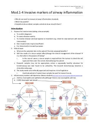
Samples/Procedures
1. Table
Sample Procedure
Blood Venepuncture (venus blood sample)
Sputum (non-invasive?) Hypertonic saline solution irritate airway to bring up the phlegm
Epithelial cells Nasal/bronchial brushings
Endobronchial biopsies Flexible or rigid bronchoscopy (lining of the airway)
Lung resection Video-assisted thorascopic surgery. Thoracotomy (cut open chest)
Radiological monitoring of inflammation (CT scan)
2. Sample processing
a. How samples are taken and processed will affect results
b. Need to put biopsies cells into medium to prevent cell degeneration
c. Need to follow standardised procedures (e.g. British Thoracic Guidelines) for collecting
(biopsy) and processing tissue
Types of ways of sampling
1. Blood
a. Clinical differential cell counts for specific diseases
i. Using an automated cell counter that can break down the different types of white
blood cells
ii. Read eosinophils for asthma or neutrophils for COPD
b. Research isolate appropriate inflammatory cell for ex vivo work
i. Isolate peripheral blood mononuclear cell (PBMC) like lymphocyte, monocyte or
macrophages through the Percoll or Ficoll method
ii. Dextran is added to whole blood to allow the separation of plasma from RBC
iii. Plasma undergoes Percoll centrifuge, allow the identification of PBMC rich fraction
and a neutrophil rich fraction
iv. After isolation of PBMC region, cells are counted to quantify number of cells per
treatment. Cells are kept in medium for survival
- LPS is added to stimulate PBMC secretion of inflammatory markers
- After 24 hours, the medium is harvested and measured for the level of TNF-α
- Measurement of other cytokines are also possible
2. Induced sputum
a. Requirements
i. Isolated room/cubicle due to possibility that patients are infectious
ii. Hypertonic saline administer through a nebuliser irritate the airway
iii. However, this could cause asthma exacerbations. Hence, spirometry is done
beforehand to ascertain if there is any form of bronchoconstriction
b. Non-invasive method of obtaining airways cells
c. Risks: Bronchoconstriction (as mentioned), infection and sample contamination
d. Able to assess response to stimulation (i) or treatment (ii)
i. Stimulating with methacholine. The more methacholine needed to drop lung
function by 20% shows that the airway is less hyper-responsive.
ii. Reduction in sputum eosinophils following 6 weeks of ICS in mild asthmatics
3. Mod.1-4 – Invasive markers of airway inflammation – 3
Ting Joe Li Yah
3. Bronchoscopy
a. Rigid: Preferred for recovery of foreign body aspiration because it allows protection of the
airway and controlling the foreign body during recovery. However, requires GA
b. Flexible: Longer and thinner, allow navigation into individual lobe. Contains a fiberoptic
system that transmits image from the tip of the instrument to a video camera at the opposite
end. It causes less discomfort, can be performed easily and safely under moderate sedation
c. Risks: Sore throat, damage vocal cords, bleeding (resulting from the instrument hitting the
side of the airway due to coughing)
d. Bronchoalveolar lavage (BAL) can be performed
i. Sterile saline (up to 240ml) is squirted into part of the lung for “washing”
ii. Fluid aspirated back to obtain airway cells for examination
- Non-smokers have healthy-looking macrophages, while smokers have
macrophage with dark-blue-purple lipid filled cytoplasmic inclusion bodies
iii. BAL can be filtered to get different cell pellets for counting
iv. Risks: Infection (stir up infection causing fever) and decreased oxygen saturation
e. Bronchoscopy brushings and biopsy
i. Brush to scrap the epithelial cell from the nasal or bronchial lining
ii. Biopsy (if for research, usually taken from right lung because it is easier): 1-3mm
f. Bronchial biopsy
i. Endobronchial (passing endoscope through the nose of mouth to visualise the upper
airway, trachea, bronchi and obtain tissue) or transbronchial (small forceps is push
into a bronchus to take lung tissue samples)
ii. Tissue preservation will influence what histological examination can be carried out
- Snap-frozen (-80), stored in paraformaldehyde or embedded in paraffin
iii. Specific staining technique to identify specific cells or structure (e.g. H&E staining for
basement membrane thickness and airway smooth muscle)
- Able to observe increased basement membrane thickness and increased airway
smooth muscle in severe asthma compared to non-severe asthma
iv. Immunohistochemistry for inflammatory cells (e.g. CD3 for lymphocytes, CD68 for
macrophages, major basic proteins for eosinophils, neutrophil elastase for
neutrophils and tryptase for mast cells)
- Add primary antibodies to the section, then add secondary antibodies that will
bind to primary antibodies
- Dye will only bind to secondary antibodies and show the cells of interest
- Able to observe an influx of eosinophils into airway 6h post allergen challenge
4. Lung resection (surgical removal of all or part of lung, due to lung cancer or other lung diseases)
a. Can be obtained from post mortem or explanted lungs
b. Able to observe dilated airspaces due to destruction of alveoli walls
5. Radiological assessment of airway inflammation
a. CT-guided biopsy, where a nodule is not accessible by bronchoscopy but close to chest wall
b. Use CT to check for gas trapping and assess thickness
c. Risks: Bleeding, pneumothorax
6. Summary
a. Invasive markers of inflammation can be used for diagnosis, assessing severity and
monitoring response to interventions
b. Benefit must outweigh any risks
c. Ethics must be in place for any clinical samples to be used for research and must comply with
Human Tissue Act regulations.