SlideShare ist ein Scribd-Unternehmen logo
1 von 3
Downloaden Sie, um offline zu lesen
GRAM	
  +ve	
  Bacteria:	
  BACILLI	
  
	
   SPORE	
  FORMERS	
   NON-­‐SPORE	
  FORMERS	
  
BACTERIA	
   Clostridium	
   Bacillis	
   Listeria	
   Erysipelothrix	
   Corynebacterium	
   Rhodoccus	
  :o	
  
C.botulinum	
   **B.anthracis	
   L.monocytogenes	
   E.rhusiophathiae	
   C.diphtheriae	
   C.pseudoTB	
   R.equi	
  
PATHOGENISIS	
   Blood	
  to	
  PNS;	
  
Blocks	
  release	
  of	
  
Ach;	
  Flaccid	
  
paralysis	
  and	
  death	
  	
  
kills	
  phagocytic	
  
cells;	
  inc	
  cap	
  
perm;	
  interfere	
  
clotting	
  cascade	


Ruminants:	
  
Meningoencephalitis	
  
Listereosis:	
  Zoonosis	
  
Intracellular:	
  
Penetrating	
  epi	
  cells;	
  
grow	
  
Survives	
  in	
  
macrophages	
  	
  
Serotypes	
  1a,	
  1b,	
  2	
  
Erysipalis	
  
Swelling	
  of	
  
capillary/venule	
  
endothelial	
  cells	
  and	
  
thrombus	
  formation	
  
Only	
  when	
  
infected	
  
w/temperate	
  
phage	
  (bacterial	
  
virus)	
  which	
  
encodes	
  toxin	
  
Caseous	
  
lymphadenitis	
  
(cheeeeese)	
  
Chronic	
  suppurative	
  
bronchopneumonia	
  in	
  
foal;	
  associated	
  
w/underdevo	
  cell-­‐med	
  im	
  
(=	
  adults	
  immune)	
  
Large	
  pulmonary	
  
abscesses	
  in	
  lungs	
  
TOXIN	
   Exotoxins	
  A-­‐H	
   Holotoxin	
   Lysteriolysin	
  to	
  escape	
  
phagosome;	
  contents	
  =	
  
host	
  cell	
  damage	
  =	
  
Antibody	
  ineffective	
  =	
  
cell-­‐med	
  im	
  resp	
  
Neurominodase?	
   Diphtheria	
  toxin	
  
(not	
  known	
  to	
  
cause	
  disease	
  in	
  
animals)	
  
Phospholypase	
  
toxin	


SPREAD	
   Bloodstream	
   Spiny/irritating	
  
vegetation	
  allows	
  
spores	
  access	
  
tissue.	
  	
  
Lymphatics	
  to	
  
bloodstream	
  
Reside	
  in	
  safe	
  
cytoplasm	
  Neutrophils	
  
can	
  kill	
  when	
  in	
  
extracellular	
  env	
  
Systemic	
  
Lymphoid	
  tissues	
  
Penetrate;	
  access	
  to	
  
bloodstream;	
  Joints	
  &	
  
Heart	
  valves	
  
Skin	
  and	
  mucous	
  
membranes	
  
Carried	
  on	
  skin	
  
of	
  sheep	


CHARACTERISTI
CS/STERILISATI
ON	
  
Resistant	
  to	
  boiling.	
  
Heat-­‐liable	
  (20min	
  
100C):	
  Cook	
  
Capsule	
  
Autoclaving	
  
(200ppm	
  
hypochlorite)	
  
Dry	
  heating	
  (160C	
  
2h)	
  
Live	
  spore	
  Sterne	
  
vaccine	
  
Antitoxin	
  
Survive	
  pH	
  4.0	
  
Can	
  grow	
  in	
  low	
  temp	
  
(<10C)	


Surface	
  capsule	
  
Non-­‐motile	
  
Adjuvanted,	
  killed	
  
serotypes	
  1&2	
  and	
  
live	
  vaccines:	
  Pigs	
  
Small,	
  tough,	
  
innocuous	
  
“Diptheroids”	
  
Facultative	
  
intracellular	
  
(survive	
  in	
  m’s!)	


Similar	
  to	
  Coryneacterium!	
  
Is	
  seen	
  as	
  short	
  rods!	


Persist	
  in	
  manure-­‐
contaminated	
  soil	
  for	
  
years.	


TRANSMISSION	
   Spores	
  germinate	
  in	
  anaerobic	
  soil	
  and	
  
sewage	
  (Saprophytes)	
  –	
  enter	
  G.I.T	
  by	
  
ingestion.	
  
Saprophytes	
  in	
  
environment;	
  silage	
  
Shed	
  intermittently	
  in	
  
faeces	
  and	
  milk	
  esp.	
  
after	
  stress	
  
Carriers	
  shed	
  in	
  env	
  
Enter	
  oral/resp	
  
route/break	
  in	
  skin	
  
Sheering	
  wounds	
   Naturally	
  found	
  in	
  soil	
  and	
  
intestines	
  of	
  horses	
  
DIAGNOSIS	
   **Sensitive	
  to	
  Penicillin	
  G	
  
MacFadyean’s	
  reaction:	
  	
  Stains	
  purple	
  
w/Polychrome	
  methylene	
  blue;	
  Bacilli	
  appear	
  as	
  
chains	
  of	
  dark	
  blue,	
  square	
  ended	
  rods;	
  
encapsulated.	
  Before	
  P.M	
  or	
  moving	
  carcass;	
  
Notifiable	
  disease	
  
Haemolytic	
  on	
  blood	
  
agar	
  (listeriolysin)	


Grows	
  on	
  blood	
  agar	
  
α-­‐haemolytic	
  
Coagulase	
  (usually)	
  
Catalase	
  -­‐ve	


   Salmon-­‐pink	
  pigment	
  
GRAM	
  +ve	
  Bacteria:	
  BACILLI	


Arcanobacterium	
   Acintomyces	
   Nocardia	
   Mycobacterium	
  
A.pyrogens	
   A.viscosus	
   A.bovis	
   N.asteroides	
   M.bovis	


  *Badgers	
  &	
  Cattle	
   Other	
  	
  
Abscesses	
  (liver)	
  
Summer	
  mastitis	
  
Ovine	
  foot	
  disease	
  
Pyolysin:	
  cytotoxic	
  for	
  
phagocytic	
  cells	
  
Actinomycosi
s	
  in	
  dogs	
  
Localised	
  
granulomato
us	
  abscess	
  of	
  
skin/thoracic	
  
lesion	
  
Lumpy	
  jaw	
  in	
  
cattle	
  
Granulomatous	
  
lesions	
  of	
  soft	
  
tissues	
  and	
  
bone	
  
Granulomatous	
  lesions;	
  
able	
  to	
  survive	
  and	
  grow	
  
in	
  macrophages	
  
Chronic	
  -­‐>	
  fatal	
  
Pneumonia	
  &	
  wasting	
  	
  
Disease	
  beings	
  in	
  lesion	
  elsewhere	
  in	
  body;	
  
slow	
  multiplication;	
  phagocytosis;	
  inhib	
  
phagosome-­‐lys	
  fusion	
  
Sensitisation	
  via	
  lymphocyte	
  recognition;	
  
infectious	
  granuloma	
  (tubercle)	
  
M.xenopi:	
  	
  Cold-­‐blooded	
  
M.avian:	
  Common	
  in	
  pigs	
  at	
  slaughter	


  Poultry	
  (wild	
  birds	
  and	
  free	
  range)	


  Deer:	
  common.	
  M.bovis	
  inc	
  
[Horses:	
  Naturally	
  resistant;	
  rare]	
  
[Dogs:	
  M.tuberculosis	
  from	
  humans]	
  
[Sheep/Goats:	
  V.rare]	
  
Several	
  extracellular	
  
pore-­‐forming	
  toxins	


   If	
  infection	
  not	
  destroyed	
  by	
  CMI	
  of	
  granuloma;	
  release	
  bacilli;	
  tubercles	
  in	
  other	
  tissues	
  and	
  
organs	
  
Upper	
  resp	
  tract	
  and	
  
genital	
  tracts	
  of	
  
domestic	
  animals	
  
Ruminal	
  wall	
  
May	
  
progress	
  into	
  
abdomen	
  
(pyothorax)	
  
	
   Lesions	
  more	
  difficult	
  to	
  
treat	
  than	
  acintom.	
  As	
  
resistant	
  to	
  number	
  of	
  
antimicrobials	
  
Cattle	
  >6months:	
  Respiratory	
  route	
  via	
  Alimentary	
  tract	
  (post-­‐natal,	
  calf	
  infection)	
  and	
  
congenital	
  route.	
  
Adult:	
  After	
  resp	
  infection	
  as	
  tuberculosis	
  bronchiolitis;	
  bronchiolar	
  spread;	
  TB	
  pneumonia;	
  
Caseous	
  liquefaction	
  &	
  bronchiectasis;	
  open	
  case	
  
Similar	
  to	
  
Coryneacteriu:	
  
Chinese	
  
characteristics	
  in	
  W,	
  
N,	
  M	
  arrangement	
  
Curved/swollen	
  ends	
  
Filamentous;	
  
branching	


Prefers	
  
anaerobic	
  but	
  
not	
  strict	
  
anaerobe	
  
Forms	
  branching	
  rods	
  
much	
  shorter	
  than	
  
actinomycetes	
  
[Isolation	
  PM:	
  inject	
  material	
  into	
  guinipig	
  (v.susceptible):	
  act	
  as	
  a	
  biological	
  filter;	
  succumb	
  
to	
  disease	
  if	
  M.bovis	
  present	
  –	
  now	
  discontinued]	
  
Relatively	
  resistant	
  to	
  drying	
  (viable	
  for	
  yrs),	
  antibiotics	
  and	
  chemical	
  agents:	
  common	
  to	
  pre-­‐
treat	
  specimens	
  w/NaOH/Oxalic	
  acid	
  to	
  destroy	
  normal	
  bacteria	
  leaving	
  mycobacteria	
  
unharmed	
  
Round-­‐ended	
  rods	
  
Strict	
  anaerobes	
  
Slow	
  growing	
  	
  
Some	
  saprophytic	
  will	
  grow	
  in	
  colonies	
  in	
  few	
  days	
  but	
  pathogenic	
  take	
  2-­‐8	
  weeks	
  
Opportunistic	
  
pathogen	
  
Soft	
  grey	
  
granules:	
  
when	
  
squashed,	
  
organism	
  
released	
  
Oral	
  bacteria;	
  
inoculated	
  into	
  
tissues	
  by	
  
trauma	
  
Soil	
  bacterium	
   Shedding	
  into	
  env	
  –	
  lesion	
  excretion;	
  ‘open	
  case’:	
  udder/uterus/resp	
  tract;	
  coughing	
  
Immunity	
  does	
  develop	
  but	
  inadequate	
  
Grows	
  slowly	
  on	
  
blood	
  agar:	
  small	
  
white	
  colonies	
  
surrounded	
  by	
  zone	
  
of	
  β-­‐haemolysis	
  after	
  
48	
  hours.	
  
Non-­‐acid	
  fast	
  
Catalase	
  +ve	
  
Partially	
  acid-­‐fast;	
  cell	
  
walls	
  contain	
  mycolic	
  acids	
  
(like	
  mycobacteria)	
  
Mycolic	
  acid	
  repels	
  aq	
  
stain	
  
Although	
  G+ve	
  
in	
  cell	
  wall	
  
structure,	
  are	
  
difficult	
  to	
  
stain;	
  Acid	
  fast	
  
Zeihl-­‐Neelsen	
  
stain	
  used	
  
instead.	
  
TESTS:	
  Single,	
  comparative	
  intradermal	
  test	
  of	
  PPD	
  –	
  TCA	
  precipitated	
  
tuberculin.	
  M.bovis	
  and	
  M.avian	
  –	
  both	
  injected	
  intradermally	
  –	
  exam	
  after	
  
72h	
  [Cattle	
  susceptible	
  to	
  M.avian	
  (localised	
  lesions);	
  PPD	
  w/M.avian	
  
would	
  cause	
  sensitisation	
  alone]	
  	
  
CMI	
  response!	
  =	
  Swelling	
  +	
  Oedema	
  =	
  +ve	
  	
  (Type	
  IV	
  reaction)	
  
No	
  Vac	
  as	
  interfere	
  w/test.	
  
IF-­‐γ	
  stimulation	
  test:	
  Collect	
  blood;	
  culture	
  lymphocytes;	
  stimulate	
  w/PPD	
  
ELISA	
  (useless	
  as	
  v.little/no	
  antibody!):	
  Measure	
  release	
  of	
  IF-­‐γ	
  
PCR/Immunofluorescence	
  –	
  look	
  for	
  bacteria	
  
GRAM	
  +ve	
  Bacteria:	
  BACILLI	


Weitere ähnliche Inhalte

Was ist angesagt? (20)

Neisseria
NeisseriaNeisseria
Neisseria
 
Streptococcus
StreptococcusStreptococcus
Streptococcus
 
15. shigella
15. shigella15. shigella
15. shigella
 
Mycobacterium tuberculosis (Practical Medical Microbiology, 14)
Mycobacterium tuberculosis (Practical Medical Microbiology, 14)Mycobacterium tuberculosis (Practical Medical Microbiology, 14)
Mycobacterium tuberculosis (Practical Medical Microbiology, 14)
 
Streptococcus & Enterococcus by Dr. Rakesh Prasad Sah
Streptococcus & Enterococcus by Dr. Rakesh Prasad SahStreptococcus & Enterococcus by Dr. Rakesh Prasad Sah
Streptococcus & Enterococcus by Dr. Rakesh Prasad Sah
 
Gram negative cocci
Gram negative cocciGram negative cocci
Gram negative cocci
 
Neisseria Meningitidis
Neisseria MeningitidisNeisseria Meningitidis
Neisseria Meningitidis
 
Laboratory diagnosis of salmonella
Laboratory diagnosis of salmonellaLaboratory diagnosis of salmonella
Laboratory diagnosis of salmonella
 
Pseudomonas
PseudomonasPseudomonas
Pseudomonas
 
Yersinia & pasteurella
Yersinia & pasteurellaYersinia & pasteurella
Yersinia & pasteurella
 
Salmonella
SalmonellaSalmonella
Salmonella
 
9 cocci
9 cocci9 cocci
9 cocci
 
Subcutaneous mycoses.ppt
Subcutaneous mycoses.pptSubcutaneous mycoses.ppt
Subcutaneous mycoses.ppt
 
Superficial mycosis
Superficial mycosisSuperficial mycosis
Superficial mycosis
 
KLEBSIELLA SLIDESHARE PRESENTATION
KLEBSIELLA SLIDESHARE PRESENTATIONKLEBSIELLA SLIDESHARE PRESENTATION
KLEBSIELLA SLIDESHARE PRESENTATION
 
Klebsiella Dr. Mahadi ppt
Klebsiella Dr. Mahadi pptKlebsiella Dr. Mahadi ppt
Klebsiella Dr. Mahadi ppt
 
Lab Diagnosis of Bacterial infections
Lab Diagnosis of Bacterial infectionsLab Diagnosis of Bacterial infections
Lab Diagnosis of Bacterial infections
 
Clostridium
ClostridiumClostridium
Clostridium
 
Medical Microbiology Laboratory (sample collection)
Medical Microbiology Laboratory (sample collection)Medical Microbiology Laboratory (sample collection)
Medical Microbiology Laboratory (sample collection)
 
Intestinal protozoa ( entamoeba, giardia, dientamoeba
Intestinal protozoa ( entamoeba, giardia, dientamoebaIntestinal protozoa ( entamoeba, giardia, dientamoeba
Intestinal protozoa ( entamoeba, giardia, dientamoeba
 

Andere mochten auch

Andere mochten auch (9)

Gram positive rods
Gram positive rodsGram positive rods
Gram positive rods
 
Antibiotic Therapy
Antibiotic TherapyAntibiotic Therapy
Antibiotic Therapy
 
Biology HL IRP
Biology HL IRPBiology HL IRP
Biology HL IRP
 
Bacillus cereus
Bacillus cereusBacillus cereus
Bacillus cereus
 
Diphtheria - Prac. Microbiology
Diphtheria - Prac. MicrobiologyDiphtheria - Prac. Microbiology
Diphtheria - Prac. Microbiology
 
Bacillus cereus
Bacillus cereusBacillus cereus
Bacillus cereus
 
Identification of bacteria, Bacterial identification, Lab identification of b...
Identification of bacteria, Bacterial identification, Lab identification of b...Identification of bacteria, Bacterial identification, Lab identification of b...
Identification of bacteria, Bacterial identification, Lab identification of b...
 
Bacteriology: Bacillus
Bacteriology: BacillusBacteriology: Bacillus
Bacteriology: Bacillus
 
Bacillus cereus microbiologia
Bacillus cereus microbiologiaBacillus cereus microbiologia
Bacillus cereus microbiologia
 

Ähnlich wie Gram positive bacilli

Bohomolets Microbiology Lecture #20
Bohomolets Microbiology Lecture #20Bohomolets Microbiology Lecture #20
Bohomolets Microbiology Lecture #20Dr. Rubz
 
Pathogenic anaerobe gram positive bls 209
Pathogenic anaerobe gram positive bls 209Pathogenic anaerobe gram positive bls 209
Pathogenic anaerobe gram positive bls 209Bruno Mmassy
 
2nd term lecture,_,vib,_helico,tb,_spiro,rick[1]
2nd term lecture,_,vib,_helico,tb,_spiro,rick[1]2nd term lecture,_,vib,_helico,tb,_spiro,rick[1]
2nd term lecture,_,vib,_helico,tb,_spiro,rick[1]عادل الحربي
 
Aerobic spore forming bacilli
Aerobic spore forming bacilliAerobic spore forming bacilli
Aerobic spore forming bacilliBruno Mmassy
 
Aerobic spore forming bacilli
Aerobic spore forming bacilliAerobic spore forming bacilli
Aerobic spore forming bacilliBruno Mmassy
 
Rickettsia & chlamydia presentation.
Rickettsia & chlamydia presentation.Rickettsia & chlamydia presentation.
Rickettsia & chlamydia presentation.Bruno Mmassy
 
Mycobacterium tuberculosis
Mycobacterium tuberculosisMycobacterium tuberculosis
Mycobacterium tuberculosisAwaaz Batazoo
 
Bacillus and Corynebacterium
Bacillus and CorynebacteriumBacillus and Corynebacterium
Bacillus and CorynebacteriumAman Ullah
 
Bacillus and Corynebacterium
Bacillus and CorynebacteriumBacillus and Corynebacterium
Bacillus and CorynebacteriumAman Ullah
 
CLS Histoplasmosis.pptx
CLS Histoplasmosis.pptxCLS Histoplasmosis.pptx
CLS Histoplasmosis.pptxYadav Raj
 
Other Gram Negative Bacilli
Other Gram Negative BacilliOther Gram Negative Bacilli
Other Gram Negative BacilliMD Specialclass
 
Other Gram Negative Bacilli
Other Gram Negative BacilliOther Gram Negative Bacilli
Other Gram Negative BacilliMD Specialclass
 
Mycobacterium Tuberculosis by Dr. Rakesh Prasad Sah
Mycobacterium Tuberculosis by Dr. Rakesh Prasad SahMycobacterium Tuberculosis by Dr. Rakesh Prasad Sah
Mycobacterium Tuberculosis by Dr. Rakesh Prasad SahDr. Rakesh Prasad Sah
 
Fat2.chapter15 (pathogenicity)
Fat2.chapter15 (pathogenicity)Fat2.chapter15 (pathogenicity)
Fat2.chapter15 (pathogenicity)Tim Husain
 

Ähnlich wie Gram positive bacilli (20)

Bohomolets Microbiology Lecture #20
Bohomolets Microbiology Lecture #20Bohomolets Microbiology Lecture #20
Bohomolets Microbiology Lecture #20
 
Pathogenic anaerobe gram positive bls 209
Pathogenic anaerobe gram positive bls 209Pathogenic anaerobe gram positive bls 209
Pathogenic anaerobe gram positive bls 209
 
2nd term lecture,_,vib,_helico,tb,_spiro,rick[1]
2nd term lecture,_,vib,_helico,tb,_spiro,rick[1]2nd term lecture,_,vib,_helico,tb,_spiro,rick[1]
2nd term lecture,_,vib,_helico,tb,_spiro,rick[1]
 
Chapter 11 micro
Chapter 11 microChapter 11 micro
Chapter 11 micro
 
Aerobic spore forming bacilli
Aerobic spore forming bacilliAerobic spore forming bacilli
Aerobic spore forming bacilli
 
Aerobic spore forming bacilli
Aerobic spore forming bacilliAerobic spore forming bacilli
Aerobic spore forming bacilli
 
Pathogenesis of infection
Pathogenesis of infectionPathogenesis of infection
Pathogenesis of infection
 
Rickettsia & chlamydia presentation.
Rickettsia & chlamydia presentation.Rickettsia & chlamydia presentation.
Rickettsia & chlamydia presentation.
 
Mycobacterium tuberculosis
Mycobacterium tuberculosisMycobacterium tuberculosis
Mycobacterium tuberculosis
 
Dimorphic fungi
Dimorphic fungiDimorphic fungi
Dimorphic fungi
 
Bacillus and Corynebacterium
Bacillus and CorynebacteriumBacillus and Corynebacterium
Bacillus and Corynebacterium
 
Bacillus and Corynebacterium
Bacillus and CorynebacteriumBacillus and Corynebacterium
Bacillus and Corynebacterium
 
Bacteriology
BacteriologyBacteriology
Bacteriology
 
CLS Histoplasmosis.pptx
CLS Histoplasmosis.pptxCLS Histoplasmosis.pptx
CLS Histoplasmosis.pptx
 
Other Gram Negative Bacilli
Other Gram Negative BacilliOther Gram Negative Bacilli
Other Gram Negative Bacilli
 
Other Gram Negative Bacilli
Other Gram Negative BacilliOther Gram Negative Bacilli
Other Gram Negative Bacilli
 
Mycobacterium Tuberculosis by Dr. Rakesh Prasad Sah
Mycobacterium Tuberculosis by Dr. Rakesh Prasad SahMycobacterium Tuberculosis by Dr. Rakesh Prasad Sah
Mycobacterium Tuberculosis by Dr. Rakesh Prasad Sah
 
Bacteriology.pptx
Bacteriology.pptxBacteriology.pptx
Bacteriology.pptx
 
Fat2.chapter15 (pathogenicity)
Fat2.chapter15 (pathogenicity)Fat2.chapter15 (pathogenicity)
Fat2.chapter15 (pathogenicity)
 
Health considerations done
Health considerations doneHealth considerations done
Health considerations done
 

Mehr von meducationdotnet

Mehr von meducationdotnet (20)

No Title
No TitleNo Title
No Title
 
Spondylarthropathy
SpondylarthropathySpondylarthropathy
Spondylarthropathy
 
Diagnosing Lung cancer
Diagnosing Lung cancerDiagnosing Lung cancer
Diagnosing Lung cancer
 
Eczema Herpeticum
Eczema HerpeticumEczema Herpeticum
Eczema Herpeticum
 
The Vagus Nerve
The Vagus NerveThe Vagus Nerve
The Vagus Nerve
 
Water and sanitation and their impact on health
Water and sanitation and their impact on healthWater and sanitation and their impact on health
Water and sanitation and their impact on health
 
The ethics of electives
The ethics of electivesThe ethics of electives
The ethics of electives
 
Intro to Global Health
Intro to Global HealthIntro to Global Health
Intro to Global Health
 
WTO and Health
WTO and HealthWTO and Health
WTO and Health
 
Globalisation and Health
Globalisation and HealthGlobalisation and Health
Globalisation and Health
 
Health Care Worker Migration
Health Care Worker MigrationHealth Care Worker Migration
Health Care Worker Migration
 
International Institutions
International InstitutionsInternational Institutions
International Institutions
 
Haemochromotosis brief overview
Haemochromotosis brief overviewHaemochromotosis brief overview
Haemochromotosis brief overview
 
Ascities overview
Ascities overviewAscities overview
Ascities overview
 
Overview of the Liver
Overview of the LiverOverview of the Liver
Overview of the Liver
 
Overview of Antidepressants
Overview of AntidepressantsOverview of Antidepressants
Overview of Antidepressants
 
Gout Presentation
Gout PresentationGout Presentation
Gout Presentation
 
Review of orthopaedic services: Prepared for the Auditor General for Scotland...
Review of orthopaedic services: Prepared for the Auditor General for Scotland...Review of orthopaedic services: Prepared for the Auditor General for Scotland...
Review of orthopaedic services: Prepared for the Auditor General for Scotland...
 
Sugammadex - a revolution in anaesthesia?
Sugammadex - a revolution in anaesthesia?Sugammadex - a revolution in anaesthesia?
Sugammadex - a revolution in anaesthesia?
 
Ophthamology Revision
Ophthamology RevisionOphthamology Revision
Ophthamology Revision
 

Gram positive bacilli

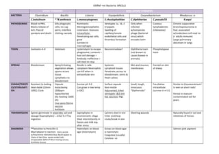
  • 1. GRAM  +ve  Bacteria:  BACILLI     SPORE  FORMERS   NON-­‐SPORE  FORMERS   BACTERIA   Clostridium   Bacillis   Listeria   Erysipelothrix   Corynebacterium   Rhodoccus  :o   C.botulinum   **B.anthracis   L.monocytogenes   E.rhusiophathiae   C.diphtheriae   C.pseudoTB   R.equi   PATHOGENISIS   Blood  to  PNS;   Blocks  release  of   Ach;  Flaccid   paralysis  and  death     kills  phagocytic   cells;  inc  cap   perm;  interfere   clotting  cascade     Ruminants:   Meningoencephalitis   Listereosis:  Zoonosis   Intracellular:   Penetrating  epi  cells;   grow   Survives  in   macrophages     Serotypes  1a,  1b,  2   Erysipalis   Swelling  of   capillary/venule   endothelial  cells  and   thrombus  formation   Only  when   infected   w/temperate   phage  (bacterial   virus)  which   encodes  toxin   Caseous   lymphadenitis   (cheeeeese)   Chronic  suppurative   bronchopneumonia  in   foal;  associated   w/underdevo  cell-­‐med  im   (=  adults  immune)   Large  pulmonary   abscesses  in  lungs   TOXIN   Exotoxins  A-­‐H   Holotoxin   Lysteriolysin  to  escape   phagosome;  contents  =   host  cell  damage  =   Antibody  ineffective  =   cell-­‐med  im  resp   Neurominodase?   Diphtheria  toxin   (not  known  to   cause  disease  in   animals)   Phospholypase   toxin         SPREAD   Bloodstream   Spiny/irritating   vegetation  allows   spores  access   tissue.     Lymphatics  to   bloodstream   Reside  in  safe   cytoplasm  Neutrophils   can  kill  when  in   extracellular  env   Systemic   Lymphoid  tissues   Penetrate;  access  to   bloodstream;  Joints  &   Heart  valves   Skin  and  mucous   membranes   Carried  on  skin   of  sheep     CHARACTERISTI CS/STERILISATI ON   Resistant  to  boiling.   Heat-­‐liable  (20min   100C):  Cook   Capsule   Autoclaving   (200ppm   hypochlorite)   Dry  heating  (160C   2h)   Live  spore  Sterne   vaccine   Antitoxin   Survive  pH  4.0   Can  grow  in  low  temp   (<10C)     Surface  capsule   Non-­‐motile   Adjuvanted,  killed   serotypes  1&2  and   live  vaccines:  Pigs   Small,  tough,   innocuous   “Diptheroids”   Facultative   intracellular   (survive  in  m’s!)     Similar  to  Coryneacterium!   Is  seen  as  short  rods!     Persist  in  manure-­‐ contaminated  soil  for   years.     TRANSMISSION   Spores  germinate  in  anaerobic  soil  and   sewage  (Saprophytes)  –  enter  G.I.T  by   ingestion.   Saprophytes  in   environment;  silage   Shed  intermittently  in   faeces  and  milk  esp.   after  stress   Carriers  shed  in  env   Enter  oral/resp   route/break  in  skin   Sheering  wounds   Naturally  found  in  soil  and   intestines  of  horses   DIAGNOSIS   **Sensitive  to  Penicillin  G   MacFadyean’s  reaction:    Stains  purple   w/Polychrome  methylene  blue;  Bacilli  appear  as   chains  of  dark  blue,  square  ended  rods;   encapsulated.  Before  P.M  or  moving  carcass;   Notifiable  disease   Haemolytic  on  blood   agar  (listeriolysin)     Grows  on  blood  agar   α-­‐haemolytic   Coagulase  (usually)   Catalase  -­‐ve       Salmon-­‐pink  pigment  
  • 2. GRAM  +ve  Bacteria:  BACILLI     Arcanobacterium   Acintomyces   Nocardia   Mycobacterium   A.pyrogens   A.viscosus   A.bovis   N.asteroides   M.bovis              *Badgers  &  Cattle   Other     Abscesses  (liver)   Summer  mastitis   Ovine  foot  disease   Pyolysin:  cytotoxic  for   phagocytic  cells   Actinomycosi s  in  dogs   Localised   granulomato us  abscess  of   skin/thoracic   lesion   Lumpy  jaw  in   cattle   Granulomatous   lesions  of  soft   tissues  and   bone   Granulomatous  lesions;   able  to  survive  and  grow   in  macrophages   Chronic  -­‐>  fatal   Pneumonia  &  wasting     Disease  beings  in  lesion  elsewhere  in  body;   slow  multiplication;  phagocytosis;  inhib   phagosome-­‐lys  fusion   Sensitisation  via  lymphocyte  recognition;   infectious  granuloma  (tubercle)   M.xenopi:    Cold-­‐blooded   M.avian:  Common  in  pigs  at  slaughter                                      Poultry  (wild  birds  and  free  range)                                      Deer:  common.  M.bovis  inc   [Horses:  Naturally  resistant;  rare]   [Dogs:  M.tuberculosis  from  humans]   [Sheep/Goats:  V.rare]   Several  extracellular   pore-­‐forming  toxins         If  infection  not  destroyed  by  CMI  of  granuloma;  release  bacilli;  tubercles  in  other  tissues  and   organs   Upper  resp  tract  and   genital  tracts  of   domestic  animals   Ruminal  wall   May   progress  into   abdomen   (pyothorax)     Lesions  more  difficult  to   treat  than  acintom.  As   resistant  to  number  of   antimicrobials   Cattle  >6months:  Respiratory  route  via  Alimentary  tract  (post-­‐natal,  calf  infection)  and   congenital  route.   Adult:  After  resp  infection  as  tuberculosis  bronchiolitis;  bronchiolar  spread;  TB  pneumonia;   Caseous  liquefaction  &  bronchiectasis;  open  case   Similar  to   Coryneacteriu:   Chinese   characteristics  in  W,   N,  M  arrangement   Curved/swollen  ends   Filamentous;   branching       Prefers   anaerobic  but   not  strict   anaerobe   Forms  branching  rods   much  shorter  than   actinomycetes   [Isolation  PM:  inject  material  into  guinipig  (v.susceptible):  act  as  a  biological  filter;  succumb   to  disease  if  M.bovis  present  –  now  discontinued]   Relatively  resistant  to  drying  (viable  for  yrs),  antibiotics  and  chemical  agents:  common  to  pre-­‐ treat  specimens  w/NaOH/Oxalic  acid  to  destroy  normal  bacteria  leaving  mycobacteria   unharmed   Round-­‐ended  rods   Strict  anaerobes   Slow  growing     Some  saprophytic  will  grow  in  colonies  in  few  days  but  pathogenic  take  2-­‐8  weeks   Opportunistic   pathogen   Soft  grey   granules:   when   squashed,   organism   released   Oral  bacteria;   inoculated  into   tissues  by   trauma   Soil  bacterium   Shedding  into  env  –  lesion  excretion;  ‘open  case’:  udder/uterus/resp  tract;  coughing   Immunity  does  develop  but  inadequate   Grows  slowly  on   blood  agar:  small   white  colonies   surrounded  by  zone   of  β-­‐haemolysis  after   48  hours.   Non-­‐acid  fast   Catalase  +ve   Partially  acid-­‐fast;  cell   walls  contain  mycolic  acids   (like  mycobacteria)   Mycolic  acid  repels  aq   stain   Although  G+ve   in  cell  wall   structure,  are   difficult  to   stain;  Acid  fast   Zeihl-­‐Neelsen   stain  used   instead.   TESTS:  Single,  comparative  intradermal  test  of  PPD  –  TCA  precipitated   tuberculin.  M.bovis  and  M.avian  –  both  injected  intradermally  –  exam  after   72h  [Cattle  susceptible  to  M.avian  (localised  lesions);  PPD  w/M.avian   would  cause  sensitisation  alone]     CMI  response!  =  Swelling  +  Oedema  =  +ve    (Type  IV  reaction)   No  Vac  as  interfere  w/test.   IF-­‐γ  stimulation  test:  Collect  blood;  culture  lymphocytes;  stimulate  w/PPD   ELISA  (useless  as  v.little/no  antibody!):  Measure  release  of  IF-­‐γ   PCR/Immunofluorescence  –  look  for  bacteria  
  • 3. GRAM  +ve  Bacteria:  BACILLI