1. 1
THE BLOOD
Blood can be considered a special type of connective tissue. Its fluid content contents
include nutrients, wastes, salts, hormones, proteins and often medically administered
substances. In addition blood transport gases, cells and heat around the body.
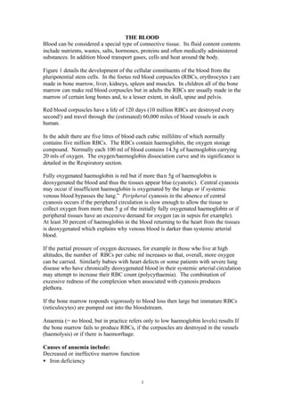
Figure 1 details the development of the cellular constituents of the blood from the
pluripotential stem cells. In the foetus red blood corpuscles (RBCs, erythrocytes ) are
made in bone marrow, liver, kidneys, spleen and muscles. In children all of the bone
marrow can make red blood corpuscles but in adults the RBCs are usually made in the
marrow of certain long bones and, to a lesser extent, in skull, spine and pelvis.
Red blood corpuscles have a life of 120 days (10 million RBCs are destroyed every
second!) and travel through the (estimated) 60,000 miles of blood vessels in each
human.
In the adult there are five litres of blood each cubic millilitre of which normally
contains five million RBCs. The RBCs contain haemoglobin, the oxygen storage
compound. Normally each 100 ml of blood contains 14.5g of haemoglobin carrying
20 mls of oxygen. The oxygen/haemoglobin dissociation curve and its significance is
detailed in the Respiratory section.
Fully oxygenated haemoglobin is red but if more than 5g of haemoglobin is
deoxygenated the blood and thus the tissues appear blue (cyanotic). Central cyanosis
may occur if insufficient haemoglobin is oxygenated by the lungs or if systemic
venous blood bypasses the lung.” Peripheral cyanosis in the absence of central
cyanosis occurs if the peripheral circulation is slow enough to allow the tissue to
collect oxygen from more than 5 g of the initially fully oxygenated haemoglobin or if
peripheral tissues have an excessive demand for oxygen (as in sepsis for example).
At least 30 percent of haemoglobin in the blood returning to the heart from the tissues
is deoxygenated which explains why venous blood is darker than systemic arterial
blood.
If the partial pressure of oxygen decreases, for example in those who live at high
altitudes, the number of RBCs per cubic ml increases so that, overall, more oxygen
can be carried. Similarly babies with heart defects or some patients with severe lung
disease who have chronically deoxygenated blood in their systemic arterial circulation
may attempt to increase their RBC count (polycythaemia). The combination of
excessive redness of the complexion when associated with cyanosis produces
plethora.
If the bone marrow responds vigorously to blood loss then large but immature RBCs
(reticulocytes) are pumped out into the bloodstream.
Anaemia (= no blood, but in practice refers only to low haemoglobin levels) results If
the bone marrow fails to produce RBCs, if the corpuscles are destroyed in the vessels
(haemolysis) or if there is haemorrhage.
Causes of anaemia include:
Decreased or ineffective marrow function
• Iron deficiency
2. 2
• Lack of vitamin B12 or folate
• A damaged marrow
• Infiltration by malignancy or other processes
Peripheral causes
• Blood loss
• Haemolysis (excessive or premature breakdown of RBCs)
• An overactive spleen (hypersplenism)
Hereditary anaemias
Causes include:
• Faulty synthesis or structure of haemoglobin (the haemoglobinopathies). Most are
geographically or racially distributed and include sickle cell disease and
thalassaemia. Both causes significant problems if sufferers are homozygous rather
than heterozygous for the relevant gene
• Defects of red blood cell enzymes. Deficiency of glucose 6-phosphate
dehydrogenase may present with haemolysis (breakdown of RBCs ) when affected
patients are given certain drugs
• Red blood cell membrane abnormalities (spherocytosis, a tendency of the red
blood corpuscles to be spherical is an example).
Causes of haemolytic anaemia
Congenital
• RBC abnormalities
• Defective forms of haemoglobin
• RBC enzyme defects
Acquired
• Immune mediated
• Non-immune mediated
• Mechanical, turbulence around artificial heart valves for example
• Infections
• Drugs effects
Red blood corpuscles may exhibit various patterns on microscopy or on staining (Fig.
2).
Iron and the blood
Iron is required for haemoglobin formation (haem contains iron and the globin is a
protein). If there is insufficient iron then smaller than usual (microcytic) RBCs are
produced which contain less haemoglobin then normal and which are therefore less
red (hypochromic). The adult male has about 4.5g of iron in his body. Women have
slightly less because of menstrual losses or because of increased requirements in
pregnancy. The diagnosis of the cause of iron deficiency anaemia (decreased iron
absorption, increased RBC breakdown or excessive blood loss) may be difficult.
In iron deficiency anaemia there is:
• Reduced haemoglobin. In men less than 135g/l, in women less than 115g/l
• Reduced mean cell volume (microcytosis)
• Reduced red blood cell redness (hypochromia)
• Reduced mean cell haemoglobin
3. 3
• Reduced mean corpuscular haemoglobin concentration
• Reduced serum ferritin
• Reduced serum iron and increased iron binding capacity
Iron deficiency of gastroenterological causation may result from:
• Diets low in meat (for example in the elderly or in vegans)
• Blood loss into the gut
• Gastric surgery which interferes with absorption
• Intestinal hurry which interferes with absorption
• Malabsorption
Anaemia of chronic disorders
Some chronic disorders, particularly those associated with chronic inflammatory
disorders, may depress the bone marrow to produce anaemia (if this involves all
cellular element of the blood this is termed pancytopenia). One major stimulus to
RBC production is erythropoetin production by the kidney. If erythropoetin
production is reduced, as it may be in kidney failure, then anaemia results.
Vitamin B12
Pernicious anaemia (pernicious because it develops insidiously and can be fatal if
untreated) is a common cause of Vitamin B12 deficiency. Vitamin B12 in the diet is
combined with an intrinsic factor secreted by the parietal cells of the stomach and the
complex absorbed by the lower small gut (the terminal ileum). If the stomach fails to
produce intrinsic factor (as in pernicious anaemia) or if the diet is defective in vitamin
B12 (in vegans for example), or if part of the stomach has been surgically removed, or
if the terminal ileum is diseased then vitamin B12 absorption is decreased and red
blood corpuscles cannot develop properly. Patients becomes anaemic and their RBCs
become bigger than normal (macrocytosis) and their bone marrow contains
megaloblastic (large nucleated) RBC precursors. Giving vitamin B12 intramuscularly
bypasses the gut and its absorption problems.
Folate deficiency
Folate deficiency may be caused by a diet poor in vegetables, malabsorption,
increased demand for folate (pregnancy) or as a drug effect. A macrocytic blood
picture and a megaloblastic marrow may result.
Breakdown of red blood corpuscles
Haemoglobin breakdown occurs in the reticuloendothelial system with formation of
bilirubin of various types.
The white blood cells (Fig. 2)
White blood cells are not white! Apart from their nuclei and granules they are
colourless. White blood cells, unlike red blood cells, can migrate from the
bloodstream into the tissues.
Granulocytes (polymorphonuclear leukocytes, “polymorphs,” pus cells, neutrophils)
have lobulated nuclei, have granules in their cytoplasm, and specialize in ingesting
(phagocytosing) invading pathogens.
Eosinophils increase as a response to allergic reactions of various sorts, including hay
fever and invasive worm infections.
4. 4
Basophils liberate inflammatory mediators such as histamine.
Lymphocytes are produced in the bone marrow, lymph nodes and spleen and are often
increased in long duration intracellular infections. Lymphocytes either form
antibodies, liberate chemical messengers “kines” including inflammatory mediators,
or enact cell mediated immunity.
Monocytes are phagocytic, usually increase in bacterial infections, and produce
cytokines.
Platelets contribute to blood clotting. A low platelet count (thrombocytopenia) can
result from decreased platelet production (including drug actions, malignancies
especially if involving the bone marrow, infections and pernicious anaemia),
increased platelet consumption, or an overactive spleen.
Although the relative proportions of the blood cellular constituents may provide
diagnostic clues it is usually the absolute numbers of each constituent that are
functionally important.
Blood groups (Fig. 3).
In the ABO blood group system the serum contains agglutinins which damage cells
not of their group. Serum of Group A RBCs contains anti-B, serum of Group B
RBCs contains anti-A, serum of Group AB contains no agglutinins (and thus can
receive cells of any ABO grouping – and are thus “universal recipients”) and serum of
Group O RBCs contains anti-A and anti-B and can thus be given to any ABO group -
the “universal donor” (the donated plasma of Group O RBCs is diluted by the
recipient’s plasma and does not cause significant problems by agglutinating the
recipient’s RBCs). Red blood corpuscles given to a patient whose serum plasma
contains agglutinins to the donated RBCs (A -> O, B -> O, A -> B, or B ->A) causes
the donated RBCs to be agglutinated to produce a systemic reaction. Transfusion
reactions caused by ABO incompatibility depend on the RBCs being given into an
environment containing antibodies to the donated RBCs. All blood grouping has
depended, classically, on reacting the recipient’s RBCs with known antiserum and
known RBCs with the recipient’s serum.
There are various other mechanisms by which blood can be grouped. Most depend on
RBCs characteristics rather than spontaneously occurring plasma agglutinins as occur
in the ABO system.
Rhesus incompatibility
If RBCs which have Rhesus positive antigens on their surface enter the bloodstream
of a Rhesus negative person then this recipient will make antibodies to the Rhesus
positive RBCs. Such an occurrence may occur if Rhesus positive blood is transfused
into a Rhesus negative person, or at the time of childbirth if Rhesus positive foetal
blood enters a maternal (Rhesus negative) circulation. If such a Rhesus negative
Normal neutrophil count 1.5-7.5 x 10⁹
/litre
Normal lymphocyte count 1.5-4.0 x 10⁹
/litre
Normal monocyte count 0.2-0.8 x 10⁹
/litre
Normal eosinophil count 0.04-0.4 x 10⁹
/litre
Normal basophil count <0.1 x 10⁹
/litre
Normal platelet count 150-400 x 109
/litre
5. 5
woman has a subsequent pregnancy then the RBCs of her second or subsequent
Rhesus positive foetus may be agglutinated in utero by maternal IgG antibody
containing rhesus antibody which crosses the placenta to cause jaundice and brain
damage in the foetus. To prevent this a Rhesus negative mother who has just given
birth to a Rhesus positive baby is given anti-D Rhesus factor antibodies which will
agglutinate the Rhesus positive foetal RBCs in her circulation before they can evoke
production of anti-Rhesus antibodies.
Blood clotting
Blood clotting is essential to prevent haemorrhage from damaged blood vessels.
Although plasma without cells can clot usually both plasma and cellular elements of
the blood are required for effective clotting.
Figure 4 shows a greatly simplified mechanism of clotting. In reality a number of
intrinsic factors, including Factor VIII (anti-haemophilic globulin) and Factor IX
(Christmas factor) are required to initiate or maintain clotting. Thrombus is clot in the
blood vessels. Thrombi form in response to changes in the blood vessel wall to which
platelets adhere with liberation of thromboplastin. This system is the intrinsic system
(intrinsic to the blood). The extrinsic system refers to clotting initiated by damage to
tissues outwith the blood vessels which release tissue thromboplastin. In reality there
is overlap between factors and systems and other factors are also required.
Calcium in blood specimens is often removed or complexed to prevent clotting in
laboratory specimens in vitro or to prevent clotting in blood required for transfusion.
Heparin (which occurs naturally in the body and which can only be given by
injection) can be used to inhibit clotting by inhibiting the formation of thrombin from
prothrombin and by inhibiting the formation of fibrin from fibrinogen. The major
action of most oral anticoagulants is to reduce the formation of prothrombin.
6. 6
Disseminated intravascular coagulation
Disseminated intravascular coagulation is a paradox. Intravascular clotting causes
consumption of clotting factors, particularly fibrin, which results in haemorrhage.
Additionally the body’s response may entail excessive attempts to dissolve fibrin in
existing clots by production of an enzyme, plasmin, which may cause haemorrhage by
reducing clotting elsewhere. In disseminated intravascular coagulation the fibrin
levels are low, fibrin degradation products are high and the platelets are low.
Fragmentation of RBCs may also be seen.
The plasma proteins
The direct antiglobulin (Coombs’) test (Fig. 5) detects antibodies on RBCs which are
found in most cases of immune mediated anaemia associated with antibodies of
various sorts which cause the red blood corpuscles to have a shortened lifespan. The
osmotic significance of plasma proteins is detailed elsewhere.
The Erythrocyte sedimentation rate (ESR)
If clotting is prevented RBCs sink, and the distance they sink at the end of one
hourcan be measured (the erythrocyte sedimentation rate) and eventually constitutes a
layer 40-47 percent of the total column height (the haematocrit or packed cell
volume). The white cells form a shallow layer on top of the red corpuscles (the buffy
coat), leaving the plasma above (Fig. 6).
Normally the slight negative charge on each RBC prevents them from clumping
together. However if they are pushed together they may then clump and form
rouleaux (stacks of RBCs) which fall down the column of plasma quicker than would
Commonly used tests of clotting
The blood count (including the platelet count) and blood film examination
The bleeding time is measured by the time to bleeding cessation after pricking of
an ear lobe with removal of blood every 15 seconds. The modern version involves
making two standardized cuts on the forearm. The usual value is 2-6 minutes. The
bleeding time is essentially a measure of platelet capacity to stop bleeding by
forming plugs in small vessels and of vessel integrity. The test is dependent on
platelet numbers, and the function and integrity of small vessels.
The clotting time (usually less than one minute) is the time that blood, kept at body
temperature, takes to stop flowing. It is essentially a test of clotting mechanisms
The activated partial thromboplastin time is a test of intrinsic clotting mechanisms.
Measurement of specific clotting factors may be necessary for a definitive
diagnosis
The prothrombin time assesses the extrinsic mechanisms
The thrombin clotting time is a measure of thrombin/fibrinogen status (the speed
of clotting depends on the concentration of thrombin added). It is useful for
detecting fibrinogen and its degradation products. D-dimer tests are used for
estimating fibrinogen degradation products.
7. 7
individual RBCs because their mass to surface area ratio increases. The erythrocyte
sedimentation rate thus depends on the viscosity of blood (which may be increased by
an increase in globulins or increases in fibrinogen). If RBCs are coated by antibody,
particularly reactive antibodies produced by the liver in response to inflammation,
their stickiness may increase and thus their propensity to from rouleaux. If RBCs
cannot easily clump together to form rouleaux (for example if there are abnormally
shaped corpuscles) then the erythrocyte sedimentation rate may remain normal in
conditions in which it is usually high.