SlideShare ist ein Scribd-Unternehmen logo
1 von 13
© 2007 WebMD, Inc. All rights reserved.                                                       ACS Surgery: Principles and Practice
 6 VASCULAR SYSTEM                                                      17 INFRAINGUINAL ARTERIAL PROCEDURES — 1


17           INFRAINGUINAL ARTERIAL
             PROCEDURES
William D. Suggs, M.D., F.A.C.S., and Frank J.Veith, M.D., F.A.C.S.


                                                                         NONINVASIVE TESTING
Since the early 1980s, there have been enormous advances in the
treatment of lower-extremity ischemia secondary to infrainguinal            Noninvasive tests are helpful in that they provide semiquantita-
arteriosclerosis. Effective interventional management strategies         tive assessment of the circulation and help confirm the diagnosis
have been developed for virtually all patterns of arteriosclerosis       made on the basis of the history and the physical examination.
underlying limb-threatening ischemia.1,2 Bypasses to the infrain-        Such test measurements include the ankle-brachial index (ABI)
guinal arteries using segments of autologous vein have become            and pulse volume recordings (PVRs).
routine for limb salvage. As this technique has evolved, the distal         The ABI is determined by dividing the ankle pressure in each
limits of revascularization have been extended. Bypasses to arteries     lower limb by the higher of the two brachial pressures. Normal cir-
near the ankle or in the foot can now be offered to patients who         culation typically yields an ABI of 1.0 to 1.2; claudication, an ABI
have no patent arteries suitable for more proximal bypasses. In          of 0.40 to 0.95; and limb-threatening ischemia, an ABI of 0 to 0.5.
addition, bypasses to distal tibial or tarsal vessels may be per-        It is vital to remember, however, that lower-extremity pressure
formed in some patients whose popliteal arteries are patent but          measurements are less reliable in patients with heavily calcified ves-
who have three-vessel distal occlusive disease and forefoot gan-         sels (e.g., diabetics and patients with end-stage renal disease). In
grene.3,4 Frequently, patients who require very distal bypasses have     these patients, ABIs are falsely elevated as a result of the higher cuff
already undergone vascular reconstruction; these patients may be         pressures required to occlude calcified vessels, which in some cases
candidates for alternative approaches, such as a popliteal-distal        are not occluded even with pressures higher than 300 mm Hg.
bypass or a tibiotibial bypass.5-7                                          PVRs are obtained by means of calibrated air-cuff plethysmog-
   Patients with limbs threatened by distal tibial occlusive disease     raphy. Standard blood pressure cuffs are placed at different levels
present an ongoing challenge to the vascular surgeon. Provided           of the lower extremity, and the increases in pressure within the
that careful attention is paid to obtaining high-quality preoperative    cuffs resulting from the volume increase during systole are record-
angiograms and that the surgeon is willing to consider alternative       ed as pulse waves. Tracings exhibiting a brisk rise during systole
approaches, it is generally possible to achieve good results from        and a dicrotic notch are characterized as normal, those exhibiting
limb salvage procedures.                                                 loss of the notch and a more prolonged downslope are character-
   It should be kept in mind that only patients with threatened          ized as moderately abnormal, and those exhibiting a flattened wave
limbs—manifested by rest pain, frank gangrene, or nonhealing             are characterized as severely abnormal. Absolute amplitudes on
ulcers—should be considered candidates for infrainguinal bypass.         PVRs are not directly comparable between patients; however, ser-
Patients who have gangrene that extends into the deeper tarsal           ial PVRs from a single patient are highly reproducible and thus are
region of the foot, who have a severe organic mental syndrome, or        quite useful for following the course of severe peripheral vascular
who are nonambulatory are not candidates for limb salvage                disease in individual cases.4 One disadvantage of PVRs is that they
surgery and should be treated with primary amputation instead.1,2        cannot differentiate proximal femoral disease from iliac occlusive
                                                                         disease.8
Preoperative Evaluation                                                  IMAGING

HISTORY AND PHYSICAL EXAMINATION                                           Duplex Scanning
   A careful history and a thorough physical examination are cru-           Duplex scanning is a useful noninvasive method of assessing the
cial for accurate assessment of the extent of the patient’s athero-      aortoiliac and infrainguinal arterial systems. Several studies have
sclerotic disease. In the course of the history, the examiner should     evaluated the ability of duplex scanning to predict iliac artery
pay particular attention to distinguishing true rest pain from other     stenosis. A 1987 trial found that duplex scanning was highly sensi-
causes of pain (e.g., arthritis and neuritis). Significant ischemic       tive (89%) and specific (90%) in predicting iliac stenosis of 50%
pain is usually associated not only with decreased pulses but also       or greater.9 Three subsequent trials corroborated these findings,
with other manifestations of ischemia (e.g., atrophy, decreased skin     reporting sensitivities ranging from 81% to 89% and specificities
temperature, marked rubor, and pain that is relieved when the foot       ranging from 88% to 99%.10-12 This noninvasive modality may be
is dangled). In the course of the physical examination, the exam-        especially useful for improving evaluation of diabetic patients
iner should look for and assess the extent of any underlying infec-      before invasive procedures (e.g., angiography and angioplasty).10
tion and should closely examine any surgical scars for clues to the         Duplex scanning has been increasingly employed in performing
nature and extent of any previous vascular operations involving the      infrainguinal bypasses to both popliteal and tibial vessels without
use of the saphenous vein. In addition, a careful pulse examination      preoperative angiography. In one study, a limb salvage rate of 86%
should be performed to assess the patient’s baseline arterial status;    was achieved with this approach, and completion arteriography
these baseline measurements provide a basis for comparison if the        matched the runoff status predicted by duplex scanning in 96% of
disease subsequently progresses and may help determine the               cases.13 In a study from our own institution, we were able to per-
approach to be used to salvage the threatened limb.                      form femoropopliteal bypasses without preoperative arteriography
© 2007 WebMD, Inc. All rights reserved.                                                      ACS Surgery: Principles and Practice
6 VASCULAR SYSTEM                                                       17 INFRAINGUINAL ARTERIAL PROCEDURES — 2


and were able to perform distal bypasses with confirmatory arte-          ever, for patients with weak or nonpalpable femoral pulses, other
riograms at the time of operation.14                                     approaches (e.g., translumbar, transbrachial, or transaxillary) may
                                                                         be preferable. These alternative approaches are associated with
  Magnetic Resonance Angiography                                         higher rates of local complications, including hematomas,
   Magnetic resonance angiography (MRA), a noninvasive modal-            pseudoaneurysms, dissections, thrombosis, and embolization.
ity that does not require contrast agents, often yields good arterial       Renal insufficiency is an important complication of angiogra-
images and may, in fact, be more sensitive than angiography in           phy: 6.5% to 8.2% of patients who undergo arteriography experi-
imaging distal lower-extremity runoff vessels.15,16 Developments         ence some degree of impairment associated with contrast
such as gadolinium enhancement, multistation examination, and            agents.26,27 Patients who have preexisting azotemia and whose
the floating table technique have further improved the resolution         baseline creatinine concentrations exceed 2.0 mg/dl are at highest
of MRA,17-19 to the point where many institutions that use current       risk for renal complications after angiography. Elderly patients typ-
forms of MRA no longer routinely obtain preoperative                     ically have lower creatinine clearances for a given serum creatinine
angiograms. The development of time-resolved imaging of con-             level and thus should always be considered at higher risk for
trast kinetics (TRICKS) MRA has further improved the accuracy            nephrotoxicity. All possible precautions should be taken to limit
of MRA for assessing the distal vasculature in preparation for a         the renal insult. There is some evidence to suggest that the use of
bypass.20 MRA, in conjunction with arterial duplex scanning, has         low-osmolar contrast agents can decrease the incidence of renal
the potential to replace contrast arteriography in the evaluation of     impairment,28,29 but the data are not unanimous on this point.30
patients with distal arterial occlusive disease.                         Adequate hydration with oral administration of acetylcysteine
                                                                         before arteriography is highly effective in diminishing the risk of
  Computed Tomographic Angiography                                       contrast nephropathy. Administration of mannitol, which has an
   Computed tomographic angiography (CTA) has become a                   osmotic diuretic effect, helps prevent contrast toxicity as well.
viable alternative to both MRA and conventional digital subtrac-         These measures, coupled with judicious use of contrast agents,
tion angiography for imaging the aortoiliac region and the lower-        should minimize renal impairment associated with arteriography.
extremity arteries. Current variants of this modality (e.g., 16-
detector row CTA) have been shown to be as accurate as conven-
tional angiography for imaging distal vessels. As a consequence,         Femoropopliteal Bypass
operative planning can be done without the need for an invasive             Patients whose limbs are clearly threatened and who have
angiographic procedure. CTA has also shown promise in the eval-          undergone arteriographic examination should undergo
uation of graft-related complications, including graft stenosis,         femoropopliteal bypass when the superficial femoral artery or the
aneurysmal changes, and arteriovenous fistulas.21-23                      popliteal artery is occluded and when arteriography indicates that
                                                                         a patent popliteal artery segment distal to the occlusion has lumi-
  Arteriography                                                          nal continuity with any of its three terminal branches (even if one
   Contrast angiography remains the gold standard for the evalu-         or more of these branches ends in an occlusion anywhere in the
ation of patients with distal arterial occlusive disease. A complete     leg). Even if the popliteal artery segment into which the graft is to
evaluation of the existing arterial disease from the aorta to the        be inserted is occluded distally, femoropopliteal bypass to this seg-
pedal vessels is necessary for diabetic patients, who frequently have    ment can be considered.31,32 If the isolated popliteal segment is
multilevel occlusive disease. Obtaining intra-arterial pressure mea-     shorter than 7 cm or if there is extensive gangrene or infection in
surements at the time of angiography significantly improves detec-        the foot, a femorodistal artery bypass or a sequential bypass is
tion of clinically significant stenosis.The systolic pressure gradient    sometimes performed in one or two stages.
across the lesion should also be measured: gradients greater than
                                                                         OPERATIVE TECHNIQUE
15 mm Hg are considered hemodynamically significant.24
   In the general population, arteriography has a complication rate        Femoropopliteal bypass may be carried out either above or
of only 1.7% to 3.3%.25 Elderly patients with severe aortoiliac or       below the knee.
infrainguinal disease must be carefully evaluated before the proce-
dure because they are more likely to experience local and systemic         Above-the-Knee Bypass
complications than patients in the general population are. For the         For above-the-knee bypass, the patient is placed in the supine
majority of patients, the transfemoral approach is the safest; how-      position with the thigh externally rotated and the knee flexed


                                                                                               Figure 1 Femoropopliteal bypass:
                                                                                               above knee. Shown is the appropriate
                                                                                               position of the leg. Interrupted skin
                                                                                               incisions are made in the thigh and
                                                                                               upper leg to permit harvesting of the
                                                                                               great saphenous vein.
© 2007 WebMD, Inc. All rights reserved.                                                      ACS Surgery: Principles and Practice
6 VASCULAR SYSTEM                                                       17 INFRAINGUINAL ARTERIAL PROCEDURES — 3


                                                                            Lymphoadipose Mass
           a                                       b                                                  c
                                                                                     Deep Fascia


                                             Sartorius


                                                                                                   Femoral Arterial
                                                                                                   Sheath


                 d                                                      e


                        Figure 2 Femoropopliteal bypass: above knee. Depicted is exposure of the femoral artery.
                        (a) A curved 10 to 12.5 cm skin incision is made slightly lateral to the pulsation of the
                        femoral artery. (b) Lymphoadipose tissue is retracted to expose the deep fascia overlying
                        the course of the femoral artery. (c) The deep fascia is incised, exposing the femoral arter-
                        ial sheath, which is then opened along its axis (d). (e) The common and superficial femoral
                        arteries are mobilized and encircled with Silastic vessel loops.


approximately 30°. This position affords easy exposure of the            through the distal end of the divided vein, and the vessel is irrigat-
femoral and popliteal arteries, as well as of the great saphenous        ed with cold Hanks solution to expel any liquid blood or clot and
vein (GSV).                                                              to locate any leaks. If a leak is found, it is repaired with 6-0
                                                                         polypropylene.
   Harvesting of great saphenous vein The GSV is harvest-
ed through intermittent skip incisions starting in the groin and pro-       Step 1: exposure of femoral artery A slightly curved skin
ceeding distally toward the knee [see Figure 1]. Multiple short skin     incision, with the concavity facing the medial aspect, is made start-
incisions heal better than a single long one and are less likely to      ing at a point slightly above the inguinal crease and extending dis-
result in skin necrosis.                                                 tally for 10 to 12.5 cm [see Figure 2a].The incision should be slight-
   Dissection of the GSV begins at the groin. This proximal inci-        ly lateral to the pulsation of the femoral artery so as to avoid the
sion is also used for exposure of the femoral artery. The saphe-         lymphatics as much as possible. Any minor bleeding or divided
nofemoral junction is carefully mobilized, and the tributaries are       lymphatic vessels should be controlled with electrocoagulation or
ligated with fine silk close to where they enter the main trunk, with     fine ligatures. Self-retaining retractors are placed both proximally
care taken not to impinge on the wall of the trunk. As dissection        and distally in the wound, and the lymphoadipose tissue is gently
continues distally, the main trunk of the GSV is progressively ele-      retracted medially [see Figure 2b].
vated, and all tributaries are identified and ligated.The vein is then       The deep fascia is opened along the femoral artery [see Figure
removed from its bed and immediately placed into a basin con-            2c], and the sheath of the artery is opened along its axis [see Figure
taining heparinized saline (or heparinized whole blood) at a tem-        2d]. The common and superficial femoral arteries are mobilized,
perature of 4° C or cold Hanks solution. A small cannula is passed       and Silastic loops are placed around them [see Figure 2e]. These
© 2007 WebMD, Inc. All rights reserved.                                                         ACS Surgery: Principles and Practice
 6 VASCULAR SYSTEM                                                        17 INFRAINGUINAL ARTERIAL PROCEDURES — 4

                      a                                                              b


                                                                                    d
                      c


                                                                          Adductor
                                                                          Magnus


                      e
                                                                                f


                   Adductor
                   Magnus                                    Highest
                   Tendon                                    Genicular
                                                             Artery


                    Figure 3 Femoropopliteal bypass: above knee. Depicted is medial exposure of the proximal
                    popliteal artery. (a) An incision is made in the lower third of the thigh, anterior to the sartorius.
                    (b) The deep fascia is incised, and the sartorius is retracted posteriorly, allowing the popliteal
                    artery to be readily palpated. (c) The popliteal arterial sheath is opened, exposing the vessel and
                    its surrounding venules. The adductor magnus tendon may be seen covering the proximal end of
                    the artery (d), and it may have to be divided (e) to provide better exposure of the artery. (f) The
                    popliteal artery, freed of the venous plexus, is mobilized between two vessel loops.


vessels are then elevated slightly, and the origin of the deep femoral      of the knee [see Figure 3a].The deep fascia anterior to the sartorius
artery comes into view lateral and posterior to the common                  is opened, and the sartorius is detached from the vastus medialis
femoral artery and just proximal to the superficial femoral artery.          and retracted posteriorly. The popliteal artery is identified by pal-
Dissection of the origin of the deep femoral artery must be done            pation; it is the most superficial structure palpable through this
carefully so as not to injure the collateral vessels coming off the         exposure [see Figure 3b].The overlying fascia is incised, and the adi-
artery at that level and the one or two branches of the satellite veins     pose tissue usually present at this level is dissected until the vascu-
that cross the anterior portion of its initial segment. If mobilization     lar bundle is reached.
of the deep femoral artery proves difficult, the satellite vein branch-         The sheath of the artery is opened [see Figure 3c]. At this level,
es can be divided and ligated.                                              there is almost always a network of venules surrounding the artery,
                                                                            which must be carefully dissected away from the arterial wall.
   Step 2: exposure of proximal popliteal artery For the                    Division of the adductor magnus tendon may be required to yield
approach to the popliteal artery, the surgeon moves to the opposite         adequate exposure of the proximal portion of the popliteal artery
side of the table. An incision is made in the lower third of the thigh      [see Figures 3d and 3e]. The venous network is separated from the
anterior to the sartorius and is extended close to the medial aspect        arterial adventitia, and the various branches are divided and ligat-
© 2007 WebMD, Inc. All rights reserved.                                                       ACS Surgery: Principles and Practice
6 VASCULAR SYSTEM                                                        17 INFRAINGUINAL ARTERIAL PROCEDURES — 5


ed.The popliteal vein is then separated from the artery—a process         a tunneler or an aortic clamp with a red rubber catheter attached
that, because of the intimate connection between the two vessels, is      to it to mark the tunnel.
sometimes quite difficult. In separating the vein from the artery,
care must be taken not to injure any of the genicular branches of            Step 4: construction of distal anastomosis to popliteal
the artery.The popliteal artery is freed for a length of approximate-     artery Heparin is routinely administered before the vascular
ly 4 to 5 cm, and vessel loops are placed around it [see Figure 3f].      clamps are applied. A longitudinal arteriotomy is made in the ante-
   If the proximal popliteal artery appears markedly sclerotic and        rior wall of the artery with a sharp No. 15 knife blade.This arteri-
unsuitable for anastomosis to the graft, the exposure must be             otomy is then enlarged with a scalpel or a Potts angled scissors.The
extended to the middle portion of the artery. To achieve this             length of the opening in the artery should be approximately twice
extended exposure, the hamstring muscles and their tendons are            the diameter of the vessel. If the edges of the arteriotomy are calci-
mobilized and retracted posteriorly, and the medial head of the           fied and the atheromatous intima overlaps the cut edge, the dis-
gastrocnemius is divided close to the medial condyle of the femur.        eased intima should be excised with arteriotomy scissors.
   Next, the sheath of the popliteal artery is opened farther distal-        The GSV segment is then brought into the field. It is reversed
ly, and the tributaries of the veins surrounding the artery are fur-      so that its proximal end becomes the distal end for the anastomo-
ther dissected away from it. Dissection of the middle portion of the      sis. This distal end is then enlarged with a longitudinal incision in
popliteal artery may be facilitated by flexing the knee; this measure      its posterior wall, and the right-angle tips of the two sides of the
relaxes the artery, thereby allowing it to be readily drawn closer to     divided posterior wall are cut away. Double-armed sutures are
the superficial level of the exposure.                                     placed through the proximal angle (or heel) of the graft, with the
                                                                          needles going from the outside of the arteriotomy to the inside and
   Step 3: creation of tunnel Implantation of the graft may be            then from the inside to the outside. Next, a similar double-armed
started in either the popliteal artery or the femoral artery; the for-    suture is passed through the distal angle (or toe) of the graft from
mer is our usual preference. Before the anastomoses are con-              the outside to the inside and then from the inside to the outside
structed, a tunnel is created under the sartorius by means of either      through the end of the arteriotomy.
                                                                             The edge of the vein is then approximated to that of the arteri-
                                                                          otomy with a continuous suture starting at the toe of the graft and
                                                                          proceeding toward the heel [see Figure 4].When half of the anasto-
                                                                          mosis has been completed, the edge of the vein graft is separated
                                                                          from the edge of the opposite side of the arteriotomy to permit
                                                                          inspection of the arterial lumen and the completed suture. The
                                                                          other half of the anastomosis is then completed by placing a sec-
                                                                          ond continuous suture, starting at the heel and proceeding toward
                                                                          the toe. Finally, the two sutures are tied together midway between
                                                                          the heel and the toe to complete the popliteal anastomosis.

                                                                             Step 5: placement of graft in tunnel The graft is distend-
                                                                          ed by injecting heparinized saline solution into it to test for leaks
                                                                          either from the vein segment itself or from the anastomotic site.
                                                                          The graft is then marked to ensure that it does not become twist-
                                                                          ed when brought through the tunnel.The graft is brought through
                                                                          the tunnel either by using an aortic clamp or by attaching it to the
                                                                          previously placed red rubber catheter.

                                                                             Step 6: construction of proximal anastomosis to femoral
                                                                          artery Before the proximal anastomosis is begun, the proper
                                                                          length of the graft should be determined to ensure that there is no
                                                                          redundancy. The proximal end of the graft is split and enlarged in
                                                                          the same fashion as the distal end, and the resulting right-angle
                                                                          corners are similarly trimmed. The graft is then anastomosed to
                                                                          the arteriotomy made in the femoral artery (which, like the
                                                                          popliteal arteriotomy, should be at least twice as long as the vessel
                                                                          is wide).The graft is attached by double-armed needles at its prox-
                                                                          imal angle and then in a similar fashion at its distal angle.The anas-
                                                                          tomosis is then completed from each end toward the center, just as
                                                                          the popliteal anastomosis was.
                                                                            Below-the-Knee Bypass
                                                                             When occlusion or marked stenosis renders the proximal and
Figure 4 Femoropopliteal bypass: above knee. Shown are details            middle portions of the popliteal artery unsuitable for graft implan-
of the anastomotic suturing, which is begun at the distal end and         tation, the lower portion of the vessel, which is often relatively free
continued to the middle portion of each side of the anastomosis of        of atherosclerosis, may be used for the distal anastomosis instead.
the artery and the GSV graft. Equal bites of all layers of each
vessel are included in each stitch, all of which are placed under            Step 1: exposure of popliteal artery With the knee moder-
direct vision.                                                            ately flexed and supported by a rolled sheet placed under it, a ver-
© 2007 WebMD, Inc. All rights reserved.                                                              ACS Surgery: Principles and Practice
 6 VASCULAR SYSTEM                                                             17 INFRAINGUINAL ARTERIAL PROCEDURES — 6


             a                                        b                                        Gracilis
                                                                                                           c
                                                     Semitendinous                             Tendon
                                                     Tendon


                                                                                         Crural
                                                                                         Fascia


              d                                                            e
                                                                                                                    Popliteus


            Soleus


                                                     Medial Head of
                                                     Gastrocnemius


                     Figure 5 Femoropopliteal bypass: below knee. Depicted is medial exposure of the distal
                     popliteal artery. (a) An incision is made just behind the posteromedial surface of the tibia.
                     (b) The crural fascia is exposed. (c) The fascia is incised, exposing the vascular bundle. (d)
                     The medial head of the gastrocnemius is retracted posteriorly, exposing the distal popliteal
                     vessels and the arcade of the soleus. (e) The distal popliteal artery is freed and mobilized
                     between vessel loops.


tical skin incision is made just behind the posteromedial surface of            femoropopliteal bypass—the distal anastomosis of the vein graft to
the tibia [see Figure 5a], exposing the crural fascia [see Figure 5b].          the distal popliteal artery, the placement of the graft in the tunnel,
Care must be taken to avoid injury to the GSV during the skin                   and the proximal anastomosis to the femoral artery—are carried
incision. When a GSV graft is to be used, the same incision can                 out in much the same way as the corresponding steps in an above-
serve both for harvesting of the vein and for exposure of the artery.           the-knee bypass. A completion angiogram should be obtained to
   The crural fascia is opened along its fibers [see Figure 5c], its dis-        confirm the adequacy of the distal anastomosis and verify the posi-
tal attachments are separated from the semitendinosus and gracilis              tion of the graft in the tunnel [see Figure 6].
tendons, and the two tendons are mobilized proximally and, if nec-
                                                                                OUTCOME EVALUATION
essary, divided. The medial head of the gastrocnemius is retracted
posteriorly [see Figure 5d] to expose the popliteal artery and vein                Femoropopliteal bypasses performed with segments of the GSV
and the posterior tibial nerve as these structures cross the popliteus          are associated with 4-year primary patency rates ranging from 68%
posteriorly [see Figure 5e].                                                    to 80% and limb salvage rates ranging from 75% to 80%.33
   It should be noted that (1) the distal popliteal artery has few              Femoropopliteal bypasses performed with polytetrafluoroethylene
branches below the inferior geniculate arteries, (2) atheromatous               (PTFE) grafts yield comparable patency and limb salvage rates
plaques are rarely present at this level, and (3) the arterial wall is          above the knee but are significantly less successful below the
often more suitable for graft implantation in this portion of the               knee.34
popliteal wall than it is above the knee.                                          Newer vein harvesting techniques may help improve outcome
                                                                                further. The use of endoscopic vein harvesting methods has been
  Step 2: exposure of femoral artery This exposure is                           shown to reduce the incidence of wound complications associated
accomplished in essentially the same way as it would be in an                   with femoropopliteal bypass.35 This approach allows above-the-
above-the-knee bypass.                                                          knee bypasses to be performed through two incisions.

   Step 3: creation of tunnel Tunneling for a below-the-knee
femoropopliteal bypass is carried out through Hunter’s canal,                   Infrapopliteal Bypass
through the upper popliteal space, and finally through the region                   Bypasses to the small arteries beyond the popliteal artery are
behind the popliteus.                                                           performed only when femoropopliteal bypass is contraindicated
                                                                                according to accepted criteria [see Femoropopliteal Bypass, above].
  Steps 4 through 6          Steps 4, 5, and 6 of a below-the-knee              Infrapopliteal bypasses are performed to the posterior tibial artery,
© 2007 WebMD, Inc. All rights reserved.                                                        ACS Surgery: Principles and Practice
6 VASCULAR SYSTEM                                                         17 INFRAINGUINAL ARTERIAL PROCEDURES — 7


the anterior tibial artery, or the peroneal artery, in that order of
preference. As a rule, a tibial artery is used only if its lumen runs
without obstruction into the foot, though bypasses to isolated tib-
ial artery segments and other disadvantaged outflow tracts have
been performed and have remained patent for more than 4 years.2,3
Generally, the peroneal artery is used only if it is continuous with
one or two of its terminal branches, which communicate with foot
arteries [see Figure 7]. Neither the absence of a plantar arch nor vas-
cular calcification is considered a contraindication to a reconstruc-
tion.2,7 With both femoropopliteal and infrapopliteal bypasses,
stenosis of less than 50% of the diameter of the vessel is acceptable
at or distal to the site chosen for the distal anastomosis.
OPERATIVE TECHNIQUE

   Bypasses to tibial arteries should be performed with autogenous
vein grafts, and either the reversed technique (as previously
described [see Femoropopliteal Bypass, above]) or the in situ tech-
nique [see In Situ Bypass, below] may be used. Placement of a
tourniquet above the knee allows the distal anastomosis to be per-
formed without extensive dissection of the tibial vessels or the
application of clamps.36 Exposure of the inflow vessel (i.e., the
femoral artery or the popliteal artery) is achieved in the same way
as in femoropopliteal bypass. Accordingly, bypasses to tibial and
peroneal arteries are best described in terms of the approaches
required for exposure of these vessels and the tunnels required for
routing the bypass conduits.
  Exposure of Posterior Tibial Artery
  The very proximal portion of the posterior tibial artery is
aproached via a below-the-knee popliteal incision. The deep fascia


                                                                           Figure 7 Infrapopliteal bypass. An arteriogram from a 65-year-
                                                                           old female with rest pain in the right foot who underwent in situ
                                                                           bypass to the middle portion of the peroneal artery shows com-
                                                                           munication of the peroneal artery with foot arteries and reconsti-
                                                                           tution of the dorsalis pedis artery.

                                                                           is incised, and the popliteal space is entered. The gastrocnemius is
                                                                           retracted posteriorly, and the soleus is separated from the posterior
                                                                           surface of the tibia. The distal portion of the posterior tibial artery
                                                                           is approached via a medial incision along the posterior edge of the
                                                                           tibia [see Figure 8]; deepening this incision along the posterior tib-
                                                                           ialis muscle and the posterior surface of the tibia allows exposure of
                                                                           the posterior tibial artery.The tunnel from the popliteal fossa to the
                                                                           distal posterior tibial artery is made just below the muscle fascia,
                                                                           ideally with a long, gently curved clamp.
                                                                             Exposure of Anterior Tibial Artery
                                                                              To expose the proximal portion of the anterior tibial artery, an
                                                                           anterolateral incision is made in the leg midway between the tibia
                                                                           and the fibula over the appropriate segment of patent artery [see
                                                                           Figure 9a]. Additional small medial incisions are also required for
                                                                           tunneling. The anterior incision is carried through the deep fascia,
                                                                           and the fibers of the anterior tibial muscle and the extensor digito-
                                                                           rum longus are separated to reveal the neurovascular bundle. The
Figure 6 Femoropopliteal bypass: below knee. A completion                  accompanying veins are mobilized and their branches divided to
arteriogram from a patient who underwent below-the-knee                    allow visualization of the anterior tibial artery, which can then be
femoropopliteal bypass for a nonhealing toe amputation site                carefully mobilized [see Figure 9b]. With the artery mobilized, fur-
shows runoff through all three tibial vessels.                             ther posterior dissection can be performed, and the interosseous
© 2007 WebMD, Inc. All rights reserved.                                                         ACS Surgery: Principles and Practice
 6 VASCULAR SYSTEM                                                         17 INFRAINGUINAL ARTERIAL PROCEDURES — 8

                                                                             Exposure of Peroneal Artery
                                                                              The peroneal artery is usually approached via the same incision
                                                                           as the posterior tibial artery [see Exposure of Posterior Tibial
                                                                           Artery, above].The artery is located and isolated just medial to the
                                                                           medial edge of the fibula. In its distal third, however, and in patients
                                                                           with stout calves and ankles, the peroneal artery should be
                                                                           approached via a lateral incision [see Figure 8], followed by excision
                                                                           of the fibula.
                                                                              For lateral exposure of the peroneal artery, a long segment of
                                                                           fibula is freed from its muscle attachments with a combination of
                                                                           blunt and sharp dissection; particular care should be taken in dis-
                                                                           secting along the medial edge of the bone because the peroneal
                                                                           vessels run just below this edge and are easily injured by instru-
Figure 8 Infrapopliteal bypass: posterior tibial artery. Shown are
                                                                           ments. Next, a finger is passed around the fibula [see Figure 10a];
incisions for bypasses to the distal regions of the leg.
                                                                           once this is done, the free edge of bone is further developed by
                                                                           forcefully pushing a right-angle clamp inferiorly and superiorly
                                                                           [see Figure 10b]. A right-angle retractor is passed behind the bone,
membrane can then be visualized and incised in a cruciate fashion.         and the fibula is divided with a power saw. The peroneal artery
   Careful blunt finger dissection via the anterior incision and from       can then be dissected free from surrounding veins and used in the
the popliteal fossa via the medial incision facilitates creation of a      construction of the distal anastomosis.Gentle blunt finger dissec-
tunnel without injury to the numerous veins in the area [see Figure        tion is required to develop a tunnel from this lateral wound to the
9c]. Alternatively, the tunnel for the bypass may be placed lateral to     distal popliteal fossa, and great care should be taken to avoid
the knee in a subcutaneous plane.                                          injury to the numerous veins in the area. Because the peroneal
   The distal anterior tibial artery is approached via an anterior inci-   artery is the least accessible of the three leg arteries used for
sion placed midway between the tibia and the fibula [see Figure 8].         infrapopliteal bypasses and normally has the poorest connections
A tunnel is made from the distal popliteal fossa across the                with the arteries of the foot, we recommend that it be used as a
interosseous membrane (like the tunnel to the peroneal artery) and         distal implantation site only when the anterior and posterior tib-
beneath the deep fascia to a point 5 to 7 cm above the lateral malle-      ial arteries are not suitable.
olus. Once the distal anastomosis is complete and the graft has been
drawn through the tunnel, any tendons that may be distorting or              Exposure of Dorsalis Pedis Artery
compressing the graft in its course around the tibia are divided; this       When no more proximal procedure is possible, a bypass to the
measure proves necessary in most low anterior tibial bypasses.             ankle region or the foot may be performed. The dorsalis pedis


        a                                         b                                           c


            Figure 9 Infrapopliteal bypass: anterior tibial artery. (a) An anterolateral incision is made midway
            between the tibia and the fibula over the artery; small medial incisions are also made for tunneling. (b)
            The anterior incision is carried through the deep fascia, the anterior tibial muscle and the extensor digi-
            torum longus are separated, the accompanying veins are mobilized and divided, and the anterior tibial
            artery is mobilized. (c) A tunnel is created with careful blunt finger dissection.
© 2007 WebMD, Inc. All rights reserved.                                                      ACS Surgery: Principles and Practice
6 VASCULAR SYSTEM                                                       17 INFRAINGUINAL ARTERIAL PROCEDURES — 9


       a                                                                OUTCOME EVALUATION

                                                                          Infrapopliteal bypasses should have 5-year primary patency rates
                                                                        ranging from 60% to 67% and limb salvage rates ranging from
                                                                        70% to 75% whether they are done with the reversed-vein tech-
                                                                        nique or with the in situ technique.38,39 For all such grafts, close
                                                                        patient follow-up and graft surveillance improve secondary paten-
                                                                        cy rates. Reduced complications and decreased length of stay have
                                                                        been reported for patients undergoing distal in situ bypasses using
                                                                        the endoscopic side-branch occlusion approach.40


                                                                        Plantar Bypass
                                                                           Extension of the standard approaches to limb salvage has led to
                                                                        the performance of bypasses to vessels below the ankle joint [see
                                                                        Figures 13 and 14]. Such bypasses are indicated when the more
                                                                        proximal tibial vessels are occluded, which frequently occurs sec-
                                                                        ondary to failure of a more proximal bypass. The technique
                                                                        required for performing bypasses to secondary branches in the foot
       b                                                                is essentially the same as that required for performing bypasses to
                                                                        major infrapopliteal vessels.
                                                                           Optimal illumination by means of head lamps is important for
                                                                        achieving technical success with plantar bypass, and loupe magni-
                                                                        fication is helpful when the vessel is less than 1.5 mm in diameter.
                                                                        In addition, visualization of perimalleolar and inframalleolar arter-
                                                                        ies requires excellent preoperative imaging studies.
                                                                           These very distal bypasses offer a viable alternative to a major
                                                                        amputation. Like infrapopliteal bypasses, they are best described in
                                                                        terms of the anatomic approaches to the distal branch vessels. In
                                                                        what follows, we outline exposure of the plantar and tarsal arteries;
                                                                        exposure of the dorsalis pedis artery is outlined elsewhere [see
Figure 10 Infrapopliteal bypass: lateral exposure of peroneal           Infrapopliteal Bypass, above].
artery. Lateral exposure of the peroneal artery typically requires
excision of part of the fibula; this is done by (a) passing a finger
behind the fibula, developing the free bone edge further with a
right-angle clamp, (b) passing a right-angle retractor behind the              a
fibula, and dividing the bone with a power saw.


artery is easily approached via a lateral incision on the dorsum of
the foot [see Figure 11a]. The incision is curved slightly and a flap
raised so that the incision will not be directly over the anastomosis
[see Figure 11b]. If the artery must be approached at the ankle, the
extensor retinaculum must be divided. Otherwise, the operation is
performed in much the same fashion as a distal anterior tibial
bypass [see Figure 12].The posterior tibial artery can be approached
down to a point several centimeters below the medial malleolus.
                                                                               b

  In Situ Bypass
   In situ bypass is an acceptable alternative to reversed vein
bypass. In situ procedures were traditionally done through long
skin incisions to access the GSV and its side branches.
Subsequently, minimally invasive techniques were developed to
reduce the wound complications encountered when an in situ
bypass is performed via long skin incisions.
   Side-branch occlusion involves the use of an endoscopic vein
harvesting system.Three skin incisions are made: two incisions for
arterial access and one 2 cm incision above the knee for insertion
of an endoscopic device to locate and clip the side branches of
the saphenous vein. Once the proximal anastomosis is complete,
the valves are lysed with a flexible valvulotome passed through the       Figure 11 Infrapopliteal bypass: dorsalis pedis artery. (a) The
distal end of the vein. Completion cineangiography is then per-          dorsalis pedis artery is approached via an incision on the dorsum
formed to confirm side-branch occlusion and to assess the entire          of the foot. (b) The incision is curved and a flap raised so that the
reconstruction.37                                                        incision is not directly over the anastomosis.
© 2007 WebMD, Inc. All rights reserved.                                                          ACS Surgery: Principles and Practice
 6 VASCULAR SYSTEM                                                          17 INFRAINGUINAL ARTERIAL PROCEDURES — 10


                                                                                                  Posterior Tibial Artery
                                                                                                     Peroneal Artery

                                                                                                        Anterior Tibial Artery

                                                                                                                        Lateral Tarsal Artery
                                                                                                                             Dorsalis Pedis


                                                                                                                                 Arcuate Artery

                                                                                                                                        Deep Plantar
                                                                                                                                        Artery


                                                                               Lateral Plantar
                                                                               Artery            Medial Plantar
                                                                                                 Artery
                                                                             Figure 13 Plantar bypass. Shown are the major arteries of the
                                                                             foot, including the two major branches of the posterior tibial
                                                                             artery.3 The lateral plantar artery is usually the larger and ends
                                                                             in the deep plantar arch.


                                                                               Exposure of the proximal 2 to 3 cm of the plantar branches is
                                                                             accomplished by incising the flexor retinaculum and the adductor
                                                                             muscle of the great toe. More distal exposure of these branches can
                                                                             be obtained by dividing the medial border of the plantar aponeu-
                                                                             rosis and the extensor digitorum brevis.
                                                                               Exposure of Deep Plantar Artery and Lateral Tarsal Artery
                                                                                The deep plantar artery and the lateral tarsal artery are
                                                                             branches of the dorsalis pedis artery. The deep plantar artery
                                                                             originates at the metatarsal level, where it descends into a fora-
                                                                             men bounded proximally by the dorsal metatarsal ligament, dis-
Figure 12 Infrapopliteal bypass: dorsalis pedis artery. Shown is             tally by the dorsal interosseous muscle ring, and medially and
an arteriogram from a 72-year-old diabetic patient who under-
went a popliteal artery–dorsalis pedis artery bypass with a
reversed GSV graft for a nonhealing great toe amputation.


OPERATIVE TECHNIQUE

  Exposure of Lateral and Medial Plantar Arteries
    The lateral and medial plantar branches are the continuation of
the posterior tibial artery in the foot [see Figure 15].The lateral plan-
tar artery forms the deep plantar arch and is larger than the medial
plantar artery. If the lateral branch is occluded, the medial branch
may enlarge and feed the plantar arch through collateral vessels.
    The initial incision is made over the termination of the posteri-
or tibial artery below the malleolus.The artery is isolated, and the
incision is extended inferiorly and laterally onto the sole. A direct
approach to the individual branches is difficult, for several reasons.
First, because the skin of the sole is not easily retracted, adequate
exposure of the lateral and medial plantar arteries is hard to obtain
if the incision does not follow their course exactly. Second, because
these arteries are small in diameter and lie deep within the foot,
they can be quite difficult to locate. Third, it is sometimes hard to
distinguish the lateral plantar artery from the medial plantar artery.       Figure 14 Plantar bypass. A completion arteriogram from a 62-
Dissection of the termination of the posterior tibial artery can help        year-old diabetic with nonhealing toe ulcers who underwent a
the surgeon make this distinction. The lateral branch is usually             popliteal artery–medial plantar artery bypass with a reversed
located inferiorly when the foot is externally rotated on the operat-        GSV graft shows the distal anastomosis, with flow visible through
ing table.                                                                   a small but patent medial plantar artery.
© 2007 WebMD, Inc. All rights reserved.                                                      ACS Surgery: Principles and Practice
6 VASCULAR SYSTEM                                                     17 INFRAINGUINAL ARTERIAL PROCEDURES — 11


                                                                        hallucis brevis is retracted laterally—or, if necessary, transected—
                                                    Lateral Plantar     and the dorsal interosseous muscle ring is split to allow better
                                                    Artery              exposure of the proximal portion of the deep plantar artery. The
                                                                        periosteum of the proximal portion of the second metatarsal bone
                                                                        is then incised and elevated. A fine-tipped rongeur is used to excise
                                                                        enough of the metatarsal shaft to permit ample exposure of the
                                                                        deep plantar artery.
                                                                        OUTCOME EVALUATION

                                                                           Bypasses to the dorsalis pedis artery and its branches have yield-
                                                                        ed results comparable to those of bypasses to more proximal tibial
                                                                        vessels, with 3-year primary patency rates ranging from 58% to
                                                                        60% and limb salvage rates ranging from 75% to 95%.41-43 In one
                                                                        review, patency rates were higher in patients who had an intact
                                                                        plantar arch than in those who did not; however, failure to visual-
                                                                        ize the plantar arch on preoperative arteriograms does not pre-
                                                                        clude the performance of these bypasses for limb salvage. With
                                                                        careful follow-up, the assisted primary patency rates for these
                                                                        grafts have been substantially improved.44 The available reports
                                            Medial Plantar Artery       emphasize the need to repair failing grafts because their secondary
                                                                        patency was much better than that of failed grafts. In one study,
  Posterior                                                             patients who required shorter bypasses or had lower preoperative
  Tibial Artery
                                                                        C-reactive protein levels experienced significantly better out-
                                                                        comes.45 In some patients, occlusion of the distal tibial vessel
Figure 15 Plantar bypass. Depicted is exposure of the distal por-
tion of the posterior tibial artery. The lateral and medial plantar
                                                                        necessitates performance of a tibiotibial bypass to achieve wound
arteries branch from this vessel and lie beneath the flexor reti-        healing.7
naculum and the abductor hallucis, which can be incised.                   Bypasses to plantar or tarsal vessels performed with vein grafts
                                                                        yield 2-year patency rates ranging from 65% to 75% and limb sal-
                                                                        vage rates higher than 80%.4,5 In one report, the primary patency
laterally by the bases of the first and second metatarsal bones.         rate for these grafts was 74% at 1 year and 67% at 2 years, and the
As the deep plantar artery exits from this tunnel, it connects          limb salvage rate was 78% at 2 years.3
with the lateral plantar artery to form the deep plantar arch [see
Figure 16].
   A slightly curved longitudinal 3 to 4 cm incision is made over       Alternative Bypasses Using More Distal Inflow Vessels
the dorsum of the middle portion of the foot, and the dorsalis            Traditionally, the femoral artery has been the inflow site of
pedis artery is dissected distally down to its bifurcation into the     choice for infrainguinal bypasses. Since the early 1980s, the super-
deep plantar and first dorsal metatarsal branches. The extensor          ficial femoral, deep femoral, popliteal, and tibial arteries have all


  Dorsalis Pedis


  Lateral
  Tarsal
  Artery


  Arcuate Artery


                                                              Deep Plantar Artery
                                                                                    Figure 16 Plantar bypass. Shown is a dorsal view
                                                                                    of the arteries of the foot. Exposure of the distal
                                                                                    dorsalis pedis artery (insert) highlights the origin of
                                                                                    the deep plantar artery and its downward course
                                                                                    between the first and second metatarsal bones. This
                                                                                    exposure is facilitated by lateral retraction of the
                                                                                    extensor hallucis brevis.
© 2007 WebMD, Inc. All rights reserved.                                                                                 ACS Surgery: Principles and Practice
 6 VASCULAR SYSTEM                                                                     17 INFRAINGUINAL ARTERIAL PROCEDURES — 12


been used as inflow sources when these vessels were relatively dis-                         tions of the GSV to be preserved for other purposes.
ease free or when the amount of autologous vein available was lim-                            An increasing number of limb salvage procedures are secondary
ited. Currently, the superficial femoral artery and the popliteal                           interventions.These secondary procedures are generally more dif-
artery are preferentially used for primary bypasses when they are                          ficult to perform because the access routes to the arteries have
free of disease.                                                                           been previously dissected and because there frequently is little
   The strategy of utilizing more distal inflow sources is particu-                         good autologous vein left. In some cases, patients present with gan-
larly applicable to inframalleolar bypasses, in which very long vein                       grene developing below a functioning bypass or after a previous
segments would be required to reach the dorsalis pedis or other                            failed bypass. Some of these patients need nothing more than a
pedal arteries from the usual more proximal inflow sites. Two                               short distal extension of their functioning bypass; others have only
studies from the latter half of the 1980s reported that the paten-                         enough vein left to make up a short graft. For such patients, a tibi-
cy rates in bypasses originating from the superficial femoral and                           otibial bypass may be an effective alternative revascularization
popliteal arteries were comparable to those in bypasses originat-                          approach.
ing from the common femoral artery.46,47 In a review of our own                               In 1994, our group reported our 11-year experience with tibi-
experience with popliteal-distal vein graft bypasses,5 we reported                         otibial bypasses, comprising 42 procedures in 41 patients.7 Ten of
a patency rate of 65% at 4 years—a figure comparable to rates                               these bypasses were performed because previous bypasses failed;
reported for femorodistal bypasses with reversed or in situ vein                           the remainder were performed because the amount of autologous
grafts (67% and 69%, respectively).38 Given these results, sur-                            vein available was limited. Approximately 50% of the bypasses
geons should not hesitate to employ either the popliteal artery or                         were to pedal or tarsal vessels. At 5 years, the patency rate for these
the superficial femoral artery as an inflow source. Use of these                             grafts was 65%, and the limb salvage rate was 73%.7 A subsequent
more distal inflow sites results in shorter grafts and allows por-                          study reported comparable results.48,49


References

  1. Veith FJ, Gupta SK, Wengerter KR, et al:                    duplex scan arterial mapping replace contrast arte-           Femoral artery pressure measurement during aor-
     Changing arteriosclerotic disease patterns and              riography as the test of choice before infrainguinal          tography. Circulation 60:120, 1979
     management strategies in lower-limb–threatening             revascularization? J Vasc Surg 29:100, 1999               25. Hessel SJ, Adams DF, Abrams HL: Complications
     ischemia. Ann Surg 212:402, 1990                        14. Ascher E, Hingorani A, Markevich N, et al. Lower              of angiography. Radiology 138:273, 1981
  2. Veith FJ, Gupta SK, Samson RH, et al: Progress in           extremity revascularization without preoperative          26. Gomes AS, Baker JD, Martin-Paredero V, et al:
     limb salvage by reconstructive arterial surgery             contrast arteriography: experience with duplex                Acute renal dysfunction after major arteriography.
     combined with new or improved adjunctive proce-             ultrasound arterial mapping in 485 cases. Ann Vasc            AJR Am J Roentgenol 145:1249, 1985
     dures. Ann Surg 194:386, 1981                               Surg 16:108, 2002
                                                                                                                           27. Martin-Paredero V, Dixon SM, Baker JD, et al:
  3. Ascer E,Veith FJV, Gupta SK: Bypasses to plantar        15. Mandolfino T, Canciglia A, D’Alfonso M, et al:                 Risk of renal failure after major angiography. Arch
     arteries and other tibial branches: an extended             Infrainguinal revascularization based on duplex               Surg 118:1417, 1983
     approach to limb salvage. J Vasc Surg 8:434, 1988           ultrasound arterial mapping. Int Angiol 25:256,
                                                                 2006                                                      28. Nikonoff T, Skau T, Berglund J, et al: Effects of
  4. Andros G: Bypass grafts to the ankle and foot: a                                                                          femoral arteriography and low osmolar contrast
     personal perspective. Surg Clin North Am 75:715,        16. Carpenter JP, Owen RS, Baum RA, et al: Magnetic
                                                                                                                               agents on renal function. Acta Radiol 34:88, 1993
     1995                                                        resonance angiography of peripheral runoff ves-
                                                                 sels. J Vasc Surg 16:807, 1992                            29. Katholi RE, Taylor GJ, Woods WT, et al:
  5. Wengerter KR,Yang PM, Veith FJ, et al: A twelve-                                                                          Nephrotoxicity of nonionic low-osmolality versus
     year experience with the popliteal-to-distal artery     17. Fenlon HM, Yucel EK: Advances in abdominal,
                                                                                                                               ionic high osmolality contrast media: a prospective
     bypass: the significance and management of prox-             aortic, and peripheral contrast-enhanced MR
                                                                                                                               double-blind randomized comparison in human
     imal disease. J Vasc Surg 15:143, 1992                      angiography. Magn Reson Imaging Clin North
                                                                                                                               beings. Radiology 186:183, 1993
                                                                 Am 7:319, 1999
  6. Veith FJ, Gupta SK, Samson RH, et al: Superficial                                                                      30. Lautin EM, Freeman NJ, Schoenfeld AH, et al:
     femoral and popliteal arteries as inflow site for dis-   18. Earls JP, Patel NH, Smith PA, et al: Gadolinium-
                                                                                                                               Radiocontrast-associated renal dysfunction: a
     tal bypasses. Surgery 90:980, 1981                          enhanced three-dimensional MR angiography of
                                                                                                                               comparison of lower-osmolality and conventional
                                                                 the aorta and peripheral arteries: evaluation of a
  7. Lyon RT, Veith FJ, Marsan BU, et al: Eleven-year                                                                          high-osmolality contrast media. AJR Am J
                                                                 multistation examination using two gadopentetate
     experience with tibiotibal bypass: an unusual but                                                                         Roentgenol 157:59, 1991
                                                                 dimeglumine infusions. AJR Am J Roentgenol
     effective solution to distal tibial artery occlusive        171:599, 1998                                             31. Kram HB, Gupta SK, Veith FJ, et al: Late results
     disease and limited autologous vein. J Vasc Surg                                                                          of two hundred seventeen femoropopliteal bypass-
                                                             19. Fellner F, Janka R, Fellner C, et al: Post occlusion
     17:1128, 1994                                                                                                             es to isolated popliteal artery segments. J Vasc Surg
                                                                 visualization of peripheral arteries with “floating
  8. Baker JD, Dix D:Variability of Doppler ankle pres-                                                                        14:386, 1991
                                                                 table” MR angiography. Magn Reson Imaging
     sures with arterial occlusive disease: an evaluation        17:1235, 1999                                             32. Veith FJ, Gupta SK, Daly V: Femoropopliteal
     of ankle index and brachial-ankle pressure gradi-                                                                         bypass to the isolated popliteal segment: is polyte-
                                                             20. Mell M,Tefera G,Thornton F, et al: Clinical utili-
     ent. Surgery 79:134, 1976                                                                                                 trafluoroethylene graft acceptable? Surgery
                                                                 ty of time-resolved imaging of contrast kinetics
  9. Langsfeld M, Nupute J, Hershey FB, et al:The use                                                                          89:296, 1981
                                                                 (TRICKS) magnetic resonance for infrageniculate
     of deep duplex scanning to predict hemodynami-              arterial occlusive disease. J Vasc Surg 45:543, 2007      33. Taylor LM, Edwards JM, Porter JM: Present sta-
     cally significant aortoiliac stenosis. J Vasc Surg                                                                         tus of reversed vein bypass grafting: five-year
                                                             21. Willmann JK, Baumert B, Schertler, et al:
     7:395, 1988                                                                                                               results of a modern series. J Vasc Surg 11:193,
                                                                 Aortoiliac and lower extremity arteries assessed
 10. Moneta GL, Yeager RA, Antonovic R, et al:                                                                                 1990
                                                                 with 16-detector row CT angiography: prospective
     Accuracy of lower extremity arterial duplex map-            comparison with digital subtraction angiography.          34. Veith FJ, Gupta SK, Ascer E, et al: Six year
     ping. J Vasc Surg 15:275, 1992                              Radiology 236:1083, 2005                                      prospective multicenter randomized comparison
 11. Legemate DA, Teeuwen C, Hoenveld H, et al:              22. Mishra A, Bhaktrahalli JN, Ehtuish EF: Imaging of             of autologous saphenous vein and expanded poly-
     Value of duplex scanning compared with angiog-              peripheral arteries by 16-row multidetector com-              tetrafluoroethylene grafts in infrainguinal arterial
     raphy and pressure measurement in the assess-               puted tomography angiography: a feasible tool?                reconstructions. J Vasc Surg 3:104, 1986
     ment of aortoiliac lesions. Br J Surg 78:1003, 1991         Eur J Radiol 61:528, 2007                                 35. Jordan WD, Voellinger DC, Schroeder PT, et al:
 12. Ascher E, Mazzariol F, Hingorani A, et al:The use       23. Willmann JD, Mayer D, Banyai M et al:                         Video-assisted saphenous vein harvest: the evalua-
     of duplex ultrasound arterial mapping as an alter-          Evaluation of peripheral arterial bypass grafts with          tion of a new technique. J Vasc Surg 26:405, 1997
     native to conventional arteriography for primary            multi-detector row CT angiography: comparison             36. Wagner WH, Treiman RL, Cossman DV, et al:
     and secondary infrapopliteal bypasses. Am J Surg            with duplex US and digital subtraction angiogra-              Tourniquet occlusion technique for tibial artery
     178:162, 1999                                               phy. Radiology 229:465, 2003                                  reconstruction. J Vasc Surg 18:637, 1993
 13. Wain RA, Berdejo GL, Delvalle WN, et al: Can            24. Brewster DC, Waltman AC, O’Hara PJ, et al:                37. Suggs WD, Sanchez LA, Woo D, et al:
© 2007 WebMD, Inc. All rights reserved.                                                                                ACS Surgery: Principles and Practice
6 VASCULAR SYSTEM                                                                     17 INFRAINGUINAL ARTERIAL PROCEDURES — 13


     Endoscopically assisted in situ lower extremity         42. Pomposelli FB, Kansal N, Hamdan AD et al: A               47. Rosenbloom JS, Walsh JJ, Schuler JJ, et al: Long-
     bypass: a preliminary report of a new minimally             Decade of experience with dorsalis pedis artery               term results of infragenicular bypasses with auto-
     invasive technique. J Vasc Surg 34:668, 2001                bypass: analysis of outcome in more than 1000                 genous vein originating from the distal superficial
 38. Bergamini TM, Towne JB, Bandyk DF, et al:                   cases. J Vasc Surg 37:307, 2003                               femoral and popliteal arteries. J Vasc Surg 7:691,
     Experience with in situ saphenous vein bypasses         43. Panayiotopoulos YP, Tyrrell MR, Arnold FJ, et al:             1988
     during 1981 to 1989: determinant factors of long-           Results and cost analysis of distal [crural/pedal]        48. Plecha EJ, Lee C, Hye RJ: Factors influencing the
     term patency. J Vasc Surg 13:137, 1991                      arterial revascularization for limb salvage in dia-           outcome of paramalleolar bypass grafts. Ann Vasc
 39. Wengerter KR, Veith FJ, Gupta SK, et al:                    betic and nondiabetic patients. Diabet Med                    Surg 10:356, 1996
     Prospective randomized multi center comparison              14:214, 199                                               49. Hughes K, Domenig CM, Hamdan AD, et al:
     of in situ and reversed vein infrapopliteal bypasses.   44. Rhodes JM, Gloviczki P, Bower TC, et al:The ben-              Bypass to plantar and tarsal arteries: an acceptable
     J Vasc Surg 13:189, 1991                                    efits of secondary interventions in patients with              approach to limb salvage. J Vasc Surg 40:1149,
 40. Piano G, Schwartz LB, Foster L, et al: Assessing            failing or failed pedal bypass grafts. Am J Surg              2004
     outcomes, costs, and benefits of emerging tech-              178:151, 1999
     nology for minimally invasive saphenous vein in         45. Biancari F, Alback A, Kantonen I, et al: Predictive
     situ distal arterial bypasses. Arch Surg 133:613,           factors for adverse outcome of pedal bypasses. Eur
     1998                                                        J Vasc Endovasc Surg 18:138, 1999                                       Acknowledgments
 41. Schneider JR, Walsh DB, McDaniel MD, et al:             46. Cantelmo NL, Snow JR, Menzoian JO, et al:
     Pedal bypass versus tibial bypass with autogenous           Successful vein bypass in patients with an ischemic      Figures 1, 4, 8 through 11, 13, 15, and 16 Tom
     vein: a comparison of outcome and hemodynamic               limb and a palpable popliteal pulse. Arch Surg           Moore. Revised and updated by Thom Graves, C.M.I.
     results. J Vasc Surg 17:1029, 1993                          121:217, 1986                                            Figures 2, 3, and 5 Alice Y. Chen.

Weitere ähnliche Inhalte

Was ist angesagt?

Localization of WPW( accessory Pathway) by surface ECG
Localization of WPW( accessory Pathway) by surface ECGLocalization of WPW( accessory Pathway) by surface ECG
Localization of WPW( accessory Pathway) by surface ECGRaghu Kishore Galla
 
Cardiovascular manifestation in systemic disease
Cardiovascular manifestation in systemic diseaseCardiovascular manifestation in systemic disease
Cardiovascular manifestation in systemic diseaseAnil Khatri
 
Abdominal Aortic Aneurysms
Abdominal Aortic AneurysmsAbdominal Aortic Aneurysms
Abdominal Aortic Aneurysmstgraphos
 
Natural history and treatment of aortic stenosis
Natural history and treatment of aortic stenosisNatural history and treatment of aortic stenosis
Natural history and treatment of aortic stenosisKunal Mahajan
 
Ecg in AMI
Ecg in AMIEcg in AMI
Ecg in AMIAdarsh
 
INTERESTING ECGS -- PART II
INTERESTING ECGS -- PART IIINTERESTING ECGS -- PART II
INTERESTING ECGS -- PART IIPraveen Nagula
 
Sylvia Otto: Impulse: How to evaluate and adress the risks of CTO PCI
Sylvia Otto: Impulse: How to evaluate and adress the risks of CTO PCISylvia Otto: Impulse: How to evaluate and adress the risks of CTO PCI
Sylvia Otto: Impulse: How to evaluate and adress the risks of CTO PCIEuro CTO Club
 
Ventricular tachycardia
Ventricular tachycardiaVentricular tachycardia
Ventricular tachycardiaPraveen Nagula
 
Vt vs svt ab copy
Vt vs svt ab   copyVt vs svt ab   copy
Vt vs svt ab copyGinu George
 
NATURAL HISTORY, HEMODYNAMICS AND SURGICAL MANAGEMENT OF AORTIC STENOSIS.pptx
NATURAL HISTORY, HEMODYNAMICS AND SURGICAL MANAGEMENT  OF AORTIC STENOSIS.pptxNATURAL HISTORY, HEMODYNAMICS AND SURGICAL MANAGEMENT  OF AORTIC STENOSIS.pptx
NATURAL HISTORY, HEMODYNAMICS AND SURGICAL MANAGEMENT OF AORTIC STENOSIS.pptxShivani Rao
 
Dynamic auscultation
Dynamic auscultationDynamic auscultation
Dynamic auscultationDIPAK PATADE
 

Was ist angesagt? (20)

Localization of WPW( accessory Pathway) by surface ECG
Localization of WPW( accessory Pathway) by surface ECGLocalization of WPW( accessory Pathway) by surface ECG
Localization of WPW( accessory Pathway) by surface ECG
 
Cardiovascular manifestation in systemic disease
Cardiovascular manifestation in systemic diseaseCardiovascular manifestation in systemic disease
Cardiovascular manifestation in systemic disease
 
ECG: Fascicular VT
ECG: Fascicular VTECG: Fascicular VT
ECG: Fascicular VT
 
Abdominal Aortic Aneurysms
Abdominal Aortic AneurysmsAbdominal Aortic Aneurysms
Abdominal Aortic Aneurysms
 
Natural history and treatment of aortic stenosis
Natural history and treatment of aortic stenosisNatural history and treatment of aortic stenosis
Natural history and treatment of aortic stenosis
 
ARVD
ARVDARVD
ARVD
 
Ecg in AMI
Ecg in AMIEcg in AMI
Ecg in AMI
 
Aortic ulcer intramural hematoma aortic dissection
Aortic ulcer intramural hematoma aortic dissectionAortic ulcer intramural hematoma aortic dissection
Aortic ulcer intramural hematoma aortic dissection
 
TAVI
TAVI TAVI
TAVI
 
Avrt and avnrt
Avrt and avnrtAvrt and avnrt
Avrt and avnrt
 
INTERESTING ECGS -- PART II
INTERESTING ECGS -- PART IIINTERESTING ECGS -- PART II
INTERESTING ECGS -- PART II
 
Sylvia Otto: Impulse: How to evaluate and adress the risks of CTO PCI
Sylvia Otto: Impulse: How to evaluate and adress the risks of CTO PCISylvia Otto: Impulse: How to evaluate and adress the risks of CTO PCI
Sylvia Otto: Impulse: How to evaluate and adress the risks of CTO PCI
 
Ventricular tachycardia
Ventricular tachycardiaVentricular tachycardia
Ventricular tachycardia
 
Abdominal Aortic Aneurysm
Abdominal Aortic AneurysmAbdominal Aortic Aneurysm
Abdominal Aortic Aneurysm
 
Ischemia Trial
Ischemia TrialIschemia Trial
Ischemia Trial
 
Vt vs svt ab copy
Vt vs svt ab   copyVt vs svt ab   copy
Vt vs svt ab copy
 
Infective endocarditis[1] (2)
Infective endocarditis[1] (2)Infective endocarditis[1] (2)
Infective endocarditis[1] (2)
 
Ventricular tachycardia_lecture
Ventricular tachycardia_lectureVentricular tachycardia_lecture
Ventricular tachycardia_lecture
 
NATURAL HISTORY, HEMODYNAMICS AND SURGICAL MANAGEMENT OF AORTIC STENOSIS.pptx
NATURAL HISTORY, HEMODYNAMICS AND SURGICAL MANAGEMENT  OF AORTIC STENOSIS.pptxNATURAL HISTORY, HEMODYNAMICS AND SURGICAL MANAGEMENT  OF AORTIC STENOSIS.pptx
NATURAL HISTORY, HEMODYNAMICS AND SURGICAL MANAGEMENT OF AORTIC STENOSIS.pptx
 
Dynamic auscultation
Dynamic auscultationDynamic auscultation
Dynamic auscultation
 

Andere mochten auch

Andere mochten auch (9)

Fem distal bypass
Fem distal bypassFem distal bypass
Fem distal bypass
 
BELOW KNEE INTERVENTIONS
BELOW KNEE INTERVENTIONSBELOW KNEE INTERVENTIONS
BELOW KNEE INTERVENTIONS
 
Mdsc 1102 pbl 3
Mdsc 1102 pbl 3Mdsc 1102 pbl 3
Mdsc 1102 pbl 3
 
Endovascular Prep Basic
Endovascular Prep BasicEndovascular Prep Basic
Endovascular Prep Basic
 
a rough guide to abdominal aortic aneurysms
a rough guide to abdominal aortic aneurysmsa rough guide to abdominal aortic aneurysms
a rough guide to abdominal aortic aneurysms
 
Cardiac mri
Cardiac mriCardiac mri
Cardiac mri
 
Infrapopliteal pad
Infrapopliteal padInfrapopliteal pad
Infrapopliteal pad
 
Varicocele
VaricoceleVaricocele
Varicocele
 
Aortic aneurysm imaging
Aortic aneurysm imagingAortic aneurysm imaging
Aortic aneurysm imaging
 

Ähnlich wie Acs0617 Infrainguinal Arterial Procedures

Acs0623 Endovascular Procedures For Renovascular Disease
Acs0623 Endovascular Procedures For Renovascular DiseaseAcs0623 Endovascular Procedures For Renovascular Disease
Acs0623 Endovascular Procedures For Renovascular Diseasemedbookonline
 
Acs0618 Varicose Vein Surgery
Acs0618 Varicose Vein SurgeryAcs0618 Varicose Vein Surgery
Acs0618 Varicose Vein Surgerymedbookonline
 
Acs0626 Medical Management Of Vascular Disease
Acs0626 Medical Management Of Vascular DiseaseAcs0626 Medical Management Of Vascular Disease
Acs0626 Medical Management Of Vascular Diseasemedbookonline
 
arteriovenous fistula AVF Presentation.pptx
arteriovenous fistula AVF Presentation.pptxarteriovenous fistula AVF Presentation.pptx
arteriovenous fistula AVF Presentation.pptxDr Tajamul Hassan
 
Acs0619 Endovascular Procedures For Lower Extremity Disease
Acs0619 Endovascular Procedures For Lower Extremity DiseaseAcs0619 Endovascular Procedures For Lower Extremity Disease
Acs0619 Endovascular Procedures For Lower Extremity Diseasemedbookonline
 
Acs0609 Surgical Treatment Of Carotid Artery Disease
Acs0609 Surgical Treatment Of Carotid Artery DiseaseAcs0609 Surgical Treatment Of Carotid Artery Disease
Acs0609 Surgical Treatment Of Carotid Artery Diseasemedbookonline
 
Acs0622 Open Procedures For Renovascular Disease
Acs0622 Open Procedures For Renovascular DiseaseAcs0622 Open Procedures For Renovascular Disease
Acs0622 Open Procedures For Renovascular Diseasemedbookonline
 
Aortoiliac aneurysms evaluation
Aortoiliac aneurysms evaluationAortoiliac aneurysms evaluation
Aortoiliac aneurysms evaluationTapish Sahu
 
New microsoft power point presentation
New microsoft power point presentationNew microsoft power point presentation
New microsoft power point presentationmohammed Assuit)
 
Venus obstructive outflow
Venus obstructive outflowVenus obstructive outflow
Venus obstructive outflowifrahjaved
 
Interventional therapies for hypertension
Interventional therapies for hypertensionInterventional therapies for hypertension
Interventional therapies for hypertensionFAARRAG
 
Focused Cardiac Ultrasound
Focused Cardiac UltrasoundFocused Cardiac Ultrasound
Focused Cardiac UltrasoundSun Yai-Cheng
 
Does Preoperative Coronary Revascularization Improve Perioperative Cardiac Ou...
Does Preoperative Coronary Revascularization Improve Perioperative Cardiac Ou...Does Preoperative Coronary Revascularization Improve Perioperative Cardiac Ou...
Does Preoperative Coronary Revascularization Improve Perioperative Cardiac Ou...Guilherme Barcellos
 
Arterio venous fistula - Reg Lagaac (Cambridge)
Arterio venous fistula - Reg Lagaac (Cambridge)Arterio venous fistula - Reg Lagaac (Cambridge)
Arterio venous fistula - Reg Lagaac (Cambridge)Cambridge University
 
upper limb veins pdf
upper limb veins pdfupper limb veins pdf
upper limb veins pdfJona Musai
 
Study of 89 Cases of Peripheral Vascular Disease by CT Angiography
Study of 89 Cases of Peripheral Vascular Disease by CT AngiographyStudy of 89 Cases of Peripheral Vascular Disease by CT Angiography
Study of 89 Cases of Peripheral Vascular Disease by CT AngiographyM A Hasnat
 

Ähnlich wie Acs0617 Infrainguinal Arterial Procedures (20)

Acs0623 Endovascular Procedures For Renovascular Disease
Acs0623 Endovascular Procedures For Renovascular DiseaseAcs0623 Endovascular Procedures For Renovascular Disease
Acs0623 Endovascular Procedures For Renovascular Disease
 
Acs0618 Varicose Vein Surgery
Acs0618 Varicose Vein SurgeryAcs0618 Varicose Vein Surgery
Acs0618 Varicose Vein Surgery
 
Acs0626 Medical Management Of Vascular Disease
Acs0626 Medical Management Of Vascular DiseaseAcs0626 Medical Management Of Vascular Disease
Acs0626 Medical Management Of Vascular Disease
 
Anu5 Doc
Anu5 DocAnu5 Doc
Anu5 Doc
 
arteriovenous fistula AVF Presentation.pptx
arteriovenous fistula AVF Presentation.pptxarteriovenous fistula AVF Presentation.pptx
arteriovenous fistula AVF Presentation.pptx
 
Acs0619 Endovascular Procedures For Lower Extremity Disease
Acs0619 Endovascular Procedures For Lower Extremity DiseaseAcs0619 Endovascular Procedures For Lower Extremity Disease
Acs0619 Endovascular Procedures For Lower Extremity Disease
 
Acs0609 Surgical Treatment Of Carotid Artery Disease
Acs0609 Surgical Treatment Of Carotid Artery DiseaseAcs0609 Surgical Treatment Of Carotid Artery Disease
Acs0609 Surgical Treatment Of Carotid Artery Disease
 
Acs0622 Open Procedures For Renovascular Disease
Acs0622 Open Procedures For Renovascular DiseaseAcs0622 Open Procedures For Renovascular Disease
Acs0622 Open Procedures For Renovascular Disease
 
Aortoiliac aneurysms evaluation
Aortoiliac aneurysms evaluationAortoiliac aneurysms evaluation
Aortoiliac aneurysms evaluation
 
New microsoft power point presentation
New microsoft power point presentationNew microsoft power point presentation
New microsoft power point presentation
 
Venus obstructive outflow
Venus obstructive outflowVenus obstructive outflow
Venus obstructive outflow
 
Interventional therapies for hypertension
Interventional therapies for hypertensionInterventional therapies for hypertension
Interventional therapies for hypertension
 
Abdelaal E - AIMRADIAL 2014 Technical - Local complications
Abdelaal E - AIMRADIAL 2014 Technical - Local complicationsAbdelaal E - AIMRADIAL 2014 Technical - Local complications
Abdelaal E - AIMRADIAL 2014 Technical - Local complications
 
Vascular access in early ckd.
Vascular access in early ckd. Vascular access in early ckd.
Vascular access in early ckd.
 
Focused Cardiac Ultrasound
Focused Cardiac UltrasoundFocused Cardiac Ultrasound
Focused Cardiac Ultrasound
 
Angioplasty in chronic lower limb ischemia
Angioplasty in chronic lower limb ischemiaAngioplasty in chronic lower limb ischemia
Angioplasty in chronic lower limb ischemia
 
Does Preoperative Coronary Revascularization Improve Perioperative Cardiac Ou...
Does Preoperative Coronary Revascularization Improve Perioperative Cardiac Ou...Does Preoperative Coronary Revascularization Improve Perioperative Cardiac Ou...
Does Preoperative Coronary Revascularization Improve Perioperative Cardiac Ou...
 
Arterio venous fistula - Reg Lagaac (Cambridge)
Arterio venous fistula - Reg Lagaac (Cambridge)Arterio venous fistula - Reg Lagaac (Cambridge)
Arterio venous fistula - Reg Lagaac (Cambridge)
 
upper limb veins pdf
upper limb veins pdfupper limb veins pdf
upper limb veins pdf
 
Study of 89 Cases of Peripheral Vascular Disease by CT Angiography
Study of 89 Cases of Peripheral Vascular Disease by CT AngiographyStudy of 89 Cases of Peripheral Vascular Disease by CT Angiography
Study of 89 Cases of Peripheral Vascular Disease by CT Angiography
 

Mehr von medbookonline

Acs0522 procedures for benign and malignant biliary tract disease-2005
Acs0522 procedures for benign and malignant biliary tract disease-2005Acs0522 procedures for benign and malignant biliary tract disease-2005
Acs0522 procedures for benign and malignant biliary tract disease-2005medbookonline
 
Acs0525 splenectomy-2005
Acs0525 splenectomy-2005Acs0525 splenectomy-2005
Acs0525 splenectomy-2005medbookonline
 
Hemigastrectomy, billroth I stapled
Hemigastrectomy, billroth I stapledHemigastrectomy, billroth I stapled
Hemigastrectomy, billroth I stapledmedbookonline
 
Hemigastrectomy, billroth I method
Hemigastrectomy, billroth I methodHemigastrectomy, billroth I method
Hemigastrectomy, billroth I methodmedbookonline
 
Closure of perforation
Closure of perforationClosure of perforation
Closure of perforationmedbookonline
 
A C S0103 Perioperative Considerations For Anesthesia
A C S0103  Perioperative  Considerations For  AnesthesiaA C S0103  Perioperative  Considerations For  Anesthesia
A C S0103 Perioperative Considerations For Anesthesiamedbookonline
 
A C S0105 Postoperative Management Of The Hospitalized Patient
A C S0105  Postoperative  Management Of The  Hospitalized  PatientA C S0105  Postoperative  Management Of The  Hospitalized  Patient
A C S0105 Postoperative Management Of The Hospitalized Patientmedbookonline
 
A C S0106 Postoperative Pain
A C S0106  Postoperative  PainA C S0106  Postoperative  Pain
A C S0106 Postoperative Painmedbookonline
 
A C S0104 Bleeding And Transfusion
A C S0104  Bleeding And  TransfusionA C S0104  Bleeding And  Transfusion
A C S0104 Bleeding And Transfusionmedbookonline
 
A C S0812 Brain Failure And Brain Death
A C S0812  Brain  Failure And  Brain  DeathA C S0812  Brain  Failure And  Brain  Death
A C S0812 Brain Failure And Brain Deathmedbookonline
 
Acs0906 Organ Procurement
Acs0906 Organ ProcurementAcs0906 Organ Procurement
Acs0906 Organ Procurementmedbookonline
 

Mehr von medbookonline (20)

Acs0522 procedures for benign and malignant biliary tract disease-2005
Acs0522 procedures for benign and malignant biliary tract disease-2005Acs0522 procedures for benign and malignant biliary tract disease-2005
Acs0522 procedures for benign and malignant biliary tract disease-2005
 
Acs0525 splenectomy-2005
Acs0525 splenectomy-2005Acs0525 splenectomy-2005
Acs0525 splenectomy-2005
 
Gastrostomy
GastrostomyGastrostomy
Gastrostomy
 
Hemigastrectomy, billroth I stapled
Hemigastrectomy, billroth I stapledHemigastrectomy, billroth I stapled
Hemigastrectomy, billroth I stapled
 
Hemigastrectomy, billroth I method
Hemigastrectomy, billroth I methodHemigastrectomy, billroth I method
Hemigastrectomy, billroth I method
 
Gastrostomy
GastrostomyGastrostomy
Gastrostomy
 
Gastrojejunostomy
GastrojejunostomyGastrojejunostomy
Gastrojejunostomy
 
Closure of perforation
Closure of perforationClosure of perforation
Closure of perforation
 
A C S0103 Perioperative Considerations For Anesthesia
A C S0103  Perioperative  Considerations For  AnesthesiaA C S0103  Perioperative  Considerations For  Anesthesia
A C S0103 Perioperative Considerations For Anesthesia
 
A C S0105 Postoperative Management Of The Hospitalized Patient
A C S0105  Postoperative  Management Of The  Hospitalized  PatientA C S0105  Postoperative  Management Of The  Hospitalized  Patient
A C S0105 Postoperative Management Of The Hospitalized Patient
 
A C S0106 Postoperative Pain
A C S0106  Postoperative  PainA C S0106  Postoperative  Pain
A C S0106 Postoperative Pain
 
A C S0104 Bleeding And Transfusion
A C S0104  Bleeding And  TransfusionA C S0104  Bleeding And  Transfusion
A C S0104 Bleeding And Transfusion
 
A C S0812 Brain Failure And Brain Death
A C S0812  Brain  Failure And  Brain  DeathA C S0812  Brain  Failure And  Brain  Death
A C S0812 Brain Failure And Brain Death
 
A C S9906
A C S9906A C S9906
A C S9906
 
Acs9903
Acs9903Acs9903
Acs9903
 
Acs9905
Acs9905Acs9905
Acs9905
 
Acs9904
Acs9904Acs9904
Acs9904
 
Acs0906 Organ Procurement
Acs0906 Organ ProcurementAcs0906 Organ Procurement
Acs0906 Organ Procurement
 
Acs9902
Acs9902Acs9902
Acs9902
 
Acs9901
Acs9901Acs9901
Acs9901
 

Acs0617 Infrainguinal Arterial Procedures

  • 1. © 2007 WebMD, Inc. All rights reserved. ACS Surgery: Principles and Practice 6 VASCULAR SYSTEM 17 INFRAINGUINAL ARTERIAL PROCEDURES — 1 17 INFRAINGUINAL ARTERIAL PROCEDURES William D. Suggs, M.D., F.A.C.S., and Frank J.Veith, M.D., F.A.C.S. NONINVASIVE TESTING Since the early 1980s, there have been enormous advances in the treatment of lower-extremity ischemia secondary to infrainguinal Noninvasive tests are helpful in that they provide semiquantita- arteriosclerosis. Effective interventional management strategies tive assessment of the circulation and help confirm the diagnosis have been developed for virtually all patterns of arteriosclerosis made on the basis of the history and the physical examination. underlying limb-threatening ischemia.1,2 Bypasses to the infrain- Such test measurements include the ankle-brachial index (ABI) guinal arteries using segments of autologous vein have become and pulse volume recordings (PVRs). routine for limb salvage. As this technique has evolved, the distal The ABI is determined by dividing the ankle pressure in each limits of revascularization have been extended. Bypasses to arteries lower limb by the higher of the two brachial pressures. Normal cir- near the ankle or in the foot can now be offered to patients who culation typically yields an ABI of 1.0 to 1.2; claudication, an ABI have no patent arteries suitable for more proximal bypasses. In of 0.40 to 0.95; and limb-threatening ischemia, an ABI of 0 to 0.5. addition, bypasses to distal tibial or tarsal vessels may be per- It is vital to remember, however, that lower-extremity pressure formed in some patients whose popliteal arteries are patent but measurements are less reliable in patients with heavily calcified ves- who have three-vessel distal occlusive disease and forefoot gan- sels (e.g., diabetics and patients with end-stage renal disease). In grene.3,4 Frequently, patients who require very distal bypasses have these patients, ABIs are falsely elevated as a result of the higher cuff already undergone vascular reconstruction; these patients may be pressures required to occlude calcified vessels, which in some cases candidates for alternative approaches, such as a popliteal-distal are not occluded even with pressures higher than 300 mm Hg. bypass or a tibiotibial bypass.5-7 PVRs are obtained by means of calibrated air-cuff plethysmog- Patients with limbs threatened by distal tibial occlusive disease raphy. Standard blood pressure cuffs are placed at different levels present an ongoing challenge to the vascular surgeon. Provided of the lower extremity, and the increases in pressure within the that careful attention is paid to obtaining high-quality preoperative cuffs resulting from the volume increase during systole are record- angiograms and that the surgeon is willing to consider alternative ed as pulse waves. Tracings exhibiting a brisk rise during systole approaches, it is generally possible to achieve good results from and a dicrotic notch are characterized as normal, those exhibiting limb salvage procedures. loss of the notch and a more prolonged downslope are character- It should be kept in mind that only patients with threatened ized as moderately abnormal, and those exhibiting a flattened wave limbs—manifested by rest pain, frank gangrene, or nonhealing are characterized as severely abnormal. Absolute amplitudes on ulcers—should be considered candidates for infrainguinal bypass. PVRs are not directly comparable between patients; however, ser- Patients who have gangrene that extends into the deeper tarsal ial PVRs from a single patient are highly reproducible and thus are region of the foot, who have a severe organic mental syndrome, or quite useful for following the course of severe peripheral vascular who are nonambulatory are not candidates for limb salvage disease in individual cases.4 One disadvantage of PVRs is that they surgery and should be treated with primary amputation instead.1,2 cannot differentiate proximal femoral disease from iliac occlusive disease.8 Preoperative Evaluation IMAGING HISTORY AND PHYSICAL EXAMINATION Duplex Scanning A careful history and a thorough physical examination are cru- Duplex scanning is a useful noninvasive method of assessing the cial for accurate assessment of the extent of the patient’s athero- aortoiliac and infrainguinal arterial systems. Several studies have sclerotic disease. In the course of the history, the examiner should evaluated the ability of duplex scanning to predict iliac artery pay particular attention to distinguishing true rest pain from other stenosis. A 1987 trial found that duplex scanning was highly sensi- causes of pain (e.g., arthritis and neuritis). Significant ischemic tive (89%) and specific (90%) in predicting iliac stenosis of 50% pain is usually associated not only with decreased pulses but also or greater.9 Three subsequent trials corroborated these findings, with other manifestations of ischemia (e.g., atrophy, decreased skin reporting sensitivities ranging from 81% to 89% and specificities temperature, marked rubor, and pain that is relieved when the foot ranging from 88% to 99%.10-12 This noninvasive modality may be is dangled). In the course of the physical examination, the exam- especially useful for improving evaluation of diabetic patients iner should look for and assess the extent of any underlying infec- before invasive procedures (e.g., angiography and angioplasty).10 tion and should closely examine any surgical scars for clues to the Duplex scanning has been increasingly employed in performing nature and extent of any previous vascular operations involving the infrainguinal bypasses to both popliteal and tibial vessels without use of the saphenous vein. In addition, a careful pulse examination preoperative angiography. In one study, a limb salvage rate of 86% should be performed to assess the patient’s baseline arterial status; was achieved with this approach, and completion arteriography these baseline measurements provide a basis for comparison if the matched the runoff status predicted by duplex scanning in 96% of disease subsequently progresses and may help determine the cases.13 In a study from our own institution, we were able to per- approach to be used to salvage the threatened limb. form femoropopliteal bypasses without preoperative arteriography
  • 2. © 2007 WebMD, Inc. All rights reserved. ACS Surgery: Principles and Practice 6 VASCULAR SYSTEM 17 INFRAINGUINAL ARTERIAL PROCEDURES — 2 and were able to perform distal bypasses with confirmatory arte- ever, for patients with weak or nonpalpable femoral pulses, other riograms at the time of operation.14 approaches (e.g., translumbar, transbrachial, or transaxillary) may be preferable. These alternative approaches are associated with Magnetic Resonance Angiography higher rates of local complications, including hematomas, Magnetic resonance angiography (MRA), a noninvasive modal- pseudoaneurysms, dissections, thrombosis, and embolization. ity that does not require contrast agents, often yields good arterial Renal insufficiency is an important complication of angiogra- images and may, in fact, be more sensitive than angiography in phy: 6.5% to 8.2% of patients who undergo arteriography experi- imaging distal lower-extremity runoff vessels.15,16 Developments ence some degree of impairment associated with contrast such as gadolinium enhancement, multistation examination, and agents.26,27 Patients who have preexisting azotemia and whose the floating table technique have further improved the resolution baseline creatinine concentrations exceed 2.0 mg/dl are at highest of MRA,17-19 to the point where many institutions that use current risk for renal complications after angiography. Elderly patients typ- forms of MRA no longer routinely obtain preoperative ically have lower creatinine clearances for a given serum creatinine angiograms. The development of time-resolved imaging of con- level and thus should always be considered at higher risk for trast kinetics (TRICKS) MRA has further improved the accuracy nephrotoxicity. All possible precautions should be taken to limit of MRA for assessing the distal vasculature in preparation for a the renal insult. There is some evidence to suggest that the use of bypass.20 MRA, in conjunction with arterial duplex scanning, has low-osmolar contrast agents can decrease the incidence of renal the potential to replace contrast arteriography in the evaluation of impairment,28,29 but the data are not unanimous on this point.30 patients with distal arterial occlusive disease. Adequate hydration with oral administration of acetylcysteine before arteriography is highly effective in diminishing the risk of Computed Tomographic Angiography contrast nephropathy. Administration of mannitol, which has an Computed tomographic angiography (CTA) has become a osmotic diuretic effect, helps prevent contrast toxicity as well. viable alternative to both MRA and conventional digital subtrac- These measures, coupled with judicious use of contrast agents, tion angiography for imaging the aortoiliac region and the lower- should minimize renal impairment associated with arteriography. extremity arteries. Current variants of this modality (e.g., 16- detector row CTA) have been shown to be as accurate as conven- tional angiography for imaging distal vessels. As a consequence, Femoropopliteal Bypass operative planning can be done without the need for an invasive Patients whose limbs are clearly threatened and who have angiographic procedure. CTA has also shown promise in the eval- undergone arteriographic examination should undergo uation of graft-related complications, including graft stenosis, femoropopliteal bypass when the superficial femoral artery or the aneurysmal changes, and arteriovenous fistulas.21-23 popliteal artery is occluded and when arteriography indicates that a patent popliteal artery segment distal to the occlusion has lumi- Arteriography nal continuity with any of its three terminal branches (even if one Contrast angiography remains the gold standard for the evalu- or more of these branches ends in an occlusion anywhere in the ation of patients with distal arterial occlusive disease. A complete leg). Even if the popliteal artery segment into which the graft is to evaluation of the existing arterial disease from the aorta to the be inserted is occluded distally, femoropopliteal bypass to this seg- pedal vessels is necessary for diabetic patients, who frequently have ment can be considered.31,32 If the isolated popliteal segment is multilevel occlusive disease. Obtaining intra-arterial pressure mea- shorter than 7 cm or if there is extensive gangrene or infection in surements at the time of angiography significantly improves detec- the foot, a femorodistal artery bypass or a sequential bypass is tion of clinically significant stenosis.The systolic pressure gradient sometimes performed in one or two stages. across the lesion should also be measured: gradients greater than OPERATIVE TECHNIQUE 15 mm Hg are considered hemodynamically significant.24 In the general population, arteriography has a complication rate Femoropopliteal bypass may be carried out either above or of only 1.7% to 3.3%.25 Elderly patients with severe aortoiliac or below the knee. infrainguinal disease must be carefully evaluated before the proce- dure because they are more likely to experience local and systemic Above-the-Knee Bypass complications than patients in the general population are. For the For above-the-knee bypass, the patient is placed in the supine majority of patients, the transfemoral approach is the safest; how- position with the thigh externally rotated and the knee flexed Figure 1 Femoropopliteal bypass: above knee. Shown is the appropriate position of the leg. Interrupted skin incisions are made in the thigh and upper leg to permit harvesting of the great saphenous vein.
  • 3. © 2007 WebMD, Inc. All rights reserved. ACS Surgery: Principles and Practice 6 VASCULAR SYSTEM 17 INFRAINGUINAL ARTERIAL PROCEDURES — 3 Lymphoadipose Mass a b c Deep Fascia Sartorius Femoral Arterial Sheath d e Figure 2 Femoropopliteal bypass: above knee. Depicted is exposure of the femoral artery. (a) A curved 10 to 12.5 cm skin incision is made slightly lateral to the pulsation of the femoral artery. (b) Lymphoadipose tissue is retracted to expose the deep fascia overlying the course of the femoral artery. (c) The deep fascia is incised, exposing the femoral arter- ial sheath, which is then opened along its axis (d). (e) The common and superficial femoral arteries are mobilized and encircled with Silastic vessel loops. approximately 30°. This position affords easy exposure of the through the distal end of the divided vein, and the vessel is irrigat- femoral and popliteal arteries, as well as of the great saphenous ed with cold Hanks solution to expel any liquid blood or clot and vein (GSV). to locate any leaks. If a leak is found, it is repaired with 6-0 polypropylene. Harvesting of great saphenous vein The GSV is harvest- ed through intermittent skip incisions starting in the groin and pro- Step 1: exposure of femoral artery A slightly curved skin ceeding distally toward the knee [see Figure 1]. Multiple short skin incision, with the concavity facing the medial aspect, is made start- incisions heal better than a single long one and are less likely to ing at a point slightly above the inguinal crease and extending dis- result in skin necrosis. tally for 10 to 12.5 cm [see Figure 2a].The incision should be slight- Dissection of the GSV begins at the groin. This proximal inci- ly lateral to the pulsation of the femoral artery so as to avoid the sion is also used for exposure of the femoral artery. The saphe- lymphatics as much as possible. Any minor bleeding or divided nofemoral junction is carefully mobilized, and the tributaries are lymphatic vessels should be controlled with electrocoagulation or ligated with fine silk close to where they enter the main trunk, with fine ligatures. Self-retaining retractors are placed both proximally care taken not to impinge on the wall of the trunk. As dissection and distally in the wound, and the lymphoadipose tissue is gently continues distally, the main trunk of the GSV is progressively ele- retracted medially [see Figure 2b]. vated, and all tributaries are identified and ligated.The vein is then The deep fascia is opened along the femoral artery [see Figure removed from its bed and immediately placed into a basin con- 2c], and the sheath of the artery is opened along its axis [see Figure taining heparinized saline (or heparinized whole blood) at a tem- 2d]. The common and superficial femoral arteries are mobilized, perature of 4° C or cold Hanks solution. A small cannula is passed and Silastic loops are placed around them [see Figure 2e]. These
  • 4. © 2007 WebMD, Inc. All rights reserved. ACS Surgery: Principles and Practice 6 VASCULAR SYSTEM 17 INFRAINGUINAL ARTERIAL PROCEDURES — 4 a b d c Adductor Magnus e f Adductor Magnus Highest Tendon Genicular Artery Figure 3 Femoropopliteal bypass: above knee. Depicted is medial exposure of the proximal popliteal artery. (a) An incision is made in the lower third of the thigh, anterior to the sartorius. (b) The deep fascia is incised, and the sartorius is retracted posteriorly, allowing the popliteal artery to be readily palpated. (c) The popliteal arterial sheath is opened, exposing the vessel and its surrounding venules. The adductor magnus tendon may be seen covering the proximal end of the artery (d), and it may have to be divided (e) to provide better exposure of the artery. (f) The popliteal artery, freed of the venous plexus, is mobilized between two vessel loops. vessels are then elevated slightly, and the origin of the deep femoral of the knee [see Figure 3a].The deep fascia anterior to the sartorius artery comes into view lateral and posterior to the common is opened, and the sartorius is detached from the vastus medialis femoral artery and just proximal to the superficial femoral artery. and retracted posteriorly. The popliteal artery is identified by pal- Dissection of the origin of the deep femoral artery must be done pation; it is the most superficial structure palpable through this carefully so as not to injure the collateral vessels coming off the exposure [see Figure 3b].The overlying fascia is incised, and the adi- artery at that level and the one or two branches of the satellite veins pose tissue usually present at this level is dissected until the vascu- that cross the anterior portion of its initial segment. If mobilization lar bundle is reached. of the deep femoral artery proves difficult, the satellite vein branch- The sheath of the artery is opened [see Figure 3c]. At this level, es can be divided and ligated. there is almost always a network of venules surrounding the artery, which must be carefully dissected away from the arterial wall. Step 2: exposure of proximal popliteal artery For the Division of the adductor magnus tendon may be required to yield approach to the popliteal artery, the surgeon moves to the opposite adequate exposure of the proximal portion of the popliteal artery side of the table. An incision is made in the lower third of the thigh [see Figures 3d and 3e]. The venous network is separated from the anterior to the sartorius and is extended close to the medial aspect arterial adventitia, and the various branches are divided and ligat-
  • 5. © 2007 WebMD, Inc. All rights reserved. ACS Surgery: Principles and Practice 6 VASCULAR SYSTEM 17 INFRAINGUINAL ARTERIAL PROCEDURES — 5 ed.The popliteal vein is then separated from the artery—a process a tunneler or an aortic clamp with a red rubber catheter attached that, because of the intimate connection between the two vessels, is to it to mark the tunnel. sometimes quite difficult. In separating the vein from the artery, care must be taken not to injure any of the genicular branches of Step 4: construction of distal anastomosis to popliteal the artery.The popliteal artery is freed for a length of approximate- artery Heparin is routinely administered before the vascular ly 4 to 5 cm, and vessel loops are placed around it [see Figure 3f]. clamps are applied. A longitudinal arteriotomy is made in the ante- If the proximal popliteal artery appears markedly sclerotic and rior wall of the artery with a sharp No. 15 knife blade.This arteri- unsuitable for anastomosis to the graft, the exposure must be otomy is then enlarged with a scalpel or a Potts angled scissors.The extended to the middle portion of the artery. To achieve this length of the opening in the artery should be approximately twice extended exposure, the hamstring muscles and their tendons are the diameter of the vessel. If the edges of the arteriotomy are calci- mobilized and retracted posteriorly, and the medial head of the fied and the atheromatous intima overlaps the cut edge, the dis- gastrocnemius is divided close to the medial condyle of the femur. eased intima should be excised with arteriotomy scissors. Next, the sheath of the popliteal artery is opened farther distal- The GSV segment is then brought into the field. It is reversed ly, and the tributaries of the veins surrounding the artery are fur- so that its proximal end becomes the distal end for the anastomo- ther dissected away from it. Dissection of the middle portion of the sis. This distal end is then enlarged with a longitudinal incision in popliteal artery may be facilitated by flexing the knee; this measure its posterior wall, and the right-angle tips of the two sides of the relaxes the artery, thereby allowing it to be readily drawn closer to divided posterior wall are cut away. Double-armed sutures are the superficial level of the exposure. placed through the proximal angle (or heel) of the graft, with the needles going from the outside of the arteriotomy to the inside and Step 3: creation of tunnel Implantation of the graft may be then from the inside to the outside. Next, a similar double-armed started in either the popliteal artery or the femoral artery; the for- suture is passed through the distal angle (or toe) of the graft from mer is our usual preference. Before the anastomoses are con- the outside to the inside and then from the inside to the outside structed, a tunnel is created under the sartorius by means of either through the end of the arteriotomy. The edge of the vein is then approximated to that of the arteri- otomy with a continuous suture starting at the toe of the graft and proceeding toward the heel [see Figure 4].When half of the anasto- mosis has been completed, the edge of the vein graft is separated from the edge of the opposite side of the arteriotomy to permit inspection of the arterial lumen and the completed suture. The other half of the anastomosis is then completed by placing a sec- ond continuous suture, starting at the heel and proceeding toward the toe. Finally, the two sutures are tied together midway between the heel and the toe to complete the popliteal anastomosis. Step 5: placement of graft in tunnel The graft is distend- ed by injecting heparinized saline solution into it to test for leaks either from the vein segment itself or from the anastomotic site. The graft is then marked to ensure that it does not become twist- ed when brought through the tunnel.The graft is brought through the tunnel either by using an aortic clamp or by attaching it to the previously placed red rubber catheter. Step 6: construction of proximal anastomosis to femoral artery Before the proximal anastomosis is begun, the proper length of the graft should be determined to ensure that there is no redundancy. The proximal end of the graft is split and enlarged in the same fashion as the distal end, and the resulting right-angle corners are similarly trimmed. The graft is then anastomosed to the arteriotomy made in the femoral artery (which, like the popliteal arteriotomy, should be at least twice as long as the vessel is wide).The graft is attached by double-armed needles at its prox- imal angle and then in a similar fashion at its distal angle.The anas- tomosis is then completed from each end toward the center, just as the popliteal anastomosis was. Below-the-Knee Bypass When occlusion or marked stenosis renders the proximal and Figure 4 Femoropopliteal bypass: above knee. Shown are details middle portions of the popliteal artery unsuitable for graft implan- of the anastomotic suturing, which is begun at the distal end and tation, the lower portion of the vessel, which is often relatively free continued to the middle portion of each side of the anastomosis of of atherosclerosis, may be used for the distal anastomosis instead. the artery and the GSV graft. Equal bites of all layers of each vessel are included in each stitch, all of which are placed under Step 1: exposure of popliteal artery With the knee moder- direct vision. ately flexed and supported by a rolled sheet placed under it, a ver-
  • 6. © 2007 WebMD, Inc. All rights reserved. ACS Surgery: Principles and Practice 6 VASCULAR SYSTEM 17 INFRAINGUINAL ARTERIAL PROCEDURES — 6 a b Gracilis c Semitendinous Tendon Tendon Crural Fascia d e Popliteus Soleus Medial Head of Gastrocnemius Figure 5 Femoropopliteal bypass: below knee. Depicted is medial exposure of the distal popliteal artery. (a) An incision is made just behind the posteromedial surface of the tibia. (b) The crural fascia is exposed. (c) The fascia is incised, exposing the vascular bundle. (d) The medial head of the gastrocnemius is retracted posteriorly, exposing the distal popliteal vessels and the arcade of the soleus. (e) The distal popliteal artery is freed and mobilized between vessel loops. tical skin incision is made just behind the posteromedial surface of femoropopliteal bypass—the distal anastomosis of the vein graft to the tibia [see Figure 5a], exposing the crural fascia [see Figure 5b]. the distal popliteal artery, the placement of the graft in the tunnel, Care must be taken to avoid injury to the GSV during the skin and the proximal anastomosis to the femoral artery—are carried incision. When a GSV graft is to be used, the same incision can out in much the same way as the corresponding steps in an above- serve both for harvesting of the vein and for exposure of the artery. the-knee bypass. A completion angiogram should be obtained to The crural fascia is opened along its fibers [see Figure 5c], its dis- confirm the adequacy of the distal anastomosis and verify the posi- tal attachments are separated from the semitendinosus and gracilis tion of the graft in the tunnel [see Figure 6]. tendons, and the two tendons are mobilized proximally and, if nec- OUTCOME EVALUATION essary, divided. The medial head of the gastrocnemius is retracted posteriorly [see Figure 5d] to expose the popliteal artery and vein Femoropopliteal bypasses performed with segments of the GSV and the posterior tibial nerve as these structures cross the popliteus are associated with 4-year primary patency rates ranging from 68% posteriorly [see Figure 5e]. to 80% and limb salvage rates ranging from 75% to 80%.33 It should be noted that (1) the distal popliteal artery has few Femoropopliteal bypasses performed with polytetrafluoroethylene branches below the inferior geniculate arteries, (2) atheromatous (PTFE) grafts yield comparable patency and limb salvage rates plaques are rarely present at this level, and (3) the arterial wall is above the knee but are significantly less successful below the often more suitable for graft implantation in this portion of the knee.34 popliteal wall than it is above the knee. Newer vein harvesting techniques may help improve outcome further. The use of endoscopic vein harvesting methods has been Step 2: exposure of femoral artery This exposure is shown to reduce the incidence of wound complications associated accomplished in essentially the same way as it would be in an with femoropopliteal bypass.35 This approach allows above-the- above-the-knee bypass. knee bypasses to be performed through two incisions. Step 3: creation of tunnel Tunneling for a below-the-knee femoropopliteal bypass is carried out through Hunter’s canal, Infrapopliteal Bypass through the upper popliteal space, and finally through the region Bypasses to the small arteries beyond the popliteal artery are behind the popliteus. performed only when femoropopliteal bypass is contraindicated according to accepted criteria [see Femoropopliteal Bypass, above]. Steps 4 through 6 Steps 4, 5, and 6 of a below-the-knee Infrapopliteal bypasses are performed to the posterior tibial artery,
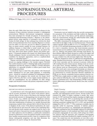
  • 7. © 2007 WebMD, Inc. All rights reserved. ACS Surgery: Principles and Practice 6 VASCULAR SYSTEM 17 INFRAINGUINAL ARTERIAL PROCEDURES — 7 the anterior tibial artery, or the peroneal artery, in that order of preference. As a rule, a tibial artery is used only if its lumen runs without obstruction into the foot, though bypasses to isolated tib- ial artery segments and other disadvantaged outflow tracts have been performed and have remained patent for more than 4 years.2,3 Generally, the peroneal artery is used only if it is continuous with one or two of its terminal branches, which communicate with foot arteries [see Figure 7]. Neither the absence of a plantar arch nor vas- cular calcification is considered a contraindication to a reconstruc- tion.2,7 With both femoropopliteal and infrapopliteal bypasses, stenosis of less than 50% of the diameter of the vessel is acceptable at or distal to the site chosen for the distal anastomosis. OPERATIVE TECHNIQUE Bypasses to tibial arteries should be performed with autogenous vein grafts, and either the reversed technique (as previously described [see Femoropopliteal Bypass, above]) or the in situ tech- nique [see In Situ Bypass, below] may be used. Placement of a tourniquet above the knee allows the distal anastomosis to be per- formed without extensive dissection of the tibial vessels or the application of clamps.36 Exposure of the inflow vessel (i.e., the femoral artery or the popliteal artery) is achieved in the same way as in femoropopliteal bypass. Accordingly, bypasses to tibial and peroneal arteries are best described in terms of the approaches required for exposure of these vessels and the tunnels required for routing the bypass conduits. Exposure of Posterior Tibial Artery The very proximal portion of the posterior tibial artery is aproached via a below-the-knee popliteal incision. The deep fascia Figure 7 Infrapopliteal bypass. An arteriogram from a 65-year- old female with rest pain in the right foot who underwent in situ bypass to the middle portion of the peroneal artery shows com- munication of the peroneal artery with foot arteries and reconsti- tution of the dorsalis pedis artery. is incised, and the popliteal space is entered. The gastrocnemius is retracted posteriorly, and the soleus is separated from the posterior surface of the tibia. The distal portion of the posterior tibial artery is approached via a medial incision along the posterior edge of the tibia [see Figure 8]; deepening this incision along the posterior tib- ialis muscle and the posterior surface of the tibia allows exposure of the posterior tibial artery.The tunnel from the popliteal fossa to the distal posterior tibial artery is made just below the muscle fascia, ideally with a long, gently curved clamp. Exposure of Anterior Tibial Artery To expose the proximal portion of the anterior tibial artery, an anterolateral incision is made in the leg midway between the tibia and the fibula over the appropriate segment of patent artery [see Figure 9a]. Additional small medial incisions are also required for tunneling. The anterior incision is carried through the deep fascia, and the fibers of the anterior tibial muscle and the extensor digito- rum longus are separated to reveal the neurovascular bundle. The Figure 6 Femoropopliteal bypass: below knee. A completion accompanying veins are mobilized and their branches divided to arteriogram from a patient who underwent below-the-knee allow visualization of the anterior tibial artery, which can then be femoropopliteal bypass for a nonhealing toe amputation site carefully mobilized [see Figure 9b]. With the artery mobilized, fur- shows runoff through all three tibial vessels. ther posterior dissection can be performed, and the interosseous
  • 8. © 2007 WebMD, Inc. All rights reserved. ACS Surgery: Principles and Practice 6 VASCULAR SYSTEM 17 INFRAINGUINAL ARTERIAL PROCEDURES — 8 Exposure of Peroneal Artery The peroneal artery is usually approached via the same incision as the posterior tibial artery [see Exposure of Posterior Tibial Artery, above].The artery is located and isolated just medial to the medial edge of the fibula. In its distal third, however, and in patients with stout calves and ankles, the peroneal artery should be approached via a lateral incision [see Figure 8], followed by excision of the fibula. For lateral exposure of the peroneal artery, a long segment of fibula is freed from its muscle attachments with a combination of blunt and sharp dissection; particular care should be taken in dis- secting along the medial edge of the bone because the peroneal vessels run just below this edge and are easily injured by instru- Figure 8 Infrapopliteal bypass: posterior tibial artery. Shown are ments. Next, a finger is passed around the fibula [see Figure 10a]; incisions for bypasses to the distal regions of the leg. once this is done, the free edge of bone is further developed by forcefully pushing a right-angle clamp inferiorly and superiorly [see Figure 10b]. A right-angle retractor is passed behind the bone, membrane can then be visualized and incised in a cruciate fashion. and the fibula is divided with a power saw. The peroneal artery Careful blunt finger dissection via the anterior incision and from can then be dissected free from surrounding veins and used in the the popliteal fossa via the medial incision facilitates creation of a construction of the distal anastomosis.Gentle blunt finger dissec- tunnel without injury to the numerous veins in the area [see Figure tion is required to develop a tunnel from this lateral wound to the 9c]. Alternatively, the tunnel for the bypass may be placed lateral to distal popliteal fossa, and great care should be taken to avoid the knee in a subcutaneous plane. injury to the numerous veins in the area. Because the peroneal The distal anterior tibial artery is approached via an anterior inci- artery is the least accessible of the three leg arteries used for sion placed midway between the tibia and the fibula [see Figure 8]. infrapopliteal bypasses and normally has the poorest connections A tunnel is made from the distal popliteal fossa across the with the arteries of the foot, we recommend that it be used as a interosseous membrane (like the tunnel to the peroneal artery) and distal implantation site only when the anterior and posterior tib- beneath the deep fascia to a point 5 to 7 cm above the lateral malle- ial arteries are not suitable. olus. Once the distal anastomosis is complete and the graft has been drawn through the tunnel, any tendons that may be distorting or Exposure of Dorsalis Pedis Artery compressing the graft in its course around the tibia are divided; this When no more proximal procedure is possible, a bypass to the measure proves necessary in most low anterior tibial bypasses. ankle region or the foot may be performed. The dorsalis pedis a b c Figure 9 Infrapopliteal bypass: anterior tibial artery. (a) An anterolateral incision is made midway between the tibia and the fibula over the artery; small medial incisions are also made for tunneling. (b) The anterior incision is carried through the deep fascia, the anterior tibial muscle and the extensor digi- torum longus are separated, the accompanying veins are mobilized and divided, and the anterior tibial artery is mobilized. (c) A tunnel is created with careful blunt finger dissection.
  • 9. © 2007 WebMD, Inc. All rights reserved. ACS Surgery: Principles and Practice 6 VASCULAR SYSTEM 17 INFRAINGUINAL ARTERIAL PROCEDURES — 9 a OUTCOME EVALUATION Infrapopliteal bypasses should have 5-year primary patency rates ranging from 60% to 67% and limb salvage rates ranging from 70% to 75% whether they are done with the reversed-vein tech- nique or with the in situ technique.38,39 For all such grafts, close patient follow-up and graft surveillance improve secondary paten- cy rates. Reduced complications and decreased length of stay have been reported for patients undergoing distal in situ bypasses using the endoscopic side-branch occlusion approach.40 Plantar Bypass Extension of the standard approaches to limb salvage has led to the performance of bypasses to vessels below the ankle joint [see Figures 13 and 14]. Such bypasses are indicated when the more proximal tibial vessels are occluded, which frequently occurs sec- ondary to failure of a more proximal bypass. The technique required for performing bypasses to secondary branches in the foot b is essentially the same as that required for performing bypasses to major infrapopliteal vessels. Optimal illumination by means of head lamps is important for achieving technical success with plantar bypass, and loupe magni- fication is helpful when the vessel is less than 1.5 mm in diameter. In addition, visualization of perimalleolar and inframalleolar arter- ies requires excellent preoperative imaging studies. These very distal bypasses offer a viable alternative to a major amputation. Like infrapopliteal bypasses, they are best described in terms of the anatomic approaches to the distal branch vessels. In what follows, we outline exposure of the plantar and tarsal arteries; exposure of the dorsalis pedis artery is outlined elsewhere [see Figure 10 Infrapopliteal bypass: lateral exposure of peroneal Infrapopliteal Bypass, above]. artery. Lateral exposure of the peroneal artery typically requires excision of part of the fibula; this is done by (a) passing a finger behind the fibula, developing the free bone edge further with a right-angle clamp, (b) passing a right-angle retractor behind the a fibula, and dividing the bone with a power saw. artery is easily approached via a lateral incision on the dorsum of the foot [see Figure 11a]. The incision is curved slightly and a flap raised so that the incision will not be directly over the anastomosis [see Figure 11b]. If the artery must be approached at the ankle, the extensor retinaculum must be divided. Otherwise, the operation is performed in much the same fashion as a distal anterior tibial bypass [see Figure 12].The posterior tibial artery can be approached down to a point several centimeters below the medial malleolus. b In Situ Bypass In situ bypass is an acceptable alternative to reversed vein bypass. In situ procedures were traditionally done through long skin incisions to access the GSV and its side branches. Subsequently, minimally invasive techniques were developed to reduce the wound complications encountered when an in situ bypass is performed via long skin incisions. Side-branch occlusion involves the use of an endoscopic vein harvesting system.Three skin incisions are made: two incisions for arterial access and one 2 cm incision above the knee for insertion of an endoscopic device to locate and clip the side branches of the saphenous vein. Once the proximal anastomosis is complete, the valves are lysed with a flexible valvulotome passed through the Figure 11 Infrapopliteal bypass: dorsalis pedis artery. (a) The distal end of the vein. Completion cineangiography is then per- dorsalis pedis artery is approached via an incision on the dorsum formed to confirm side-branch occlusion and to assess the entire of the foot. (b) The incision is curved and a flap raised so that the reconstruction.37 incision is not directly over the anastomosis.
  • 10. © 2007 WebMD, Inc. All rights reserved. ACS Surgery: Principles and Practice 6 VASCULAR SYSTEM 17 INFRAINGUINAL ARTERIAL PROCEDURES — 10 Posterior Tibial Artery Peroneal Artery Anterior Tibial Artery Lateral Tarsal Artery Dorsalis Pedis Arcuate Artery Deep Plantar Artery Lateral Plantar Artery Medial Plantar Artery Figure 13 Plantar bypass. Shown are the major arteries of the foot, including the two major branches of the posterior tibial artery.3 The lateral plantar artery is usually the larger and ends in the deep plantar arch. Exposure of the proximal 2 to 3 cm of the plantar branches is accomplished by incising the flexor retinaculum and the adductor muscle of the great toe. More distal exposure of these branches can be obtained by dividing the medial border of the plantar aponeu- rosis and the extensor digitorum brevis. Exposure of Deep Plantar Artery and Lateral Tarsal Artery The deep plantar artery and the lateral tarsal artery are branches of the dorsalis pedis artery. The deep plantar artery originates at the metatarsal level, where it descends into a fora- men bounded proximally by the dorsal metatarsal ligament, dis- Figure 12 Infrapopliteal bypass: dorsalis pedis artery. Shown is tally by the dorsal interosseous muscle ring, and medially and an arteriogram from a 72-year-old diabetic patient who under- went a popliteal artery–dorsalis pedis artery bypass with a reversed GSV graft for a nonhealing great toe amputation. OPERATIVE TECHNIQUE Exposure of Lateral and Medial Plantar Arteries The lateral and medial plantar branches are the continuation of the posterior tibial artery in the foot [see Figure 15].The lateral plan- tar artery forms the deep plantar arch and is larger than the medial plantar artery. If the lateral branch is occluded, the medial branch may enlarge and feed the plantar arch through collateral vessels. The initial incision is made over the termination of the posteri- or tibial artery below the malleolus.The artery is isolated, and the incision is extended inferiorly and laterally onto the sole. A direct approach to the individual branches is difficult, for several reasons. First, because the skin of the sole is not easily retracted, adequate exposure of the lateral and medial plantar arteries is hard to obtain if the incision does not follow their course exactly. Second, because these arteries are small in diameter and lie deep within the foot, they can be quite difficult to locate. Third, it is sometimes hard to distinguish the lateral plantar artery from the medial plantar artery. Figure 14 Plantar bypass. A completion arteriogram from a 62- Dissection of the termination of the posterior tibial artery can help year-old diabetic with nonhealing toe ulcers who underwent a the surgeon make this distinction. The lateral branch is usually popliteal artery–medial plantar artery bypass with a reversed located inferiorly when the foot is externally rotated on the operat- GSV graft shows the distal anastomosis, with flow visible through ing table. a small but patent medial plantar artery.
  • 11. © 2007 WebMD, Inc. All rights reserved. ACS Surgery: Principles and Practice 6 VASCULAR SYSTEM 17 INFRAINGUINAL ARTERIAL PROCEDURES — 11 hallucis brevis is retracted laterally—or, if necessary, transected— Lateral Plantar and the dorsal interosseous muscle ring is split to allow better Artery exposure of the proximal portion of the deep plantar artery. The periosteum of the proximal portion of the second metatarsal bone is then incised and elevated. A fine-tipped rongeur is used to excise enough of the metatarsal shaft to permit ample exposure of the deep plantar artery. OUTCOME EVALUATION Bypasses to the dorsalis pedis artery and its branches have yield- ed results comparable to those of bypasses to more proximal tibial vessels, with 3-year primary patency rates ranging from 58% to 60% and limb salvage rates ranging from 75% to 95%.41-43 In one review, patency rates were higher in patients who had an intact plantar arch than in those who did not; however, failure to visual- ize the plantar arch on preoperative arteriograms does not pre- clude the performance of these bypasses for limb salvage. With careful follow-up, the assisted primary patency rates for these grafts have been substantially improved.44 The available reports Medial Plantar Artery emphasize the need to repair failing grafts because their secondary patency was much better than that of failed grafts. In one study, Posterior patients who required shorter bypasses or had lower preoperative Tibial Artery C-reactive protein levels experienced significantly better out- comes.45 In some patients, occlusion of the distal tibial vessel Figure 15 Plantar bypass. Depicted is exposure of the distal por- tion of the posterior tibial artery. The lateral and medial plantar necessitates performance of a tibiotibial bypass to achieve wound arteries branch from this vessel and lie beneath the flexor reti- healing.7 naculum and the abductor hallucis, which can be incised. Bypasses to plantar or tarsal vessels performed with vein grafts yield 2-year patency rates ranging from 65% to 75% and limb sal- vage rates higher than 80%.4,5 In one report, the primary patency laterally by the bases of the first and second metatarsal bones. rate for these grafts was 74% at 1 year and 67% at 2 years, and the As the deep plantar artery exits from this tunnel, it connects limb salvage rate was 78% at 2 years.3 with the lateral plantar artery to form the deep plantar arch [see Figure 16]. A slightly curved longitudinal 3 to 4 cm incision is made over Alternative Bypasses Using More Distal Inflow Vessels the dorsum of the middle portion of the foot, and the dorsalis Traditionally, the femoral artery has been the inflow site of pedis artery is dissected distally down to its bifurcation into the choice for infrainguinal bypasses. Since the early 1980s, the super- deep plantar and first dorsal metatarsal branches. The extensor ficial femoral, deep femoral, popliteal, and tibial arteries have all Dorsalis Pedis Lateral Tarsal Artery Arcuate Artery Deep Plantar Artery Figure 16 Plantar bypass. Shown is a dorsal view of the arteries of the foot. Exposure of the distal dorsalis pedis artery (insert) highlights the origin of the deep plantar artery and its downward course between the first and second metatarsal bones. This exposure is facilitated by lateral retraction of the extensor hallucis brevis.
  • 12. © 2007 WebMD, Inc. All rights reserved. ACS Surgery: Principles and Practice 6 VASCULAR SYSTEM 17 INFRAINGUINAL ARTERIAL PROCEDURES — 12 been used as inflow sources when these vessels were relatively dis- tions of the GSV to be preserved for other purposes. ease free or when the amount of autologous vein available was lim- An increasing number of limb salvage procedures are secondary ited. Currently, the superficial femoral artery and the popliteal interventions.These secondary procedures are generally more dif- artery are preferentially used for primary bypasses when they are ficult to perform because the access routes to the arteries have free of disease. been previously dissected and because there frequently is little The strategy of utilizing more distal inflow sources is particu- good autologous vein left. In some cases, patients present with gan- larly applicable to inframalleolar bypasses, in which very long vein grene developing below a functioning bypass or after a previous segments would be required to reach the dorsalis pedis or other failed bypass. Some of these patients need nothing more than a pedal arteries from the usual more proximal inflow sites. Two short distal extension of their functioning bypass; others have only studies from the latter half of the 1980s reported that the paten- enough vein left to make up a short graft. For such patients, a tibi- cy rates in bypasses originating from the superficial femoral and otibial bypass may be an effective alternative revascularization popliteal arteries were comparable to those in bypasses originat- approach. ing from the common femoral artery.46,47 In a review of our own In 1994, our group reported our 11-year experience with tibi- experience with popliteal-distal vein graft bypasses,5 we reported otibial bypasses, comprising 42 procedures in 41 patients.7 Ten of a patency rate of 65% at 4 years—a figure comparable to rates these bypasses were performed because previous bypasses failed; reported for femorodistal bypasses with reversed or in situ vein the remainder were performed because the amount of autologous grafts (67% and 69%, respectively).38 Given these results, sur- vein available was limited. Approximately 50% of the bypasses geons should not hesitate to employ either the popliteal artery or were to pedal or tarsal vessels. At 5 years, the patency rate for these the superficial femoral artery as an inflow source. Use of these grafts was 65%, and the limb salvage rate was 73%.7 A subsequent more distal inflow sites results in shorter grafts and allows por- study reported comparable results.48,49 References 1. Veith FJ, Gupta SK, Wengerter KR, et al: duplex scan arterial mapping replace contrast arte- Femoral artery pressure measurement during aor- Changing arteriosclerotic disease patterns and riography as the test of choice before infrainguinal tography. Circulation 60:120, 1979 management strategies in lower-limb–threatening revascularization? J Vasc Surg 29:100, 1999 25. Hessel SJ, Adams DF, Abrams HL: Complications ischemia. Ann Surg 212:402, 1990 14. Ascher E, Hingorani A, Markevich N, et al. Lower of angiography. Radiology 138:273, 1981 2. Veith FJ, Gupta SK, Samson RH, et al: Progress in extremity revascularization without preoperative 26. Gomes AS, Baker JD, Martin-Paredero V, et al: limb salvage by reconstructive arterial surgery contrast arteriography: experience with duplex Acute renal dysfunction after major arteriography. combined with new or improved adjunctive proce- ultrasound arterial mapping in 485 cases. Ann Vasc AJR Am J Roentgenol 145:1249, 1985 dures. Ann Surg 194:386, 1981 Surg 16:108, 2002 27. Martin-Paredero V, Dixon SM, Baker JD, et al: 3. Ascer E,Veith FJV, Gupta SK: Bypasses to plantar 15. Mandolfino T, Canciglia A, D’Alfonso M, et al: Risk of renal failure after major angiography. Arch arteries and other tibial branches: an extended Infrainguinal revascularization based on duplex Surg 118:1417, 1983 approach to limb salvage. J Vasc Surg 8:434, 1988 ultrasound arterial mapping. Int Angiol 25:256, 2006 28. Nikonoff T, Skau T, Berglund J, et al: Effects of 4. Andros G: Bypass grafts to the ankle and foot: a femoral arteriography and low osmolar contrast personal perspective. Surg Clin North Am 75:715, 16. Carpenter JP, Owen RS, Baum RA, et al: Magnetic agents on renal function. Acta Radiol 34:88, 1993 1995 resonance angiography of peripheral runoff ves- sels. J Vasc Surg 16:807, 1992 29. Katholi RE, Taylor GJ, Woods WT, et al: 5. Wengerter KR,Yang PM, Veith FJ, et al: A twelve- Nephrotoxicity of nonionic low-osmolality versus year experience with the popliteal-to-distal artery 17. Fenlon HM, Yucel EK: Advances in abdominal, ionic high osmolality contrast media: a prospective bypass: the significance and management of prox- aortic, and peripheral contrast-enhanced MR double-blind randomized comparison in human imal disease. J Vasc Surg 15:143, 1992 angiography. Magn Reson Imaging Clin North beings. Radiology 186:183, 1993 Am 7:319, 1999 6. Veith FJ, Gupta SK, Samson RH, et al: Superficial 30. Lautin EM, Freeman NJ, Schoenfeld AH, et al: femoral and popliteal arteries as inflow site for dis- 18. Earls JP, Patel NH, Smith PA, et al: Gadolinium- Radiocontrast-associated renal dysfunction: a tal bypasses. Surgery 90:980, 1981 enhanced three-dimensional MR angiography of comparison of lower-osmolality and conventional the aorta and peripheral arteries: evaluation of a 7. Lyon RT, Veith FJ, Marsan BU, et al: Eleven-year high-osmolality contrast media. AJR Am J multistation examination using two gadopentetate experience with tibiotibal bypass: an unusual but Roentgenol 157:59, 1991 dimeglumine infusions. AJR Am J Roentgenol effective solution to distal tibial artery occlusive 171:599, 1998 31. Kram HB, Gupta SK, Veith FJ, et al: Late results disease and limited autologous vein. J Vasc Surg of two hundred seventeen femoropopliteal bypass- 19. Fellner F, Janka R, Fellner C, et al: Post occlusion 17:1128, 1994 es to isolated popliteal artery segments. J Vasc Surg visualization of peripheral arteries with “floating 8. Baker JD, Dix D:Variability of Doppler ankle pres- 14:386, 1991 table” MR angiography. Magn Reson Imaging sures with arterial occlusive disease: an evaluation 17:1235, 1999 32. Veith FJ, Gupta SK, Daly V: Femoropopliteal of ankle index and brachial-ankle pressure gradi- bypass to the isolated popliteal segment: is polyte- 20. Mell M,Tefera G,Thornton F, et al: Clinical utili- ent. Surgery 79:134, 1976 trafluoroethylene graft acceptable? Surgery ty of time-resolved imaging of contrast kinetics 9. Langsfeld M, Nupute J, Hershey FB, et al:The use 89:296, 1981 (TRICKS) magnetic resonance for infrageniculate of deep duplex scanning to predict hemodynami- arterial occlusive disease. J Vasc Surg 45:543, 2007 33. Taylor LM, Edwards JM, Porter JM: Present sta- cally significant aortoiliac stenosis. J Vasc Surg tus of reversed vein bypass grafting: five-year 21. Willmann JK, Baumert B, Schertler, et al: 7:395, 1988 results of a modern series. J Vasc Surg 11:193, Aortoiliac and lower extremity arteries assessed 10. Moneta GL, Yeager RA, Antonovic R, et al: 1990 with 16-detector row CT angiography: prospective Accuracy of lower extremity arterial duplex map- comparison with digital subtraction angiography. 34. Veith FJ, Gupta SK, Ascer E, et al: Six year ping. J Vasc Surg 15:275, 1992 Radiology 236:1083, 2005 prospective multicenter randomized comparison 11. Legemate DA, Teeuwen C, Hoenveld H, et al: 22. Mishra A, Bhaktrahalli JN, Ehtuish EF: Imaging of of autologous saphenous vein and expanded poly- Value of duplex scanning compared with angiog- peripheral arteries by 16-row multidetector com- tetrafluoroethylene grafts in infrainguinal arterial raphy and pressure measurement in the assess- puted tomography angiography: a feasible tool? reconstructions. J Vasc Surg 3:104, 1986 ment of aortoiliac lesions. Br J Surg 78:1003, 1991 Eur J Radiol 61:528, 2007 35. Jordan WD, Voellinger DC, Schroeder PT, et al: 12. Ascher E, Mazzariol F, Hingorani A, et al:The use 23. Willmann JD, Mayer D, Banyai M et al: Video-assisted saphenous vein harvest: the evalua- of duplex ultrasound arterial mapping as an alter- Evaluation of peripheral arterial bypass grafts with tion of a new technique. J Vasc Surg 26:405, 1997 native to conventional arteriography for primary multi-detector row CT angiography: comparison 36. Wagner WH, Treiman RL, Cossman DV, et al: and secondary infrapopliteal bypasses. Am J Surg with duplex US and digital subtraction angiogra- Tourniquet occlusion technique for tibial artery 178:162, 1999 phy. Radiology 229:465, 2003 reconstruction. J Vasc Surg 18:637, 1993 13. Wain RA, Berdejo GL, Delvalle WN, et al: Can 24. Brewster DC, Waltman AC, O’Hara PJ, et al: 37. Suggs WD, Sanchez LA, Woo D, et al:
  • 13. © 2007 WebMD, Inc. All rights reserved. ACS Surgery: Principles and Practice 6 VASCULAR SYSTEM 17 INFRAINGUINAL ARTERIAL PROCEDURES — 13 Endoscopically assisted in situ lower extremity 42. Pomposelli FB, Kansal N, Hamdan AD et al: A 47. Rosenbloom JS, Walsh JJ, Schuler JJ, et al: Long- bypass: a preliminary report of a new minimally Decade of experience with dorsalis pedis artery term results of infragenicular bypasses with auto- invasive technique. J Vasc Surg 34:668, 2001 bypass: analysis of outcome in more than 1000 genous vein originating from the distal superficial 38. Bergamini TM, Towne JB, Bandyk DF, et al: cases. J Vasc Surg 37:307, 2003 femoral and popliteal arteries. J Vasc Surg 7:691, Experience with in situ saphenous vein bypasses 43. Panayiotopoulos YP, Tyrrell MR, Arnold FJ, et al: 1988 during 1981 to 1989: determinant factors of long- Results and cost analysis of distal [crural/pedal] 48. Plecha EJ, Lee C, Hye RJ: Factors influencing the term patency. J Vasc Surg 13:137, 1991 arterial revascularization for limb salvage in dia- outcome of paramalleolar bypass grafts. Ann Vasc 39. Wengerter KR, Veith FJ, Gupta SK, et al: betic and nondiabetic patients. Diabet Med Surg 10:356, 1996 Prospective randomized multi center comparison 14:214, 199 49. Hughes K, Domenig CM, Hamdan AD, et al: of in situ and reversed vein infrapopliteal bypasses. 44. Rhodes JM, Gloviczki P, Bower TC, et al:The ben- Bypass to plantar and tarsal arteries: an acceptable J Vasc Surg 13:189, 1991 efits of secondary interventions in patients with approach to limb salvage. J Vasc Surg 40:1149, 40. Piano G, Schwartz LB, Foster L, et al: Assessing failing or failed pedal bypass grafts. Am J Surg 2004 outcomes, costs, and benefits of emerging tech- 178:151, 1999 nology for minimally invasive saphenous vein in 45. Biancari F, Alback A, Kantonen I, et al: Predictive situ distal arterial bypasses. Arch Surg 133:613, factors for adverse outcome of pedal bypasses. Eur 1998 J Vasc Endovasc Surg 18:138, 1999 Acknowledgments 41. Schneider JR, Walsh DB, McDaniel MD, et al: 46. Cantelmo NL, Snow JR, Menzoian JO, et al: Pedal bypass versus tibial bypass with autogenous Successful vein bypass in patients with an ischemic Figures 1, 4, 8 through 11, 13, 15, and 16 Tom vein: a comparison of outcome and hemodynamic limb and a palpable popliteal pulse. Arch Surg Moore. Revised and updated by Thom Graves, C.M.I. results. J Vasc Surg 17:1029, 1993 121:217, 1986 Figures 2, 3, and 5 Alice Y. Chen.