Weitere ähnliche Inhalte
Ähnlich wie Acs0607 Diabetic Foot
Ähnlich wie Acs0607 Diabetic Foot (20)
Mehr von medbookonline (20)
Acs0607 Diabetic Foot
- 1. © 2007 WebMD, Inc. All rights reserved. ACS Surgery: Principles and Practice
6 VASCULAR SYSTEM 7 DIABETIC FOOT — 1
7 DIABETIC FOOT
Cameron M. Akbari, M.D., F.A.C.S., and Frank W LoGerfo, M.D., F.A.C.S.
.
Evaluation and Management of the Diabetic Foot
Surgeons caring for diabetic patients are faced with a diverse spec- ment of infection, or, most commonly, correction of arterial insuf-
trum of foot disease.1,2 The clinical presentation may range from ficiency. In some cases, the present ulcer has already healed at least
the asymptomatic patient who requires nothing more than preven- once, and the current episode represents a relapse. A history of
tive foot care to the unstable and critically ill patient in whom both intermittent healing followed by relapse raises the possibility of
loss of limb and death are imminent threats. This wide range of underlying untreated infection (e.g., recurrent osteomyelitis) or an
disease severity, coupled with inappropriate and untimely use of uncorrected architectural abnormality (e.g., a bony pressure point
diagnostic testing, contributes to the clinical confusion that often or a varus deformity).
leads to delays in diagnosis and treatment and, ultimately, to limb In view of the many vague and unsupported therapies advocat-
loss. It is important, therefore, that surgeons caring for diabetic ed for the diabetic foot, it is helpful to know the type and duration
patients develop a simple but comprehensive and orderly of any treatments the patient has already undergone for the cur-
approach to diabetic foot problems that (1) can be implemented rent problem. For example, a patient with an ischemic ulceration
for any such problem, (2) recognizes the pathogenic roles of neu- may have completed several different courses of antibiotics with-
ropathy, ischemia, and infection, and (3) emphasizes the initial out success. Thus, if antibiotic therapy is contemplated for a cur-
clinical assessment at the bedside.3 rent infection, possible antibiotic resistance must be taken into
account in choosing the appropriate agent. It is also helpful to
know which treatments were not previously offered to the patient
Clinical Evaluation and to look critically at why they were not offered. For example,
Evaluation of any diabetic foot many diabetic patients with correctable foot ulceration and limb
problem begins with a complete histo- ischemia are told that their only option is limb amputation, usual-
ry and a careful physical examination. ly because of the physician’s inherited pessimism or his or her
Broadly speaking, such evaluation inadequate knowledge of the advances made in limb and foot sal-
should address the healing potential vage. No patient should be denied an opportunity for limb salvage
of the foot, the details of the foot prob- on the basis of a previous medical or surgical opinion without a
lem (e.g., ulcer, gangrene, infection, new comprehensive evaluation being performed.
or osteomyelitis), the systemic conse- Inquiries should be made about any previous foot and limb
quences of diabetes, and any immedi- problems—for instance, whether the patient had other ulcers on
ate threats to life or limb.With this information in hand, the surgeon the same foot that healed spontaneously, how long such healing
can usually make an accurate diagnosis and start a comprehensive took, and whether foot surgery was ever performed on that side. A
treatment plan without having to order further diagnostic tests, history of recent ipsilateral ulceration or foot surgery that healed
which are liable to be both costly and time-consuming. in a timely and uncomplicated manner suggests, but does not
prove, that the arterial supply is adequate. Problems and proce-
HISTORY
dures more remote in time, however, are less useful indicators. A
The history of the foot problem can yield valuable insights into history of previous leg revascularization (including percutaneous
the potential for healing, the presence of coexisting infection or therapies) is also an important clue to possible underlying arterial
arterial occlusive disease, and the need for further treatment. insufficiency. Because of the predilection for mirror image–type
Whenever a patient presents with a foot ulceration or gangrene, atherosclerotic occlusive disease, the contralateral leg must be
possible underlying arterial insufficiency should immediately be considered as well: previous revascularization in the opposite leg is
suspected, even if neuropathy or infection is present. It is helpful to often associated with arterial insufficiency on the affected side.
be aware of the event that incited the foot problem. In a patient Other cardiovascular risk factors, such as cigarette smoking and
with diabetes and arterial insufficiency, the inciting event for a foot hyperlipidemia, must also be taken into account: their presence
ulcer may be a seemingly benign action, such as cutting a toenail, increases the likelihood that ischemia is contributing to the current
soaking the foot in a warm bath, or applying a heating pad. Unfor- foot problem.
tunately, because of the broad microneurovascular and macrovas- Although claudication and rest pain have traditionally been
cular abnormalities associated with diabetes, these relatively inno- associated with vascular disease, they may be obscured by diabet-
cuous actions can progress to a nonhealing ulcer and gangrene. ic neuropathy; hence, their absence in the diabetic patient certain-
Similarly, failure to heal after any podiatric procedure is strongly ly does not rule out ischemia. Because even moderate ischemia
suggestive of underlying unrecognized arterial insufficiency. precludes healing in the diabetic foot, the absence of rest pain is
The duration of the ulcer also provides important clues, in that not a reliable indicator of an adequate arterial blood supply; more-
a long-standing, nonhealing ulcer is strongly suggestive of over, many patients may not be walking long enough distances for
ischemia. Certainly, an ulcer or gangrenous area that has been pre- vasculogenic claudication to develop. Conversely, some patients
sent for several months is unlikely to heal without some further with true ischemic rest pain are dismissed for years as having
treatment, whether it be offloading of weight-bearing areas, treat- “painful neuropathy.”
- 2. © 2007 WebMD, Inc. All rights reserved. ACS Surgery: Principles and Practice
6 VASCULAR SYSTEM 7 DIABETIC FOOT — 2
Diabetic patient has foot problem
Obtain history: inciting event, duration, healing, previous
treatment (including surgery), vascular disease and risk
factors, overall health, functional status.
Evaluation and Management Perform physical examination: signs of infection, diabetic
neuropathy, pulses, arterial perfusion.
of the Diabetic Foot Assess clinical findings to determine direction of
subsequent workup and treatment.
Infection is absent Infection is present, and patient
is medically stable
Start antibiotics, correct metabolic abnormalities,
and drain and debride as indicated.
Assess salvageability of foot.
Foot is salvageable
Evaluate foot for ischemia.
Ischemia is absent Ischemia is present
Obtain arteriogram of entire lower extremity.
Perform revascularization.
Provide continued wound care: dressings, debridement as
necessary, adjunctive measures, correction of metabolic
abnormalities, antibiotics as needed.
Determine whether secondary (definitive) foot surgery is needed.
Secondary foot surgery is indicated
Proceed with foot operation.
Foot is not healed Foot is healed
Reevaluate for infection (see above), Provide preventive foot care.
and run through algorithm again.
- 3. © 2007 WebMD, Inc. All rights reserved. ACS Surgery: Principles and Practice
6 VASCULAR SYSTEM 7 DIABETIC FOOT — 3
Patient is in a septic state and is
medically unstable
Guillotine amputation is not indicated Guillotine amputation is indicated
Correct metabolic abnormalities, and
stabilize patient.
Foot is not salvageable
Perform formal below- or above-the-knee
amputation.
Secondary foot surgery is not indicated
Continue wound care.
- 4. © 2007 WebMD, Inc. All rights reserved. ACS Surgery: Principles and Practice
6 VASCULAR SYSTEM 7 DIABETIC FOOT — 4
a b
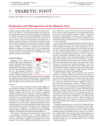
Figure 1 Shown is a benign-appearing gangrenous eschar (a) on the foot of a diabetic patient. Plain
x-ray (b) reveals extensive subcutaneous air in soft tissue, consistent with severe necrotizing fasciitis.
Both the systemic effects of the foot problem and the systemic ambulatory and rehabilitative potential. Many different methods
consequences of diabetes itself should be assessed. Because unrec- of quantifying functional status have been suggested. One simple
ognized infection in the diabetic patient may rapidly progress to a approach is to classify functional status as a point on a continuum.
life-threatening condition, attention should be directed toward de- One end of the continuum might be a fully ambulatory patient;
tecting the subtle manifestations of an infected foot ulcer. The pa- almost all surgeons would recognize that such a patient should be
tient should be asked about worsening hyperglycemia, recent errat- offered every attempt at limb salvage. The other end might be a
ic blood glucose control, and higher insulin requirements. As a con- completely bedridden patient with multiple comorbid conditions;
sequence of the microvascular and neuropathic abnormalities in most surgeons would adopt a far less aggressive approach to such
the diabetic foot, classic symptoms of infection (e.g., chills and pain) a patient. In practice, many patients fall somewhere between these
are often absent, and hyperglycemia is often the sole presenting symp- two extremes, in which case a more thorough evaluation of func-
tom of undrained infection. Faced with ongoing infection and hyper- tional and social status becomes imperative before any firm deci-
glycemia, the surgeon should strongly suspect impending ketoaci- sions can be made regarding limb salvage.
dosis or nonketotic hyperglycemic hyperosmolar coma, with the atten-
PHYSICAL EXAMINATION
dant symptoms of weakness, confusion, and altered mental status.
Because a patient with a diabetic foot problem often needs some Fever and tachycardia are strongly suggestive of deep or undrained
type of operative intervention, the history should also include a infection, with hypotension being a late manifestation of ongoing
comprehensive assessment of overall health status to help stratify sepsis. It is important to remember, however, that these signs may
perioperative risk [see ECP:6 Risk Stratification, Preoperative Testing, be absent in diabetic patients with impending or progressive infec-
and Operative Planning]. For example, knowledge of previous car- tion. A focused cardiopulmonary examination helps confirm the
diac events (e.g., myocardial infarction or revascularization) and presence or absence of congestive heart failure, valvular abnormal-
current cardiac status (e.g., New York Heart Association [NYHA] ities, or rhythm disturbances, which must be recognized in diabet-
class or anginal severity) can help determine whether perioperative ic patients with poor underlying medical reserve.
cardiac monitoring or preoperative cardiac testing is indicated and Evaluation of the diabetic foot ulcer should include a strong
what form such monitoring or testing should take. Similarly, in a suspicion of infection and a thorough search for it. In a patient
patient with suspected infection and ischemia, a history of wors- with cellulitis, the entire foot, including the web spaces and the
ening renal function or impending need for hemodialysis can help nail beds, should be examined for any potential portals of entry,
determine the choice and dosage of antibiotics and may alter plans such as a puncture wound or an interdigital (“kissing”) ulcer.
for standard contrast arteriography.The essential point is that dia- Encrusted and heavily calloused areas over the ulceration should
betes may affect virtually every organ system, often in an indolent be unroofed and the wound thoroughly inspected to determine
pattern; thus, in the evaluation of any diabetic patient with foot the extent of involvement. A benign-appearing dry gangrenous
disease, attention must be paid to all of these systems. eschar often hides an undrained infectious collection [see Figure
Functional status also becomes an important consideration at 1]. Cultures should be taken from the base of the ulcer; super-
this point, and the history should carefully determine the patient’s ficial swabs may yield only colonizing organisms.
- 5. © 2007 WebMD, Inc. All rights reserved. ACS Surgery: Principles and Practice
6 VASCULAR SYSTEM 7 DIABETIC FOOT — 5
Findings consistent with infection include purulent drainage, tip of a digit), ulceration unassociated with an exostosis or a
crepitus, tenderness, mild erythema, and sinus formation [see weight-bearing area, and gangrene are all strongly consistent with
Figure 2], though these findings may be entirely absent in the neu- underlying ischemia [see Figure 4].The presence of multiple ulcer-
ropathic foot. With more advanced and deep infection, edema ations or gangrenous areas on the foot, the absence of granulation
may be present as a result of elevated pressures within one or more tissue, and the lack of bleeding with debridement of the ulcer
of the plantar compartments [see Figure 3]. If left untreated, this should immediately be taken as signals of possible underlying
process may spread proximally along tendon sheaths to involve arterial insufficiency. Other signs suggestive of ischemia are pallor
the ankle or even the calf. Close inspection of the ulcer and the with elevation, fissures (particularly at the heel), and the absence
use of a sterile probe may also confirm the presence of osteomye- of hair growth. Poor skin condition and hyperkeratosis, though
litis, which occurs commonly even in conjunction with benign- not always good indicators of arterial disease, should be noted
appearing ulcers. If bone is detected with gentle probing, osteo- because they may help confirm initial clinical impressions.
myelitis is presumed present. The pulse examination, including the status of the foot pulses, is
Because of its prevalence and its causative role in diabetic foot the single most important component of the physical examination.
ulceration and limb loss, neuropathy should be assessed in every In the absence of a palpable pulse, ischemia is always presumed to
diabetic patient, and appropriate preventive measures should be be present. Although an accurate pulse examination of the lower
taken to guard against foot ulceration in the high-risk neuropath- extremities is not difficult, it is an acquired skill, and time should
ic foot. Protective sensation may be evaluated by pressing a be devoted to practicing and perfecting the technique.
Semmes-Weinstein 5.07 monofilament against the skin; inability The femoral pulse is palpated midway between the superior
to feel the monofilament correlates well with an increased risk of iliac spine and the pubic tubercle, just below the inguinal liga-
foot ulceration. Advanced sensorimotor neuropathy will lead to ment. The popliteal pulse is palpated with both hands and with
the presence of a so-called claw foot (from gradual atrophy of the the knee flexed no more than 15°. Palpation of the foot pulses is
intrinsic muscles) or to Charcot degeneration with bone and joint somewhat more demanding, requiring close attention and a good
destruction at the midfoot. Both of these conditions give rise to knowledge of the usual locations of the native arteries. The dor-
abnormal pressure points on the plantar prominences and the salis pedis is located between the first and second metatarsal
potential for foot ulceration. bones, just lateral to the extensor hallucis longus tendon, and its
Assessment of the arterial perfusion in the diabetic foot is a fun- pulse is palpated with the pads of the fingers as the hand is par-
damental consideration, in that the diabetic foot needs maximal tially wrapped around the foot [see Figure 5]. If the pulse cannot
perfusion to heal. In the presence of ischemia, all efforts at limb be palpated, the fingers may be moved a few millimeters in each
salvage will fail.Therefore, the physical examination must include direction; the dorsalis pedis occasionally follows a slightly aberrant
a systematic approach to the assessment of arterial insufficiency. course. A common mistake is to place a single finger at one loca-
Simple inspection of the leg and foot, including the ulcer, often tion on the dorsum of the foot. The posterior tibial artery is typi-
provides suggestive clues. For example, distal ulceration (on the cally located in the hollow curve just behind the medial malleolus,
Figure 2 Purulent drainage is seen from a sub- Figure 3 Shown is wet gangrene with edema and
metatarsal ulcer on the plantar surface of the foot of undrained severe infection in the foot of a diabetic patient
a diabetic patient. presenting with hyperglycemia and ketoacidosis.
- 6. © 2007 WebMD, Inc. All rights reserved. ACS Surgery: Principles and Practice
6 VASCULAR SYSTEM 7 DIABETIC FOOT — 6
environment (e.g., hospitalized or outpatient). Certain general
assumptions can be made about likely causative organisms. Mild
localized and superficial ulcerations, particularly in outpatients,
are usually caused by aerobic gram-positive cocci (e.g.,
Staphylococcus aureus and streptococci). In contrast, deeper ulcers
and generalized limb-threatening infections are usually polymicro-
bial. In addition to gram-positive cocci, potential causative organ-
isms include gram-negative bacilli (e.g., Escherichia coli, Klebsiella,
Enterobacter aerogenes, Proteus mirabilis, and Pseudomonas aerugi-
nosa) and anaerobes (e.g., Bacteroides fragilis and peptostreptococ-
ci). Enterococci may also be isolated from the wound, notably in
hospitalized patients; in the absence of other cultured virulent
organisms, they should probably be considered pathogenic.
Currently, it is clear that resistant organisms, particularly methi-
cillin-resistant S. aureus (MRSA), are playing a growing role in the
development of skin and soft tissue infections.Traditionally arising
in patients who had previously been hospitalized and those who
had previously received antibiotic therapy, MRSA-associated
infections are now frequently encountered in outpatient settings.
Indeed, in many U.S. cities, these so-called community-acquired
MRSA infections are the most common skin and soft tissue infec-
tions seen in patients presenting to the emergency department.6
Accordingly, in both outpatient and inpatient settings, it is advis-
able to assume that MRSA is present in a patient with a diabetic
foot infection until the culture data suggest otherwise. Awareness
Figure 4 Shown is dry gangrene of several toes in a of the increasing prevalence of resistant organisms is critical for
diabetic patient with femoropopliteal and tibial arterial current management of diabetic foot infections, especially with
occlusive disease. respect to the initiation of antibiotic coverage.
Initially, the choice of antibiotics is made empirically on basis of
approximately halfway between the malleolus and the Achilles ten- these general assumptions. When the results of the initial cultures
don.The examiner’s hand should be contralateral to the examined become available, antibiotic coverage may be broadened or nar-
foot (i.e., the right hand should be used to palpate the left foot, and rowed as appropriate. In a compliant patient with a small ulcer
vice versa), so that the curvature of the hand naturally follows the and no evidence of deep space involvement or systemic infection,
contours of the ankle [see Figure 6]. treatment may be delivered on an outpatient basis. A dual-antibi-
otic regimen (pending culture results) is instituted, typically con-
sisting of a cephalosporin or a β-lactam antibiotic (for activity
Assessment of Clinical Findings against staphylococci and streptococci) and trimethoprim-sul-
Once the clinical evaluation is famethoxazole or a tetracycline (for activity against MRSA). A
complete, the next step is to assess dual regimen consisting of fluoroquinolone and linezolid is an
the findings from the history and the acceptable alternative that also provides adequate coverage. In
physical examination so as to deter- addition, the patient is instructed to offload weight from the
mine the course and urgency of the
subsequent workup and treatment.
This assessment is made at the bed-
side, focusing on three main con-
cerns: (1) the presence and severity
of infection, (2) the salvageability of the limb, and (3) the presence
of ischemia.
PRESENCE OF INFECTION
Evaluation for and treatment of infection is the first priority in
the management of any diabetic foot problem.4,5 Although radi-
ographic tests may confirm initial clinical suspicions, the determi-
nation of the severity of infection is almost always made on the
basis of clinical findings. Infection in the diabetic foot may range
from a minimal superficial infection to fulminant sepsis with ex-
tensive necrosis and destruction of foot tissue. Accordingly, the
treatment plan should address the choice of antibiotic (which
requires knowledge of the microbiology), the need for drainage,
the option of local or even guillotine amputation, and the patient’s
overall medical condition.
The microbiology of the diabetic foot varies according to the Figure 5 Illustrated is the correct technique for palpation of the
depth and severity of the infection and the nature of the patient’s dorsalis pedis pulse.
- 7. © 2007 WebMD, Inc. All rights reserved. ACS Surgery: Principles and Practice
6 VASCULAR SYSTEM 7 DIABETIC FOOT — 7
those with limb-threatening or life-threatening infections, appro-
priate empirical antibiotic options include (1) vancomycin plus a
β-lactam antibiotic with a β-lactamase inhibitor (e.g., piperacillin-
tazobactam) and (2) vancomycin plus metronidazole plus a
quinolone (in cases of penicillin allergy). Subsequent culture
results then dictate further antibiotic coverage, if any. In the
absence of osteomyelitis, antibiotics should be continued until the
wound appears clean and all surrounding cellulitis has resolved
(typically, 10 to 14 days). If osteomyelitis is present, treatment
should include both surgical debridement and a prolonged (4- to
6-week) course of antibiotic therapy (though the course may be
abbreviated if the entire infected bone has been removed, as with
digital or transmetatarsal amputation). Heel lesions often present
with some degree of calcaneal destruction, and determination of
osteomyelitis may be made by clinical examination either alone or
in conjunction with other radiographic tests (e.g., magnetic reso-
nance imaging) [see Figure 7].
In the presence of an abscess or deep space infection, immedi-
ate incision and drainage of all infected tissue planes is mandato-
Figure 6 Illustrated is the correct technique for finding the pos- ry. Incisions should be chosen with an eye to the normal anatomy
terior tibial pulse.
of the foot (including the various compartments) and the need for
subsequent secondary (foot salvage) procedures [see Figure 8].
involved extremity and is taught appropriate methods for chang- Drainage should be complete, with incisions placed to allow for
ing wound dressings. Frequent follow-up is vital, and guidelines dependent drainage, and all necrotic tissue must be debrided.
should be imparted by which improvement or worsening of the Repeat cultures (including both aerobes and anaerobes) should be
lesion can be determined. obtained from the deep tissues. Drainage incisions on the dorsum
A more common presentation, unfortunately, is a patient with of the foot should be avoided. Abscesses in the medial, central, or
ulceration or gangrene who has a deep infection affecting tendon lateral compartment should be drained via longitudinal incisions
or bone and possible systemic involvement. For such patients, made in the direction of the neurovascular bundle and extending
immediate hospitalization is indicated, including bed rest with ele- the entire length of the abscess. The medial and central compart-
vation of the infected foot, correction of any systemic abnormali- ments are drained through a medial incision, and the lateral com-
ties, and broad-spectrum I.V. antibiotic therapy (which may be partment is drained through a lateral incision; both of these inci-
focused more tightly once culture results are complete). Because sions are made just above the plantar surface of the forefoot [see
the clinical findings of impending sepsis may be subtle, these Figure 9]. Web space infections may be drained similarly through
patients should undergo a complete laboratory workup aimed at the plantar aspect of the foot. In some instances, open amputation
detecting and correcting electrolyte and acid-base imbalances. of the foot (e.g., an open toe or transmetatarsal amputation) may
The choice of antibiotic agent and the duration of therapy are be necessary to allow complete drainage and resection of necrot-
dependent on the extent of the infection. For patients with deep ic tissue. Strict adherence to textbook amputations may lead to
or chronic recurrent ulcers, which are typically polymicrobial, or unnecessary soft tissue removal and possibly to a higher amputa-
a b
Figure 7 Shown is heel ulceration with eschar (a) in a
diabetic patient. MRI findings (b) are consistent with
calcaneal osteomyelitis.
- 8. © 2007 WebMD, Inc. All rights reserved. ACS Surgery: Principles and Practice
6 VASCULAR SYSTEM 7 DIABETIC FOOT — 8
tion during future closure; therefore, all viable tissue should be
conserved.
A patient with an ongoing undrained infection may present
with an unsalvageable foot and fulminant sepsis, manifested by
hemodynamic instability, bacteremia, and severe acid-base and
electrolyte abnormalities. Such patients should undergo prompt
open (guillotine) below-the-knee amputation [see 6:20 Lower-
Extremity Amputation for Ischemia].This type of amputation is usu-
ally performed at the ankle level, with the aim of removing the sep- Metatarsal
tic source while allowing for revision and closure at a later date. Medial Head
Administration of I.V. antibiotics, correction of dehydration and Intermuscular
electrolyte abnormalities, and continuous cardiac monitoring are Septum
absolutely essential throughout treatment.
Once the infection has been drained and tissues debrided, con-
tinued wound inspection and management are essential. Ongoing Vascular Lateral
Arch Compartment
necrosis should raise the possibility of undrained infection or
untreated ischemia, in which case further debridement and treat-
ment may be necessary. Avoidance of weight-bearing should be
Lateral
continued. Neither whirlpool therapy nor soaks are beneficial. Medial
Intermuscular
Compartment
Septum
Medical Stabilization
Concomitant with the measures outlined above to control Central
infection, medical stabilization of the diabetic patient must be car- Compartment
ried out, and the surgeon must be directly involved in this process.
Hyperglycemia is almost always seen when infection is present; it
should be gradually corrected. More advanced hyperglycemia
leads to ketotic or nonketotic hyperosmolar states, which carry a Calcaneus
10% to 25% mortality. Serum concentrations of electrolytes, mag-
nesium, and creatinine should be obtained and osmolality deter-
mined at frequent intervals; any abnormalities should be correct-
ed[see 8:24 Disorders of Water and Sodium Balance and 8:8 Acid-Base
Figure 8 The foot has three plantar compartments: medial, cen-
Disorders]. Dehydration is common in hyperglycemic patients and
tral, and lateral. The intrinsic muscles of the great toe are in the
should be corrected. A urinary catheter is mandatory to help guide medial compartment, and those of the fifth toe are in the lateral
the response to fluid therapy; in unstable patients, a central venous compartment. The central compartment contains the intrinsic
pressure catheter or a pulmonary arterial catheter may be needed muscles of the second through fourth toes, the extensor flexor
[see 8:4 Cardiovascular Monitoring]. Continuous cardiac monitor- tendons of the toes, the plantar nerves, and the plantar vascular
ing is essential in patients with the hyperglycemic hyperosmolar structures. The floor of each compartment is the rigid plantar
syndrome or ketoacidosis. fascia; the roof is composed of the metatarsal bones and
interosseous fascia. The medial and central compartments are
SALVAGEABILITY OF LIMB separated by the medial intermuscular septum, which extends
While the infection is being treat- from the medial calcaneal tuberosity to the head of the first
metatarsal. The lateral and central compartments are separated
ed and controlled, the surgeon
by the lateral intermuscular septum, which extends from the cal-
should determine whether limb sal- caneus to the head of the fifth metatarsal.
vage is feasible.This determination is
based largely on the patient’s func-
tional status and the degree of foot
PRESENCE OF ISCHEMIA
destruction. For example, primary
limb amputation may be considered Evaluation of the diabetic foot for
in a nonambulatory, bedridden pa- ischemia begins with the history and
tient or in a patient with severe Charcot the physical examination. By the
destruction and degeneration, for whom no further reconstructive conclusion of the clinical evaluation,
foot surgery is possible. Poor medical condition, by itself, is not the surgeon can usually make an
necessarily an indication for primary limb amputation, given the accurate assessment of the adequacy
high perioperative morbidity associated with amputation. More- of the arterial circulation to the foot.
over, it is often possible to improve the patient’s overall medical As noted [see Clinical Evaluation,
status while he or she is being treated for infection and evaluated Physical Examination, above], the
for ischemia. absence of a palpable foot pulse strongly suggests ischemia unless
Assessment of limb salvageability should be carried out simul- proved otherwise.
taneously with treatment of infection because appropriate Noninvasive arterial testing plays only a limited role in the assess-
drainage and antibiotics can dramatically change the appearance ment of diabetic patients with foot ulceration and should not be
and viability of the foot. If limb salvage is not deemed possible, the employed in place of the bedside evaluation. Certainly, for patients
patient should undergo formal below-the-knee or above-the-knee with poor healing or gangrene and absent foot pulses, noninvasive
amputation [see 6:20 Lower-Extremity Amputation for Ischemia]. testing adds little additional useful information to the initial clinical
- 9. © 2007 WebMD, Inc. All rights reserved. ACS Surgery: Principles and Practice
6 VASCULAR SYSTEM 7 DIABETIC FOOT — 9
evaluation and typically serves only to further delay vascular recon- soon as infection has started to resolve and signs of systemic toxic-
struction. For selected patients, however, noninvasive testing, in ity have disappeared; prolonged delays in making this decision may
conjunction with the clinical findings, may be useful. Patients who result in further tissue loss and make salvage impossible. For a
might benefit from such testing include those with absent foot puls- complete assessment, the arteriogram should include the foot ves-
es who have a superficial ulcer with evidence of healing or a previ- sels in both lateral and anterior views.
ous history of a healed foot ulcer and those without any foot lesions Concern over possible contrast-induced renal failure should not
who are scheduled to undergo elective foot surgery. be considered a contraindication to high-quality angiography of the
The presence of diabetes imposes limitations on all of the nonin- entire lower extremity.The incidence of contrast nephropathy is not
vasive arterial tests currently available. Medial arterial calcinosis higher in diabetic patients without preexisting renal disease, even
occurs frequently and unpredictably in patients with diabetes and when ionic contrast agents are employed. Selective use of magnet-
can result in noncompressible arteries and artificially elevated seg- ic resonance angiography (MRA), carbon dioxide angiography, and
mental systolic pressures and ankle-brachial indices. Because calcifi- duplex scanning may help minimize the contrast dye load.
cation levels tend to be lower in the toe vessels, toe systolic pressures In patients with preexisting renal disease, use of a nonionic con-
are sometimes obtained, but their value is often limited by the prox- trast agent, in conjunction with appropriate hydration and judi-
imity of the foot ulcer to the cuff site. Segmental Doppler waveforms cious administration of kidney-protecting agents, should minimize
and pulsed volume recordings are unaffected by medial calcification, the risk of contrast-induced nephrotoxicity. N-acetylcysteine, 600
but evaluation of these waveforms is primarily qualitative rather than mg twice daily, should be started on the day before the arterio-
quantitative. In addition, the quality of the waveforms is affected by gram and continued for 48 hours, and 0.45 N saline should be
peripheral edema, and cuff placement is commonly affected by the given I.V. at a rate of 1 mg/kg/hr, beginning 12 hours before the
presence of ulceration. Regional transcutaneous oximetry measure- scheduled arteriogram.7
ments are also unaffected by medial calcinosis, and this modality
appears to be reliable for predicting ulcer healing and amputation
levels. However, transcutaneous oximetry measurements are actual- Management
ly higher in patients with diabetes and foot ulcers than in nondiabet-
REVASCULARIZATION
ic patients, possibly because of the effects of arteriovenous shunting.
Other limitations, including lack of equipment standardization, user The goals of arterial reconstruc-
variability, and a large gray area in the interpretation of the measure- tion are to restore maximal perfusion
ments, further limit the ability of this test to predict ischemia.There- to the foot and, ideally, to restore a
fore, although these measurements have been used to predict heal- palpable foot pulse. Possible ap-
ing in patients without diabetes, high values may not correlate with proaches include endovascular tech-
healing potential in the presence of diabetes. niques (angioplasty and stenting) [see
When a patient presents with absent foot pulses in conjunction 6:19 Endovascular Procedures for
with gangrene, nonhealing ulceration, or significant tissue loss, it Lower-Extremity Disease], bypass grafting (with autogenous or pros-
can be assumed that ischemia is present and must be corrected if thetic grafts), and some combination of the two. Ultimately, the
the limb is to be salvaged. In such patients, arteriography of the choice of procedure in any given case is based on the individual
entire lower extremity should be performed. The decision to per- patient’s anatomy, the comorbid conditions present, and the results
form arteriography and vascular reconstruction should be made as of the preoperative assessment, with the aim being to provide the
a b
Figure 9 Shown is an infection in the central compartment of the foot from a submetatarsal ulcer
that has extended proximally to include the medial compartment (a). A plantar incision through the
ulcer and both compartments (including the septum) allows complete dependent drainage (b).
- 10. © 2007 WebMD, Inc. All rights reserved. ACS Surgery: Principles and Practice
6 VASCULAR SYSTEM 7 DIABETIC FOOT — 10
most durable procedure with the least risk. For example, in rare the diabetic foot is usually attributable to unrecognized ischemia,
patients with isolated iliac artery stenoses, angioplasty may be effec- ongoing infection, or poor conventional wound care—not, as a rule,
tive by itself, but in patients with multilevel disease, it may have to to the absence of more sophisticated wound therapy.
be combined with an infrainguinal bypass [see 6:17 Infrainguinal Hyperglycemia and malnutrition are common in hospitalized
Arterial Procedures]. In patients who have previously undergone sur- diabetic patients with foot ulceration, and both adversely affect
gical revascularization, careful consideration should be given to the wound healing. Correction of these abnormalities should begin
availability of an autogenous conduit (e.g., an arm vein or the lesser early and continue throughout the wound-healing period.
saphenous vein), given the superiority of autogenous vein over pros- Attention should also be directed toward preventing new foot
thetic grafts. lesions (e.g., decubitus ulcers on the heel from prolonged bed
As a rule, arterial bypass grafting is required for restoration of rest). Heel splints, air mattresses, and leg pillows are all valuable
the foot pulse.8 Proximal bypass to either the popliteal artery or in this regard.
the tibial and peroneal arteries may restore foot pulses, and pref-
SECONDARY FOOT PROCEDURES
erence should be given to these vessels if they are in continuity
with the foot. Often, however, because of the presence of more dis- After successful revasculariza-
tal obstructive disease, bypass grafting to the popliteal or even the tion, secondary procedures on the
tibial artery will not restore the foot pulse. In such cases, restora- foot may be performed for maxi-
tion of pulsatile flow to the foot may be accomplished with auto- mal foot salvage, with the aim of
genous vein bypass grafts to the paramalleolar or inframalleolar addressing both the acute prob-
arteries (e.g., the dorsalis pedis).The vein graft can be prepared as lem and the underlying cause.
an in situ graft, a reversed graft, or a nonreversed graft, without The basic goals of such proce-
any significant difference in outcome; the choice of approach dures are (1) to remove infected
should be based on the patient’s particular vascular anatomy. bone (if present), (2) to restore
As noted (see above), the absence of an ipsilateral greater saphe- functional stability, and (3) to
nous vein is not a contraindication to pedal bypass: comparable reduce the risk of subsequent ulceration.
results may be obtained by using arm vein or lesser saphenous vein In the forefoot, digital, single metatarsal (ray), or transmetatarsal
grafts. Prosthetic material should seldom, if ever, be used for dor- amputation [see 6:20 Lower-Extremity Amputation for Ischemia] may
salis pedis or other extreme distal (inframalleolar) bypass grafts. be performed, depending on the location of the ulcer. Tissue
Active infection in the foot is not a contraindication to pedal should be handled gently, forceps should not be used, and incisions
bypass, provided that the proximal dorsum of the foot is free of should be carefully placed. Care must be taken to prevent dead
infection and that incisions can be placed in clean tissue planes. spaces along tendon sheaths, bone splintering, and areas of resid-
The foot should be free of cellulitis, lymphangiitis, and edema ual necrosis, all of which can lead to failure of the procedure. If
before any inframalleolar bypass. there is persistent infection or an abscess, the wound should be left
Current advances in endovascular therapy are allowing this open. Every effort, however, should be made to perform a closed
approach to be used more frequently and successfully in the amputation so as to avoid the time and cost associated with open
peripheral vasculature. Lesions determined to be favorable on the wound care. This is a particularly important consideration in the
basis of arteriography (e.g., short-segment stenoses) may be treat- occasional diabetic patient with unreconstructible foot ischemia,
ed with angioplasty and, possibly, stenting. Similarly, endovascular because open amputations rarely heal in the presence of ischemia.
therapy may be preferred to surgical bypass grafting in high-risk Underlying bony structural abnormalities in the diabetic foot
patients and patients who have no suitable autogenous conduit. are often the cause of ulceration. Such abnormalities may be cor-
Ultimately, the goal of endovascular intervention should be the rected with hallux arthroplasty, metatarsal head resection,
same as that of bypass grafting: to restore the foot pulse and max- metatarsal osteotomy, or sesamoidectomy; if ulceration is present,
imal blood flow to the foot. these procedures may be combined with ulcer excision. Similarly,
ulceration on a previous transmetatarsal amputation may be the
CONTINUED WOUND CARE
result of an equinovarus deformity (from disrupted tendons and
Once the foot is fully revascular- a decrease in calcaneal inclination).This problem should be treat-
ized—or once it is clear that the foot ed with revision of the transmetatarsal amputation (perhaps in
was adequately perfused to begin conjunction with ulcer excision) and biomechanical correction
with—care of the foot wound should (e.g., Achilles tendon lengthening or, in more severe cases, poste-
be continued. Revascularization of a rior tibial tendon release).
severely ischemic foot ulcer may result Heel lesions are particularly formidable, and there is consider-
in an immediate paradoxical worsen- able confusion regarding how best to manage them. Generally, dry
ing of the infection, and frequent in- eschars with no evidence of deep infection or abscess may be treat-
spection is mandatory postoperatively. ed with offloading alone so as to allow healing beneath the eschar
New-onset cellulitis, hyperglycemia, or fever should prompt concern in the fully revascularized foot. In patients with chronic ulceration
regarding potential worsening of the foot infection. Frequent de- or osteomyelitis, partial calcanectomy may be considered.The pres-
bridement to healthy, bleeding tissue is required more often than not. ence of calcaneal osteomyelitis may be determined by means of
Wounds should be kept moist, with wet-to-dry dressings avoided probing or adjunctive studies such as MRI [see Figure 7]. Primary
in favor of normal saline wet-to-wet dressings. Various adjunctive closure is occasionally possible, but given the relatively fixed nature
wound treatments are available, including chemical enzymatic of the heel, either secondary healing or some type of flap coverage
debriding agents, growth factors, and hyperbaric oxygen therapy. is usually indicated. Both local and free flaps may be used in the
Some of these may offer a slight additional benefit in terms of fully revascularized foot.
improved healing, but they are expensive; blanket use of these cost- After any type of surgery of the diabetic foot, frequent post-
ly modalities is therefore discouraged. Failure of wound healing in operative observation of the wound is mandatory. Delays in
- 11. © 2007 WebMD, Inc. All rights reserved. ACS Surgery: Principles and Practice
6 VASCULAR SYSTEM 7 DIABETIC FOOT — 11
healing or wound breakdown should immediately raise the sandals, and using caustic over-the-counter foot medications
possibility of infection or unrecognized ischemia and should should all be strongly discouraged.
trigger the appropriate workup. Neuropathy continues to be the most common cause of diabet-
ic foot ulceration, and its presence is a strong predictor of the like-
PREVENTIVE FOOT CARE
lihood of future ulceration. As noted (see above), inability to feel a
Once the foot has healed, preven- Semmes-Weinstein 5.07 monofilament when it is pressed to the
tive measures should be initiated to skin correlates well with an increased risk of foot ulceration. In
prevent future ulceration. Foremost addition, abnormal pressure points (secondary to mechanical
among these measures is patient deformity of the foot and motor neuropathy) can be identified by
education, focusing on general means of pedobarography, a procedure in which plantar pressure
hygiene (e.g., daily washing and is measured on specialized contour plots as the patient walks on a
moisturizer use) and daily inspection pressure-sensitive platform. Identification of any high-pressure
of the feet. Walking barefoot, em- areas can facilitate the construction of specific, custom-molded
ploying heat pads, wearing thong orthotics and insoles to help in offloading.
References
1. LoGerfo FW, Coffman JD: Vascular and microvas- 13:671, 1996 Engl J Med 343:180, 2000
cular disease of the foot in diabetes. N Engl J Med 5. Joshi N, Caputo GM, Weitekamp MR, et al:
311:1615, 1984 8. Akbari CM, LoGerfo FW: Distal bypasses in the
Infections in patients with diabetes mellitus. N Engl
diabetic patient. Current Techniques in Vascular
2. Akbari CM, LoGerfo FW: Diabetes and peripheral J Med 341:1906, 1999
vascular disease. J Vasc Surg 30:373, 1999 Surgery. Yao JST, Pearce WH, Eds. McGraw-Hill,
6. Moran GJ, Krishnadasan A, Gorwitz RJ, et al:
3. Caputo GM, Cavanagh PR, Ulbrecht JS, et al: Methicillin-resistant S. aureus infections among New York, 2001, p 285
Assessment and management of foot disease in patients in the emergency department. N Engl J
patients with diabetes. N Engl J Med 331:854, 1994 Med 355:666, 2006
4. Fox HR, Karchmer AW: Management of diabetic 7. Tepel M, van der Giet M, Schwarzfeld C, et al: Acknowledgment
foot infections, including the use of home intra- Prevention of radiographic-contrast-agent-induced
venous antibiotic therapy. Clin Podiatr Med Surg reductions in renal function by acetylcysteine. N Figure 8 Tom Moore.