Weitere ähnliche Inhalte
Ähnlich wie Sgarboza (20)
Mehr von piratadelcielo@hotmail.com challenger (20)
Kürzlich hochgeladen (20)
Sgarboza
- 1. Copyright, 1996, by the Massachusetts Medical Society
Volume 334 FEBRUARY 22, 1996 Number 8
ELECTROCARDIOGRAPHIC DIAGNOSIS OF EVOLVING ACUTE MYOCARDIAL INFARCTION
IN THE PRESENCE OF LEFT BUNDLE-BRANCH BLOCK
ELENA B. SGARBOSSA, M.D., SERGIO L. PINSKI, M.D., ALEJANDRO BARBAGELATA, M.D.,
DONALD A. UNDERWOOD, M.D., KATHY B. GATES, ERIC J. TOPOL, M.D., ROBERT M. CALIFF, M.D.,
AND G ALEN S. W AGNER , M.D., FOR THE GUSTO-1 (G LOBAL U TILIZATION OF S TREPTOKINASE
AND T ISSUE P LASMINOGEN A CTIVATOR FOR O CCLUDED C ORONARY A RTERIES ) I NVESTIGATORS
branch block. The three electrocardiographic criteria with
Abstract Background. The presence of left bundle-
independent value in the diagnosis of acute infarction in
branch block on the electrocardiogram may conceal the
these patients were ST-segment elevation of 1 mm or
changes of acute myocardial infarction, which can delay
more that was concordant with (in the same direction as)
both its recognition and treatment. We tested electrocar-
the QRS complex; ST-segment depression of 1 mm or
diographic criteria for the diagnosis of acute infarction in
more in lead V1, V2, or V3; and ST-segment elevation of
the presence of left bundle-branch block.
Methods. The base-line electrocardiograms of patients 5 mm or more that was discordant with (in the opposite
enrolled in the GUSTO-1 (Global Utilization of Streptoki- direction from) the QRS complex. We used these three
nase and Tissue Plasminogen Activator for Occluded criteria to develop a scoring system (0 to 5), which al-
Coronary Arteries) trial who had left bundle-branch block lowed a highly specific diagnosis of acute myocardial in-
and acute myocardial infarction confirmed by enzyme farction to be made.
studies were blindly compared with the electrocardio- Conclusions. We developed and validated a clinical
grams of control patients who had chronic coronary prediction rule based on a set of electrocardiographic cri-
artery disease and left bundle-branch block. The elec- teria for the diagnosis of acute myocardial infarction in
trocardiographic criteria for the diagnosis of infarction patients with chest pain and left bundle-branch block.
were then tested in an independent sample of patients The use of these criteria, which are based on simple ST-
presenting with acute chest pain and left bundle-branch segment changes, may help identify patients with acute
block. myocardial infarction, who can then receive appropriate
Results. Of 26,003 North American patients, 131 (0.5 treatment. (N Engl J Med 1996;334:481-7.)
1996, Massachusetts Medical Society.
percent) with acute myocardial infarction had left bundle-
T HE optimal use of coronary reperfusion therapies signs have gained widespread acceptance. Many physi-
relies on a rapid diagnosis of evolving myocardial cians believe that acute myocardial injury cannot be
infarction.1,2 For most patients presenting with cardiac detected accurately in patients with left bundle-branch
block.10 We examined the value of the standard electro-
chest pain, the electrocardiogram is a powerful aid in
cardiogram for the diagnosis of acute myocardial in-
diagnosing the cause of the pain and selecting the ap-
propriate therapy.2 In patients who present with con- farction in the presence of left bundle-branch block in
a large population of patients.
comitant left bundle-branch block, however, the elec-
trocardiographic manifestations of acute myocardial
METHODS
injury may be masked. During the past five decades,
Derivation Sample
several electrocardiographic signs have been proposed
to aid in the diagnosis of infarction in such patients, but Criteria for the diagnosis of acute myocardial infarction in the
because of methodologic limitations,3-9 none of these presence of left bundle-branch block were developed from two pop-
ulations (the study and control groups), which constituted the deri-
vation sample. The study group consisted of the subgroup of North
American patients enrolled in the GUSTO-1 (Global Utilization of
From the Cleveland Clinic Foundation, Cleveland (E.B.S., S.L.P., D.A.U.,
Streptokinase and Tissue Plasminogen Activator for Occluded Cor-
E.J.T.); the Fundación Favaloro, Buenos Aires, Argentina (A.B.); and Duke Uni-
onary Arteries) trial11 who had acute myocardial infarction docu-
versity Medical Center, Durham, N.C. (K.B.G., R.M.C., G.S.W.). Address reprint
mented by serum enzyme changes and evidence of complete left bun-
requests to Dr. Sgarbossa at the Department of Cardiology, Desk M-24, Cleve-
dle-branch block on their base-line electrocardiograms. The control
land Clinic Foundation, Cleveland, OH 44195.
group was assembled by randomly selecting from the Duke Data-
Supported by grants from Bayer, Genentech, CIBA–Corning, ICI Pharmaceu-
ticals, and Sanofi Pharmaceuticals. bank for Cardiovascular Disease an equal number of patients with
Downloaded from www.nejm.org on March 20, 2009 . Copyright © 1996 Massachusetts Medical Society. All rights reserved.
- 2. 482 THE NEW ENGLAND JOURNAL OF MEDICINE Feb. 22, 1996
stable, angiographically documented coronary artery disease and com- Table 2. Selected Characteristics of Patients with Left Bundle-
plete left bundle-branch block.12 The patients in the control group Branch Block.*
did not have acute chest pain at the time of the electrocardiographic
recording. PATIENTS WITH CONTROL
ACUTE MI PATIENTS
Electrocardiograms were digitally obtained at 100 Hz with a speed
CHARACTERISTIC (N 131) (N 131) P VALUE
of 25 mm per second and an amplification of 10 mm per millivolt.
Median age — yr (25th, 75th 68.5 (62, 76) 68.0 (62, 74) 0.63
Definition of Left Bundle-Branch Block percentiles)
Male sex — no. (%) 84 (64) 78 (60) 0.52
The normal sequence of ventricular activation (depolarization and
repolarization) can be altered by both left bundle-branch block and Left-axis deviation — no. (%)† 37 (28) 63 (48) 0.002
acute myocardial injury. To achieve a reasonable compromise be- Previous MI — no. (%) 34 (26) 77 (59) 0.001
tween the electrocardiographic changes inherent in the two condi- Median time to randomization — 120 (90, 180) — —
tions,13 we used the following definition of left bundle-branch block: a min (25th, 75th percentiles)
QRS duration of at least 0.125 second in the presence of sinus or su-
praventricular rhythm, a QS or rS complex in lead V1, and an R-wave *MI denotes myocardial infarction.
peak time of at least 0.06 second in lead I, V5, or V6 associated with †A left-axis deviation was defined as a QRS axis of at least 30 degrees.
the absence of a Q wave in the same lead.7,14-16 Patients with electro-
cardiograms showing intermittent left bundle-branch block were ex-
cluded from the study. agreement was at least 0.7 and the univariate analysis showed statis-
tical significance (P 0.1) were included in a stepwise logistic-regres-
Interpretation of Electrocardiograms sion model to identify independent criteria that were significant pre-
dictors of acute infarction (P 0.05). A scoring system for the diagnosis
All the electrocardiograms were analyzed for signs of myocardial
of infarction was developed from the coefficients assigned by the logis-
injury by one of four investigators who were unaware of both the pa-
tic model for each independent criterion, on a scale of 0 to 5. Receiv-
tient’s identity and the clinical variables. The signs examined 3-9,16-20
er-operating-characteristic curves, kappa statistics, and correlation
are shown in Table 1. The ST-segment deviation was measured at the
coefficients were estimated with True Epistat software.23 Chi-square
J point. To assess interobserver variability, a random sample of 10
tests and logistic-regression analyses were performed with Egret soft-
percent of the electrocardiograms were read by all four investigators.
ware.24
The diagnostic performance of the model was then tested in an in-
Statistical Analysis
dependent population of 45 patients with left bundle-branch block,
Interobserver agreement, as estimated with the kappa statistic, acute chest pain, and a high likelihood of coronary artery disease (the
was greater than 0.85 for both QRS-complex and T-wave polarities. validation sample). Included in this sample were patients in the
For both the terminal S wave in leads V5 and V6 and notching of at GUSTO-1 registry and the GUSTO-2A (Global Use of Strategies to
least 0.05 second of the S wave in lead V3, the agreement was 0.77, Open Occluded Coronary Arteries) study and the patients from
but it was low ( 0.55) for lead V4, as well as for notching of at least GUSTO-1 subsequently found to have normal creatine kinase MB
0.05 second of the R wave in lead I, aVL, V5 , or V6. Measurements values.
of ST-segment deviations were closely correlated among the four in-
vestigators (r 0.9 for all comparisons, by Pearson’s correlation coef- RESULTS
ficient).
Of the base-line electrocardiograms from the 26,003
All electrocardiographic criteria were screened by univariate anal-
ysis (chi-square tests) to identify those associated with acute myocar- North American patients, 145 met the criteria for left
dial infarction. For ST-segment elevations, the receiver-operating-
bundle-branch block. The diagnosis of acute myocar-
characteristic curves were constructed to evaluate the diagnostic
dial infarction was confirmed by studies showing elevat-
performance of different combinations of the degree of elevation and
ed creatine kinase MB levels in 131 of the patients. The
number of leads exhibiting such elevation. Because a high specificity
is desirable for the diagnosis of acute myocardial infarction,21,22 we de- electrocardiograms from these 131 patients were ana-
termined prospectively that the optimal cutoff point would corre-
lyzed for the presence of the aforementioned diagnos-
spond to the most sensitive degree of ST-segment elevation that had
tic criteria. Selected base-line characteristics of the pa-
a specificity of at least 90 percent. Criteria for which the interobserver
tients with acute myocardial infarction and the control
patients are shown in Table 2.
Table 1. Electrocardiographic Criteria Analyzed in Patients with Univariate Analysis
Left Bundle-Branch Block.
On the basis of the receiver-operating-characteris-
tic curves, the maximal sensitivity with the target
ST-segment elevation and its concordance or discordance with the QRS complex
in every lead
specificity ( 90 percent) was achieved when at least
ST-segment depression in every lead
one lead exhibited ST-segment elevation of at least
Presence of Q waves in two contiguous precordial leads or in two limb leads
1 mm that was concordant with (in the same direction
(Q waves were considered present if they were 20 msec in lead V4
as) the QRS complex or at least 5 mm that was dis-
or 30 msec in lead I, II, aVL, V5, or V6, regardless of width in
leads V1 to V3)
cordant with (in the opposite direction from) the QRS
R-wave regression from leads V1 to V4
complex.
QS pattern from leads V1 to V4
Electrocardiographic criteria with statistical signifi-
Positive T waves in lead V5 or V6
cance for the diagnosis of acute myocardial infarction
Notching 0.05 sec in the ascending limb of the S wave in lead V3 or V4 (sign
and their sensitivities, specificities, and likelihood ratios
of Cabrera)
are listed in Table 3. The likelihood ratios indicate to
Notching 0.05 sec in the ascending limb of R waves in lead I, aVL, V5, or V6
(sign of Chapman)
what extent a particular criterion will increase or de-
Terminal S wave in lead V5 or V6
crease the probability of infarction. The electrocardio-
Left-axis deviation
graphic criterion with the highest likelihood ratio was
Downloaded from www.nejm.org on March 20, 2009 . Copyright © 1996 Massachusetts Medical Society. All rights reserved.
- 3. Vol. 334 No. 8 LEFT BUNDLE-BRANCH BLOCK AND DIAGNOSIS OF MYOCARDIAL INFARCTION 483
an index score 3) in both samples
Table 3. Results of the Univariate Analysis of Electrocardiographic Criteria.*
are shown in Table 5.
POSITIVE NEGATIVE
DISCUSSION
SENSITIVITY SPECIFICITY LIKELIHOOD LIKELIHOOD
(95% CI) (95% CI)
CRITERION RATIO (95% CI) RATIO (95% CI)
With the advent of effective re-
percent
perfusion therapies, a rapid and ac-
ST-segment elevation 1 mm 73 (64–80) 92 (86–96) 9.54 (3.1–17.3) 0.3 (0.22–0.39) curate diagnosis of acute myocardial
and concordant with QRS
infarction has become essential.22,25
complex
Thrombolytic agents are most bene-
ST-segment depression 1 mm 25 (18–34) 96 (91–99) 6.58 (2.6–16.1) 0.78 (0.7–0.87)
ficial when administered promptly,
in lead V1, V2, or V3
ST-segment elevation 5 mm 31 (23–39) 92 (85–96) 3.63 (2.0–6.8) 0.75 (0.67–0.86) and their erroneous use in patients
and discordant with QRS
with unstable angina or noncardi-
complex
ac chest pain is potentially danger-
Positive T wave in lead V5 26 (19–34) 92 (86–96) 3.42 (0.18–65) 0.8 (0.72–0.9)
ous.11,26,27 Algorithms and predictive
or V6
models developed to identify patients
Left-axis deviation 72 (63–79) 48 (39–57) 1.38 (1.13–9.8) 0.59 (0.25–1.39)
with ongoing myocardial ischemia or
*CI denotes confidence interval.
infarction28-34 do not apply to patients
with left bundle-branch block. The
ST-segment elevation of at least 1 mm in leads with a new appearance of bundle-branch block in a patient with
QRS complex in the same direction. Similarly, the ab- acute chest pain is highly suggestive of infarction, and is-
sence of this criterion was associated with the lowest chemic changes superimposed on a pattern of chronic
likelihood ratio. left bundle-branch block are easy to recognize when a
previous electrocardiogram is available for comparison.
Multivariate Analysis and Index Scores for the Independent The timely availability of a previous electrocardiogram,
Electrocardiographic Criteria
however, is the exception rather than the rule. Physicians
All five electrocardiographic criteria associated with usually must decide to administer appropriate treatment
acute infarction in the presence of left bundle-branch or perform further testing on the basis of only the most
block were included in a stepwise multiple logistic- recent electrocardiographic information. Our study sug-
regression model along with the variable “previous gests that in patients with left bundle-branch block, an
myocardial infarction.” The model identified three in- examination of the electrocardiogram obtained at the
dependent predictors of acute myocardial infarction time of presentation allows a diagnosis of acute myocar-
(Table 4 and Fig. 1). Figure 2 shows the predicted prob- dial infarction to be made with a high degree of con-
ability of infarction for each combination of criteria. A fidence.
more accurate estimate of the probability of infarction We found that ST-segment deviation was the only
can be obtained when the weight of each criterion, as electrocardiographic finding that was useful in the diag-
determined by the logistic model, is used to develop a nosis of acute myocardial infarction in the presence of
simple index score (Table 4). left bundle-branch block. Previously proposed electro-
The operating characteristics of the scoring system cardiographic signs involving the QRS complex were not
for the independent criteria provide a high discrimina- useful. These discrepancies may be due to the fact that
tive power for the diagnosis of infarction (area under we analyzed electrocardiograms obtained on admission,
the curve, 0.874) (Fig. 3). For an accurate diagnosis, a thus detecting changes in the ST-segment (an early man-
specificity of 90 percent requires a minimal total score ifestation of acute coronary occlusion) rather than chang-
of 3. Patients presenting with ST-segment elevation of at es in the QRS complex (which indicate necrosis). Fur-
least 5 mm in leads with a QRS complex in the oppo- thermore, the GUSTO-1 trial included patients with
site direction (score, 2) should therefore probably un- acute myocardial infarction, whereas previous studies of
dergo further testing. patients with left bundle-branch block have not always
differentiated between acute and chronic infarction.6-8
Testing of the Electrocardiographic Criteria in the Validation Uncomplicated left bundle-branch block is character-
Sample
ized by secondary repolarization changes in a direction
The criteria derived from the model and their index
scores were tested in the validation sample, which in- Table 4. Odds Ratios and Scores for Independent Electrocardio-
cluded 22 patients with enzymatic evidence of acute ne- graphic Criteria.
crosis and 23 with only unstable angina (the control CRITERION ODDS RATIO (95% CI) SCORE
group). As compared with the results in the derivation
ST-segment elevation 1 mm and concordant 25.2 (11.6–54.7) 5
sample, the operating characteristics of the scoring sys-
with QRS complex
tem for the three criteria combined had a lower dis- ST-segment depression 1 mm in lead V1, V2, 6.0 (1.9–19.3) 3
criminative power for the diagnosis of infarction (area or V3
ST-segment elevation 5 mm and discordant 4.3 (1.8–10.6) 2
under the curve, 0.7). The sensitivities, specificities, with QRS complex
likelihood ratios, and predictive values of the criteria (for
Downloaded from www.nejm.org on March 20, 2009 . Copyright © 1996 Massachusetts Medical Society. All rights reserved.
- 4. 484 THE NEW ENGLAND JOURNAL OF MEDICINE Feb. 22, 1996
opposite that of the main QRS de-
flection.9 In leads with a predomi-
nantly negative QRS complex, the
I aVR V1 V4
result is an electrocardiographic pat-
tern of ST-segment elevation with
positive T waves, which is similar to
the current of injury observed dur-
ing acute coronary occlusion. Studies
of patients with left bundle-branch
II aVL V2 V5
block that examined serial electro-
cardiographic changes during either
acute myocardial infarction35-37 or oc-
clusion of a coronary artery by an
angioplasty balloon38,39 have shown
III aVF V3 V6 that further ST-segment elevation oc-
curs in those leads.
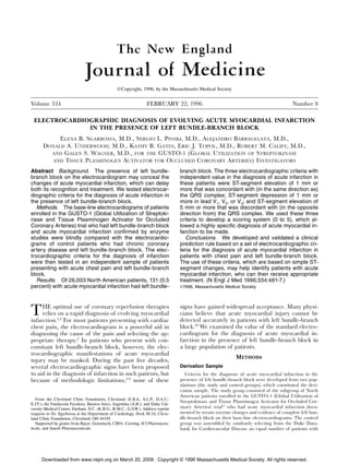
Figure 1. Electrocardiogram Meeting All Three Independent Criteria for the Diagnosis Our challenge was to determine
of Acute Myocardial Infarction in a Patient from the GUSTO Trial with Left Bundle-
the cutoff point for the ST-segment
Branch Block.
elevation that would most effective-
The electrocardiogram shows ST-segment elevation of at least 1 mm that is concord-
ly discriminate between patients with
ant with the QRS complex (lead II), ST-segment depression of at least 1 mm in leads
acute myocardial infarction and those
V2 and V3, and ST-segment elevation of at least 5 mm that is discordant with the QRS
complex (leads III and aVF). without infarction, in the absence
of information from previous or seri-
al electrocardiographic tracings. We
found that for leads with a predomi-
Patient with chest pain and LBBB
nantly negative QRS complex, ST-
segment elevation of at least 5 mm
identified patients with evolving in-
Is there ST-segment depression 1 mm that
farction. On the other hand, ST-seg-
is concordant with the QRS complex?
ment elevation in the same direction
as that of the QRS complex is not
expected in patients with uncompli-
Yes No
cated left bundle-branch block. We
found that any degree of ST-segment
Is there ST-segment elevation 1 mm in elevation in a lead with a positive
lead V1, V2, or V3? QRS complex was a highly specific
sign of acute myocardial infarction.
Likewise, ST-segment depression in
lead V1, V2, or V3 should not be
Yes No Yes No
present in patients with uncomplicat-
ed left bundle-branch block, since the
QRS complex is predominantly nega-
Is there ST-segment elevation 5 mm that
tive in those leads. In our study, ST-
is discordant with the QRS complex?
segment depression in lead V1, V2, or
V3 was also an independent marker
of acute myocardial infarction. The
mechanism for this finding is unclear;
Yes No Yes No Yes No Yes No
it could be a manifestation of true
posterior-wall infarcts (i.e., due to oc-
4/0 22/2
No. of 26/2 43/6 1/0 6/3 9/9 20/109
patients/
clusion of the left circumflex artery)40
controls
or infarcts associated with ST-seg-
Probability 100 92 93 88 100 66 50 16
of MI (%) ment depression (subendocardial in-
Total score 10 8 7 5 5 3 2 0
farcts).41
The presence of left bundle-branch
block in patients with acute myocar-
Figure 2. Flow Chart for the Prediction of Acute Myocardial Infarction in the Pres-
ence of Left Bundle-Branch Block, with the Use of All Possible Combinations of the dial infarction is associated with an
Three Independent Electrocardiographic Criteria.
increased risk of complications and
The discriminatory power of each combination of criteria for the diagnosis of acute
death.42 When it is new, left bundle-
myocardial infarction is indicated by the total score at the bottom, with higher scores
branch block is correlated with the
indicating better discriminatory power. LBBB denotes left bundle-branch block, and
occlusion of the proximal left anteri-
MI myocardial infarction.
Downloaded from www.nejm.org on March 20, 2009 . Copyright © 1996 Massachusetts Medical Society. All rights reserved.
- 5. Vol. 334 No. 8 LEFT BUNDLE-BRANCH BLOCK AND DIAGNOSIS OF MYOCARDIAL INFARCTION 485
graphic change is observed. On the other hand, the
1.0
sole presence of ST-segment elevation of at least 5 mm
2
0.9
3 that is discordant with the QRS complex (with a score
0.8
5 of 2) indicates a moderate-to-high probability of myo-
0.7
cardial infarction, and further procedures should be un-
Sensitivity
0.6
dertaken to confirm the diagnosis.
0.5 The index score correctly classified 84 percent of the
7
0.4 patients in the derivation sample but only 67 percent of
Area under curve 0.874 the patients in the validation sample. A poorer per-
0.3
formance of diagnostic criteria in the validation sample
8
0.2
is not unexpected49 and in our study is related to the
0.1
relatively low sensitivity of the criteria. One reason for
10
0.0
the decreased sensitivity is the difference in the two pa-
0.0 0.2 0.4 0.6 0.8 1.0
tient populations. Analysis of data from the GUSTO-
1 Specificity
1 sample (the source of the derivation sample) yielded
Figure 3. Receiver-Operating-Characteristic Curve for the Com- inflated sensitivities, because the study included only
bined Score for the Three Independent Electrocardiographic
patients with electrocardiographic signs of acute myocar-
Criteria.
dial infarction, whereas the GUSTO-2A sample and the
The zone to the left of the broken line indicates a high probability
GUSTO-1 registry (the sources of most of the patients
of acute myocardial infarction. The numbers along the curve are
in the validation sample) included patients with other
scores.
ischemic syndromes as well.13
It should be noted that the sensitivity of each individ-
or descending artery and a large amount of jeopardized ual electrocardiographic criterion in our study is also
myocardium.43 On the other hand, a prior left bundle- low but is similar to the sensitivity of ST-segment
branch block is a powerful marker of depressed left changes in patients with normal intraventricular con-
ventricular systolic function,44,45 and any additional loss duction.25,39 Our purpose was to improve the identifica-
of myocardium is likely to result in cardiogenic shock. tion of patients with acute infarction, because they may
It is therefore not surprising that subgroup analyses in benefit from thrombolytic therapy; to this end, a high
specificity (rather than a high sensitivity) is required.21
trials of thrombolytic therapy show a benefit of treat-
ment in patients with bundle-branch block.46 The Fibri- The high specificity of our index score in the validation
sample (96 percent) may have a medicolegal benefit.50
nolytic Therapy Trialists’ Collaborative Group analyzed
the results of nine randomized studies and reported a Electrocardiograms that are misread or considered non-
dramatic 25 percent decrease in mortality at 35 days diagnostic may result in a failure to diagnose infarc-
tion,51,52 and claims of a missed infarction account for
among 2032 patients with right or left bundle-branch
block treated with thrombolysis.26 a substantial proportion of malpractice claims involv-
ing emergency departments.51 Highly specific criteria
The relatively small number of patients with left
bundle-branch block enrolled in these trials, however, may help physicians rapidly diagnose and treat acute in-
attests to the prevailing diagnostic uncertainty. The farction in patients with left bundle-branch block.
National Registry of Myocardial Infarction has report- There are several potential limitations of our study.
ed that patients with nondiagnostic electrocardio- We did not attempt to distinguish between previous
grams (a category that presumably included those with and newly developed left bundle-branch block, since
left bundle-branch block) were less likely to receive this information was not available in the GUSTO-1
thrombolytic therapy than patients with diagnostic study. The absence of such a distinction is probably
electrocardiograms.47 In the comprehensive GUSTO-1 typical of clinical practice, since patients rarely present
registry of 637 patients hospitalized for acute myocar- to the emergency department with their previous elec-
dial infarction, 219 (34 percent) did not receive throm-
bolytic therapy because their electrocardiograms were
Table 5. Predictive Value of Criteria with an Index
considered nondiagnostic. Left bundle-branch block
Score of at Least 3 in the Derivation and Validation
was present in 20 of these patients and was thus re- Samples.
sponsible for 9 percent of the electrocardiogram-based
exclusions (unpublished observations). In a study that DERIVATION VALIDATION
did not involve thrombolytic therapy, the prevalence of SAMPLE SAMPLE
CRITERIA SCORE 3 (N 262) (N 45)
WITH
left bundle-branch block among patients with acute
chest pain was approximately 10 percent,48 which is Sensitivity (%) 78 36
Specificity (%) 90 96
similar to the prevalence before thrombolytic therapy
Likelihood ratio for positive result 7.8 9.0
was available.8
Likelihood ratio for negative result 0.2 0.7
ST-segment elevation of at least 1 mm that is con- Positive predictive value (%) 89 88
cordant with the QRS complex or ST-segment depres- Negative predictive value (%) 80 61
sion of at least 1 mm in lead V1, V2, or V3 is a specific Misclassification rate (%) 16 33
marker of infarction, even when no other electrocardio-
Downloaded from www.nejm.org on March 20, 2009 . Copyright © 1996 Massachusetts Medical Society. All rights reserved.
- 6. 486 THE NEW ENGLAND JOURNAL OF MEDICINE Feb. 22, 1996
trocardiograms. It should nonetheless be noted that the 13. Ransohoff DF, Feinstein AR. Problems of spectrum and bias in evaluating
the efficacy of diagnostic tests. N Engl J Med 1978;299:926-30.
effects of the conduction defect on repolarization are 14. Willems JL, de Medina EOR, Bernard R, et al. Criteria for intraventricular
not expected to vary over time; our proposed ST-seg- conduction disturbances and pre-excitation. J Am Coll Cardiol 1985;5:
1261-75.
ment criteria probably apply to both old and new left
15. McAnulty JH, Rahimtoola SH. Bundle branch block. Prog Cardiovasc Dis
bundle-branch block.40,41 1984;26:333-54.
Because the GUSTO-1 sample did not include a 16. Hindman NB, Schocken DD, Widmann M, et al. Evaluation of a QRS scor-
ing system for estimating myocardial infarct size. V. Specificity and method
large group of patients with chest pain, left bundle- of application of the complete system. Am J Cardiol 1985;55:1485-
branch block, and normal creatine kinase MB values, 90.
we used controls without evidence of acute coronary 17. Moia B, Acevedo HJ. El diagnóstico electrocardiográfico del infarto de
miocardio complicado por bloqueo de rama. Rev Argent Cardiol 1945;11:
events. This could have resulted in an increased speci- 341-58.
ficity of the electrocardiographic signs of infarction.13 18. Pantridge JF. Observations on the electrocardiogram and ventricular gradi-
ent in complete left bundle branch block. Circulation 1951;3:589-99.
The high interobserver agreement with respect to
19. Flowers NC. Left bundle branch block: a continuously evolving concept.
our ST-segment measurements may be due to the fact J Am Coll Cardiol 1987;9:684-97.
that all the investigators who evaluated the electrocar- 20. Chapman MG, Pearce ML. Electrocardiographic diagnosis of myocardial
infarction in the presence of left bundle-branch block. Circulation 1957;16:
diograms are cardiologists. The interpretive accuracy 558-71.
may be poorer among general practitioners, emergency 21. Califf RM, Ohman EM. The diagnosis of acute myocardial infarction. Chest
department physicians, or paramedics.25 Our criteria, 1992;101:Suppl:106S-115S.
22. Lee TH, Weisberg MC, Brand DA, Rouan GW, Goldman L. Candidates for
however, rely on the identification of signs that can be thrombolysis among emergency room patients with acute chest pain: poten-
interpreted by computerized electrocardiographic algo- tial true- and false-positive rates. Ann Intern Med 1989;110:957-62.
23. Gustafson T. True Epistat reference manual, version 5.0. Richardson, Tex.:
rithms,31 and it should be feasible to incorporate the
Epistat Services, 1994.
signs into these algorithms, ensuring an accurate inter- 24. EGRET reference manual. Seattle: Statistics and Epidemiological Research
pretation even in the nonhospital setting. Corporation, 1990.
25. Bren GB, Wasserman AG, Ross AM. The electrocardiogram in patients un-
Although the diagnostic criteria were tested in pa- dergoing thrombolysis for myocardial infarction. Circulation 1987;76:Suppl
tients presenting to the emergency room with both left II:II-18–II-24.
bundle-branch block and chest pain, our validation sam- 26. Fibrinolytic Therapy Trialists’ (FTT) Collaborative Group. Indications for
fibrinolytic therapy in suspected acute myocardial infarction: collaborative
ple may not have been sufficiently large. The criteria de- overview of early mortality and major morbidity results from all random-
rived from our model should be validated prospectively ised trials of more than 1000 patients. Lancet 1994;343:311-22. [Erratum,
Lancet 1994;343:742.]
in a larger cohort, and the effect of the criteria on patient
27. Ohman EM, Sigmon KN, Califf RM. Is diagnostic certainty essential for the
care should also be examined. Meanwhile, the systemat- use of thrombolytic therapy during myocardial infarction in the 1990s? Cir-
ic use of these highly specific electrocardiographic signs culation 1990;82:1073-5.
28. Schor S, Behar S, Modan B, Barell V, Drory J, Kariv I. Disposition of pre-
of acute myocardial infarction in patients with chest pain sumed coronary patients from an emergency room: a follow-up study.
and left bundle-branch block should facilitate timely in- JAMA 1976;236:941-3.
tervention, particularly with thrombolytic therapy. 29. Pozen MW, D’Agostino RB, Selker HP, Sytkowski PA, Hood WB Jr. A pre-
dictive instrument to improve coronary-care-unit admission practices in
acute ischemic heart disease: a prospective multicenter clinical trial. N Engl
REFERENCES
J Med 1984;310:1273-8.
1. Kleiman NS, White HD, Ohman EM, et al. Mortality within 24 hours of 30. Kudenchuk PJ, Ho MT, Weaver WD, et al. Accuracy of computer-interpret-
thrombolysis for myocardial infarction: the importance of early reperfusion. ed electrocardiography in selecting patients for thrombolytic therapy. J Am
Circulation 1994;90:2658-65. Coll Cardiol 1991;17:1486-91.
2. Muller DW, Topol EJ. Selection of patients with acute myocardial infarction 31. Tierney WM, Roth BJ, Psaty B, et al. Predictors of myocardial infarction in
for thrombolytic therapy. Ann Intern Med 1990;113:949-60. emergency room patients. Crit Care Med 1985;13:526-31.
3. Cabrera E, Friedland C. La onda de activación ventricular en el bloqueo de 32. Lee TH, Juarez G, Cook EF. Ruling out acute myocardial infarction: a pro-
rama izquierda con infarto: un nuevo signo electrocardiográfico. Arch Inst spective multicenter validation of a 12-hour strategy for patients at low risk.
Cardiol Mex 1953;23:441-60. N Engl J Med 1991;324:1239-46.
4. Besoaín-Santander M, Gómez-Ebensperguer G. Electrocardiographic diag- 33. Puleo PR, Meyer D, Wathen C, et al. Use of a rapid assay of subforms of
nosis of myocardial infarction in cases of complete left bundle branch creatine kinase MB to diagnose or rule out acute myocardial infarction.
block. Am Heart J 1960;60:886-97. N Engl J Med 1994;331:561-6.
5. Doucet P, Walsh TJ, Massie E. A vectorcardiographic and electrocardio- 34. Rude RE, Poole WK, Muller JE, et al. Electrocardiographic and clinical cri-
graphic study of left bundle branch block with myocardial infarction. Am J teria for recognition of acute myocardial infarction based on analysis of
Cardiol 1966;17:171-9. 3,697 patients. Am J Cardiol 1983;52:936-42.
6. Weiner R, Makam S, Gooch AS. Identification of myocardial infarction in 35. Kennamer R, Prinzmetal M. Myocardial infarction complicated by left bun-
the presence of left bundle-branch block: correlation of electrocardiogra- dle branch block. Am Heart J 1956;51:78-90.
phy, vectorcardiography, and angiography. J Am Osteopath Assoc 1983;83: 36. Wackers FJ. Complete left bundle branch block: is the diagnosis of myocar-
119-24. dial infarction possible? Int J Cardiol 1983;2:521-9.
7. Wackers FJ. The diagnosis of myocardial infarction in the presence of left 37. Sclarovsky S, Sagie A, Strasberg B, et al. Ischemic blocks during early
bundle branch block. Cardiol Clin 1987;5:393-401. phase of anterior myocardial infarction: correlation with ST-segment shift.
8. Hands ME, Cook EF, Stone PH, et al. Electrocardiographic diagnosis of Clin Cardiol 1988;11:757-62.
myocardial infarction in the presence of complete left bundle branch block. 38. Cannon A, Freedman SB, Bailey BP, Bernstein L. ST-segment changes dur-
Am Heart J 1988;116:23-31. ing transmural myocardial ischemia in chronic left bundle branch block.
9. Schamroth L. Myocardial infarction associated with left bundle branch Am J Cardiol 1989;64:1216-7.
block. In: Schamroth L, ed. The 12 lead electrocardiogram. I. Cambridge, 39. Stark KS, Krucoff MW, Schryver B, Kent KM. Quantification of ST-seg-
Mass.: Blackwell Scientific, 1989:193-201. ment changes during coronary angioplasty in patients with left bundle
10. Schweitzer P. The electrocardiographic diagnosis of acute myocardial in- branch block. Am J Cardiol 1991;67:1219-22.
farction in the thrombolytic era. Am Heart J 1990;119:642-54. 40. Boden WE, Kleiger RE, Gibson RS, et al. Electrocardiographic evolution
11. The GUSTO Investigators. An international randomized trial comparing of posterior acute myocardial infarction: importance of early precordial ST-
four thrombolytic strategies for acute myocardial infarction. N Engl J Med segment depression. Am J Cardiol 1987;59:782-7.
1993;329:673-82. 41. Cook RW, Edwards JE, Pruitt RD. Electrocardiographic changes in acute
12. Pryor DB, Califf RM, Harrell FE Jr, et al. Clinical data bases: accomplish- subendocardial infarction. I. Large subendocardial and large nontransmural
ments and unrealized potential. Med Care 1985;23:623-47. infarcts. Circulation 1958;18:603-12.
Downloaded from www.nejm.org on March 20, 2009 . Copyright © 1996 Massachusetts Medical Society. All rights reserved.
- 7. Vol. 334 No. 8 LEFT BUNDLE-BRANCH BLOCK AND DIAGNOSIS OF MYOCARDIAL INFARCTION 487
42. Hindman MC, Wagner GS, JaRo M, et al. The clinical significance of bundle 47. Rogers WJ, Bowlby LJ, Chandra NC, et al. Treatment of myocardial infarc-
branch block complicating acute myocardial infarction. 1. Clinical character- tion in the United States (1990 to 1993): observations from the National
istics, hospital mortality, and one-year follow-up. Circulation 1978;58:679-88. Registry of Myocardial Infarction. Circulation 1994;90:2103-14.
43. Opolski G, Kraska T, Ostrzycki A, Zielinski T, Korewicki J. The effect of 48. Sabia P, Afrookteh A, Touchstone DA, Keller MW, Esquivel L, Kaul S. Val-
infarct size on atrioventricular and intraventricular conduction disturbances ue of regional wall motion abnormality in the emergency room diagnosis of
in acute myocardial infarction. Int J Cardiol 1986;10:141-7. acute myocardial infarction: a prospective study using two-dimensional
44. Freedman RA, Alderman EL, Sheffield LT, Saporito M, Fisher LD. Bundle echocardiography. Circulation 1991;84:Suppl I:I-85–I-92.
branch block in patients with chronic coronary artery disease: angiograph- 49. Wasson JH, Sox HC, Neff RK, Goldman L. Clinical prediction rules: appli-
ic correlates and prognostic significance. J Am Coll Cardiol 1987;10:73- cations and methodological standards. N Engl J Med 1985;313:799.
80. 50. Pelberg AL. Missed myocardial infarction in the emergency room. Qual As-
45. Hamby RI, Weissman RH, Prakash MN, Hoffman I. Left bundle branch sur Util Rev 1989;4:39-42.
block: a predictor of poor left ventricular function in coronary heart dis- 51. Rusnak RA, Stair TO, Hansen K, Fastow JS. Litigation against the emergen-
ease. Am Heart J 1983;106:471-7. cy physician: common features in cases of missed myocardial infarction.
46. ISIS-2 (Second International Study of Infarct Survival) Collaborative Ann Emerg Med 1989;18:1029-34.
Group. Randomised trial of intravenous streptokinase, oral aspirin, both, or 52. McCarthy BD, Beshansky JR, D’Agostino RB, Selker HP. Missed diag-
neither among 17 187 cases of suspected acute myocardial infarction: ISIS- noses of acute myocardial infarction in the emergency department: results
2. Lancet 1988;2:349-60. from a multicenter study. Ann Emerg Med 1993;22:579-82.
Downloaded from www.nejm.org on March 20, 2009 . Copyright © 1996 Massachusetts Medical Society. All rights reserved.
- 8. New England Journal of Medicine
CORRECTION
Electrocardiographic Diagnosis of Evolving Acute
Myocardial Infarction in the Presence of Left
Bundle-Branch Block
Electrocardiographic Diagnosis of Evolving Acute Myocardial Infarc-
tion in the Presence of Left Bundle-Branch Block . On page 484, in
Figure 2, the first question in the flow chart should have read, ``Is
there ST-segment elevation >1 mm that is concordant with the QRS
complex?´´ and the second question should have read, ``Is there ST-
segment depression >1 mm in lead V1, V , or V ?´´ The corrected
2 3
figure appears below. We regret the errors.
Figure 2. Flow Chart for the Prediction of Acute Myocardial Infarction
in the Presence of Left Bundle-Branch Block, with the Use of All Possi-
ble Combinations of the Three Independent Electrocardiographic Cri-
teria.
The discriminatory power of each combination of criteria for the diag-
nosis of acute myocardial infarction is indicated by the total score at
the bottom, with higher scores indicating better discriminatory power.
LBBB denotes left bundle-branch block, and MI myocardial infarction.
N Engl J Med 1996;334:931
Downloaded from www.nejm.org on March 20, 2009 . Copyright © 1996 Massachusetts Medical Society. All rights reserved.